Copyright
©The Author(s) 2025.
World J Gastroenterol. Feb 7, 2025; 31(5): 102210
Published online Feb 7, 2025. doi: 10.3748/wjg.v31.i5.102210
Published online Feb 7, 2025. doi: 10.3748/wjg.v31.i5.102210
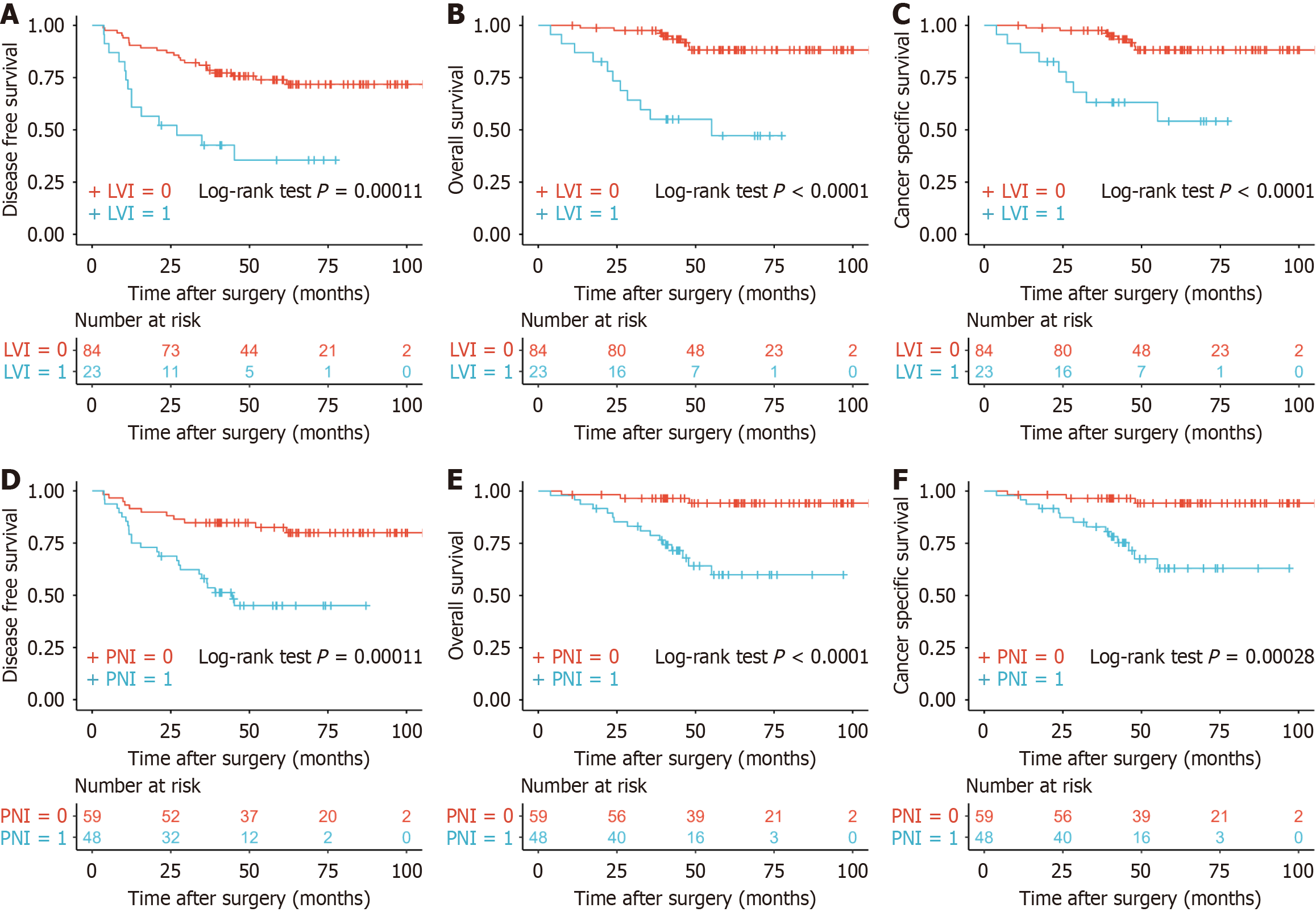
Figure 1 Groups divided according to lymphovascular invasion or perineural invasion had different prognosis.
A: Lymphovascular invasion (LVI) predicting disease-free survival (DFS) (P = 0.00011); B: LVI predicting overall survival (OS) (P < 0.0001); C: LVI predicting cancer-specific survival (CSS) (P < 0.0001); D: Perineural invasion (PNI) predicting DFS (P = 0.00011); E: PNI predicting OS (P < 0.0001); F: PNI predicting CSS (P = 0.00028). LVI: Lymphovascular invasion; PNI: Perineural invasion.
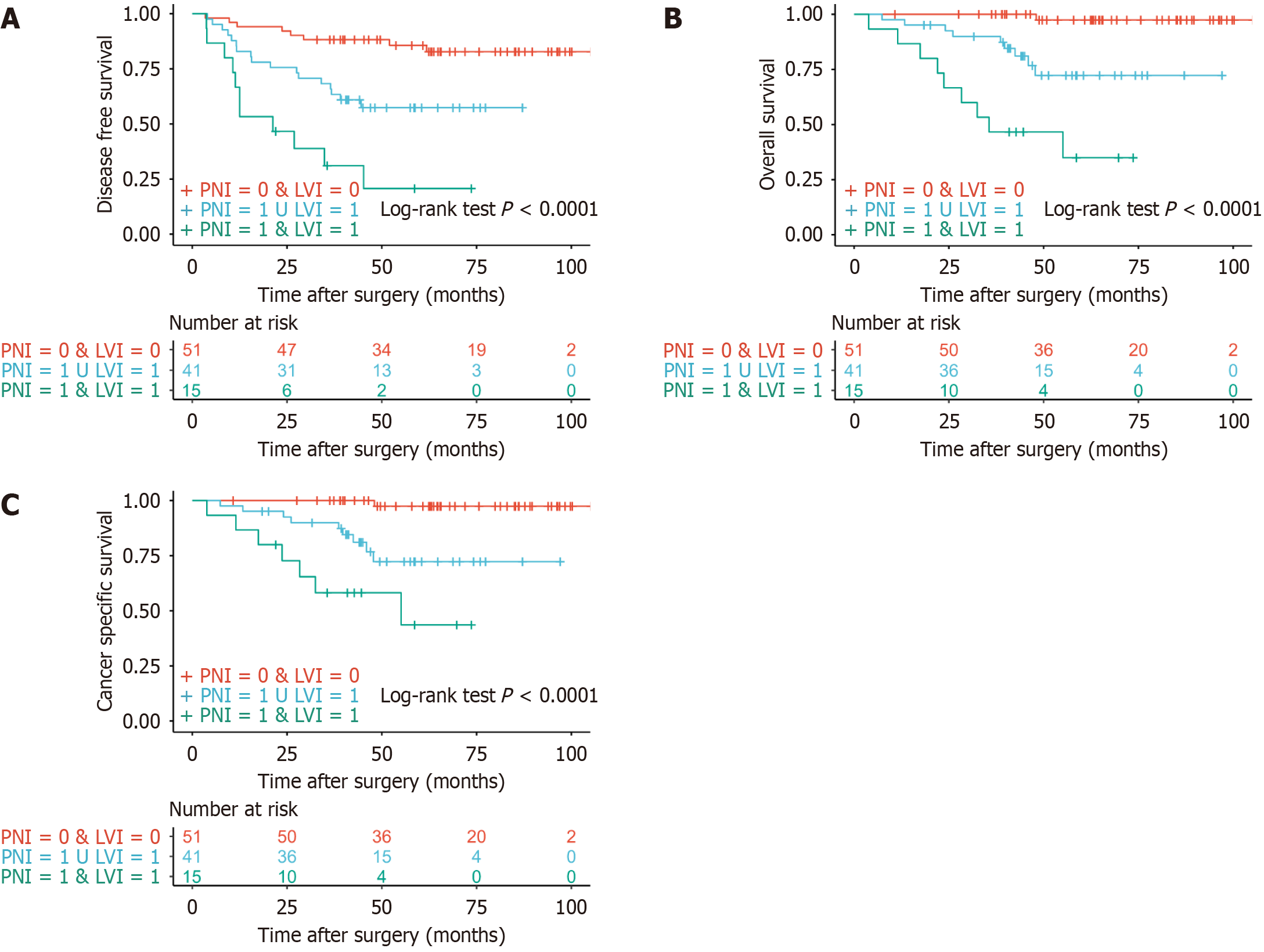
Figure 2 Three groups divided according to presence of lymphovascular invasion and perineural invasion had different prognosis.
A: Predicting disease-free survival (P < 0.0001); B: Predicting overall survival (P < 0.0001); C: Predicting cancer-specific survival (P < 0.0001). LVI: Lymphovascular invasion; PNI: Perineural invasion.
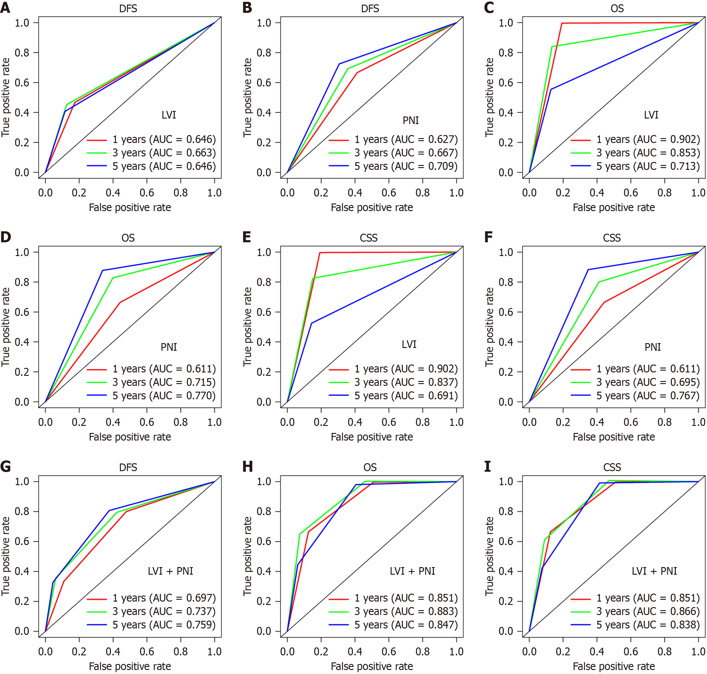
Figure 3 Receiver operating characteristic curve on the predictive prognosis.
A: Lymphovascular invasion (LVI) predicting disease-free survival (DFS); B: Perineural invasion (PNI) predicting DFS; C: LVI predicting overall survival (OS); D: PNI predicting OS; E: LVI predicting cancer-specific survival (CSS); F: PNI predicting CSS; G: Three groups predicting DFS; H: Three groups predicting OS; I: Three groups predicting CSS. LVI: Lymphovascular invasion; DFS: Disease-free survival; OS: Overall survival; CSS: Cancer-specific survival; AUC: Area under the receiver operating characteristic; PNI: Perineural invasion.
- Citation: Sun ZG, Chen SX, Sun BL, Zhang DK, Sun HL, Chen H, Hu YW, Zhang TY, Han ZH, Wu WX, Hou ZY, Yao L, Jie JZ. Important role of lymphovascular and perineural invasion in prognosis of colorectal cancer patients with N1c disease. World J Gastroenterol 2025; 31(5): 102210
- URL: https://www.wjgnet.com/1007-9327/full/v31/i5/102210.htm
- DOI: https://dx.doi.org/10.3748/wjg.v31.i5.102210


