Copyright
©The Author(s) 2025.
World J Gastroenterol. Apr 28, 2025; 31(16): 105188
Published online Apr 28, 2025. doi: 10.3748/wjg.v31.i16.105188
Published online Apr 28, 2025. doi: 10.3748/wjg.v31.i16.105188
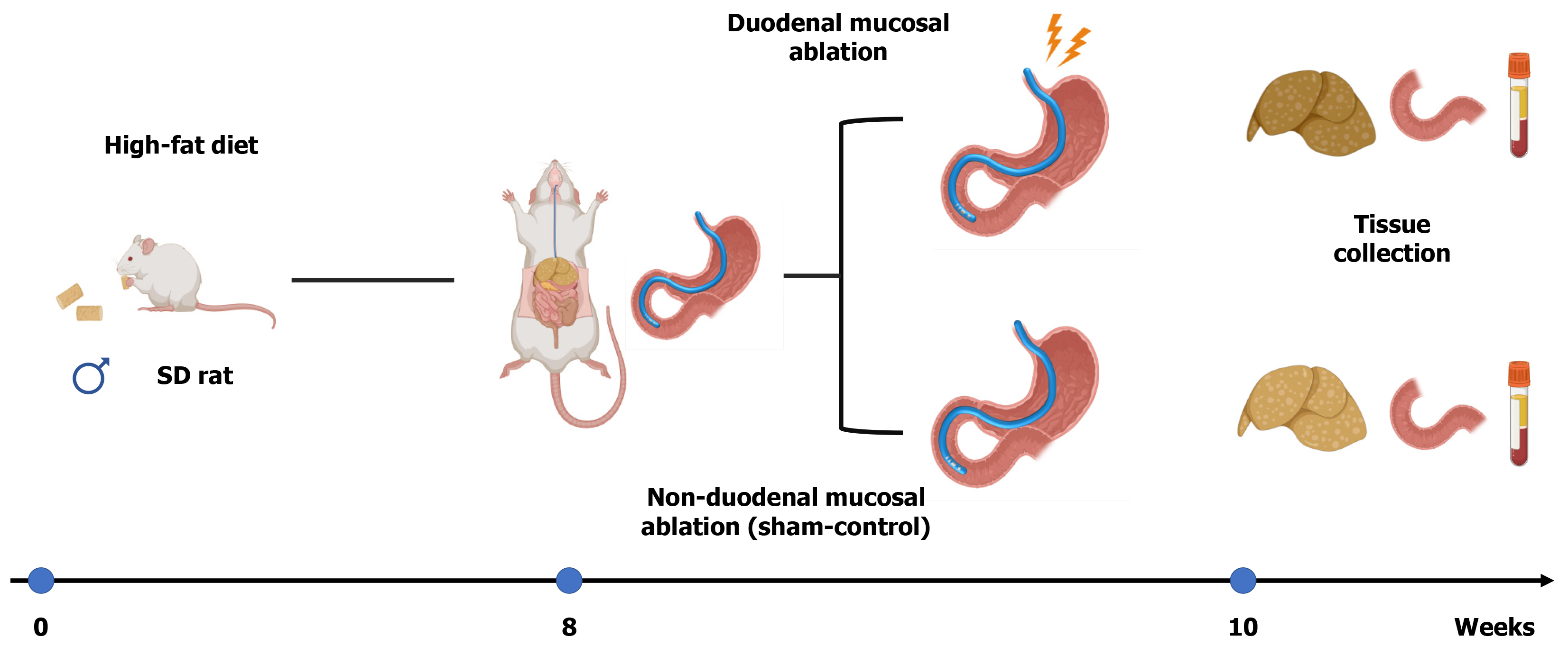
Figure 1
Experimental design scheme.
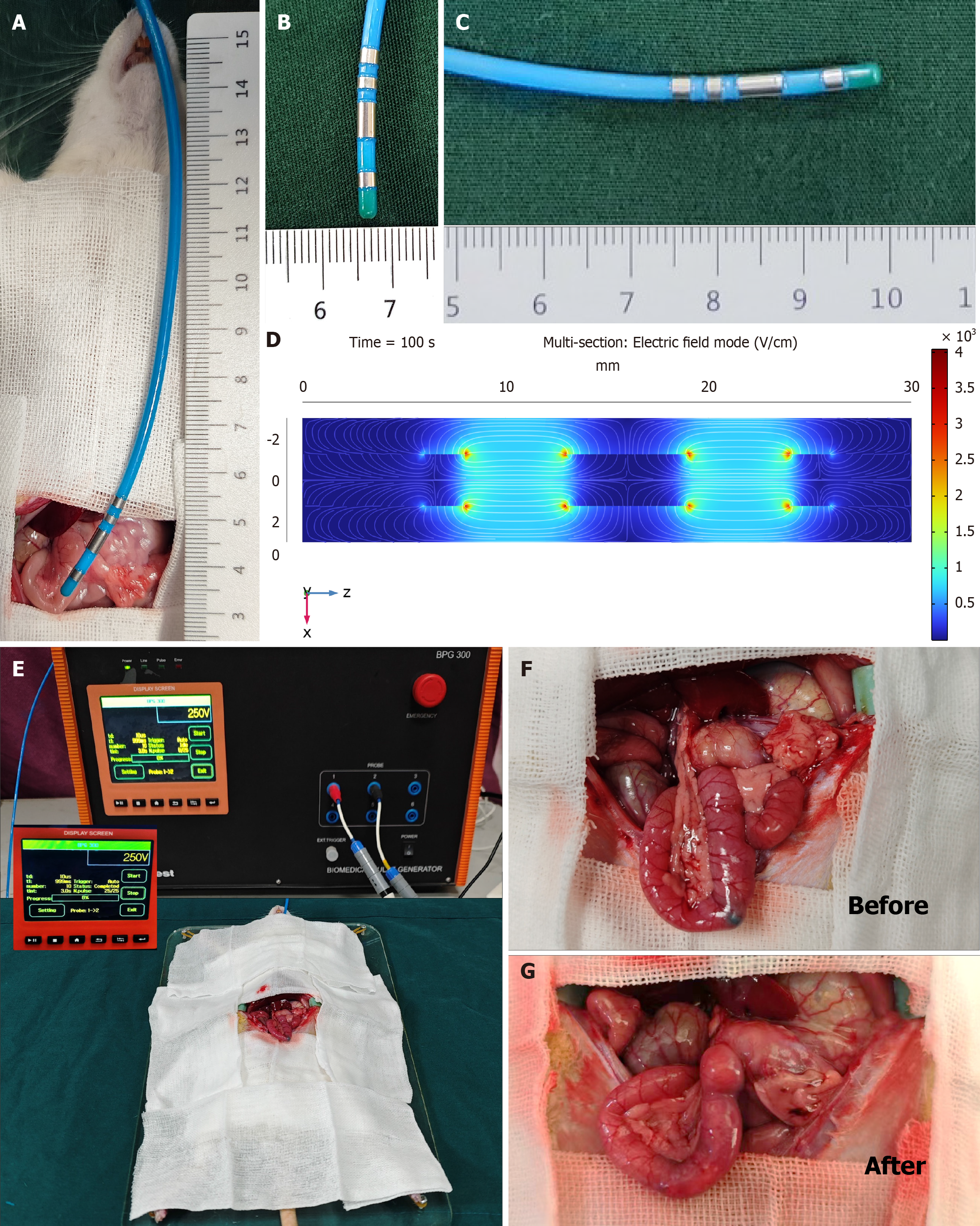
Figure 2 Operation of duodenal mucosal ablation using irreversible electroporation in non-alcoholic fatty liver disease rats.
A-C: Ablation catheter; D: Finite element analysis of electrode electric field distribution during operation. Duodenal mucosal ablation was performed using electroporation induced by a 250 V/cm electric field strength (pulse duration: 100 μs, pulse number: 90, and frequency: 1 Hz); E: Rats receiving duodenal mucosal ablation using irreversible electroporation; F and G: Macroscopic observation of the duodenum preoperatively and postoperatively.
Figure 3 Non-alcoholic fatty liver disease rat characteristics between the two groups.
A: Body weight; B: Food intake; C: Coefficient of the liver. Data are expressed as the mean ± SD (n = 6). aP < 0.05 vs sham-control group. DMA: Duodenal mucosal ablation; COL: Coefficient of the liver.
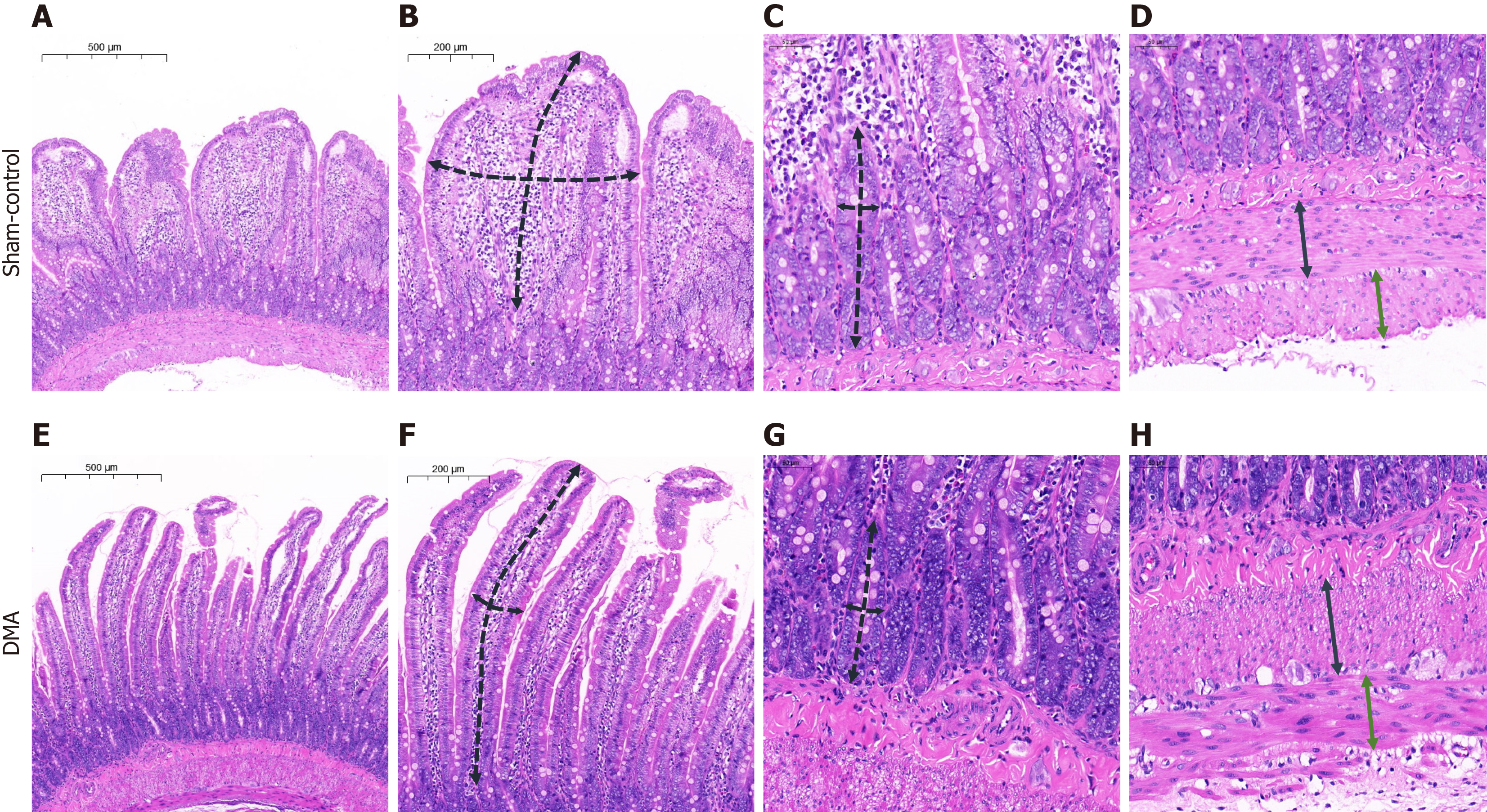
Figure 4 Histological examination of the duodenal tissue.
A: Complete layer of the duodenal wall, sham-control group; B: Measurement scheme of the length (a long dashed black line) and thickness (a short dashed black line) of the villus, sham-control group; C: Measurement scheme of the depth (a long dashed black line) and thickness (a short dashed black line) of the crypt, sham-control group; D: Measurement scheme of the longitudinal (a continuous black line) and circular (a continuous green line) lamina, sham-control group; E: Complete layer of the duodenal wall, duodenal mucosal ablation group; F: Measurement scheme of the length (a long dashed black line) and thickness (a short dashed black line) of the villus, duodenal mucosal ablation group; G: Measurement scheme of the depth (a long dashed black line) and thickness (a short dashed black line) of the crypt, duodenal mucosal ablation group; H: Measurement scheme of the longitudinal (a continuous black line) and circular (a continuous green line) lamina, duodenal mucosal ablation group. DMA: Duodenal mucosal ablation.
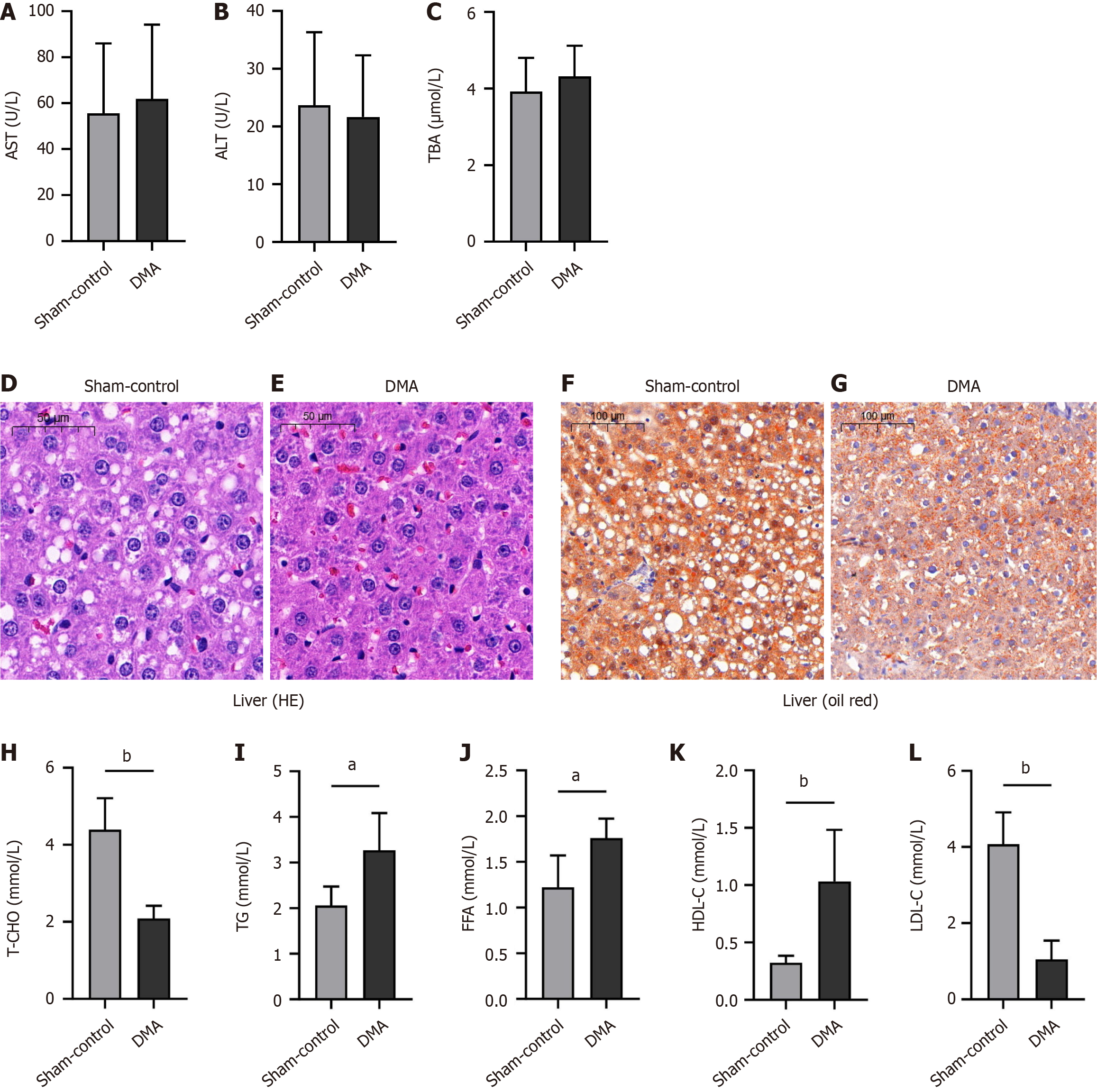
Figure 5 Liver function parameter test, histological examination, and serum lipid parameter test among different groups.
A: Serum aspartate aminotransferase levels; B: Alanine aminotransferase; C: Total bile acids; D-G: Histological examination of the liver tissue via hematoxylin and eosin staining and oil red O staining from the sham-control group and from the duodenal mucosal ablation group; H-L: Serum total cholesterol, triacylglycerol, free fatty acid, high-density lipoprotein cholesterol, and low-density lipoprotein cholesterol levels. Data are expressed as the mean ± SD (n = 6). aP < 0.05 vs sham-control group, bP < 0.01 vs sham-control group. DMA: Duodenal mucosal ablation; AST: Aspartate aminotransferase; ALT: alanine aminotransferase; TBA: Total bile acid; HE: Hematoxylin and eosin; T-CHO: Total cholesterol; TG: Triacylglycerol; FFA: Free fatty acid; HDL-C: High-density lipoprotein cholesterol; LDL-C: Low-density lipoprotein cholesterol.
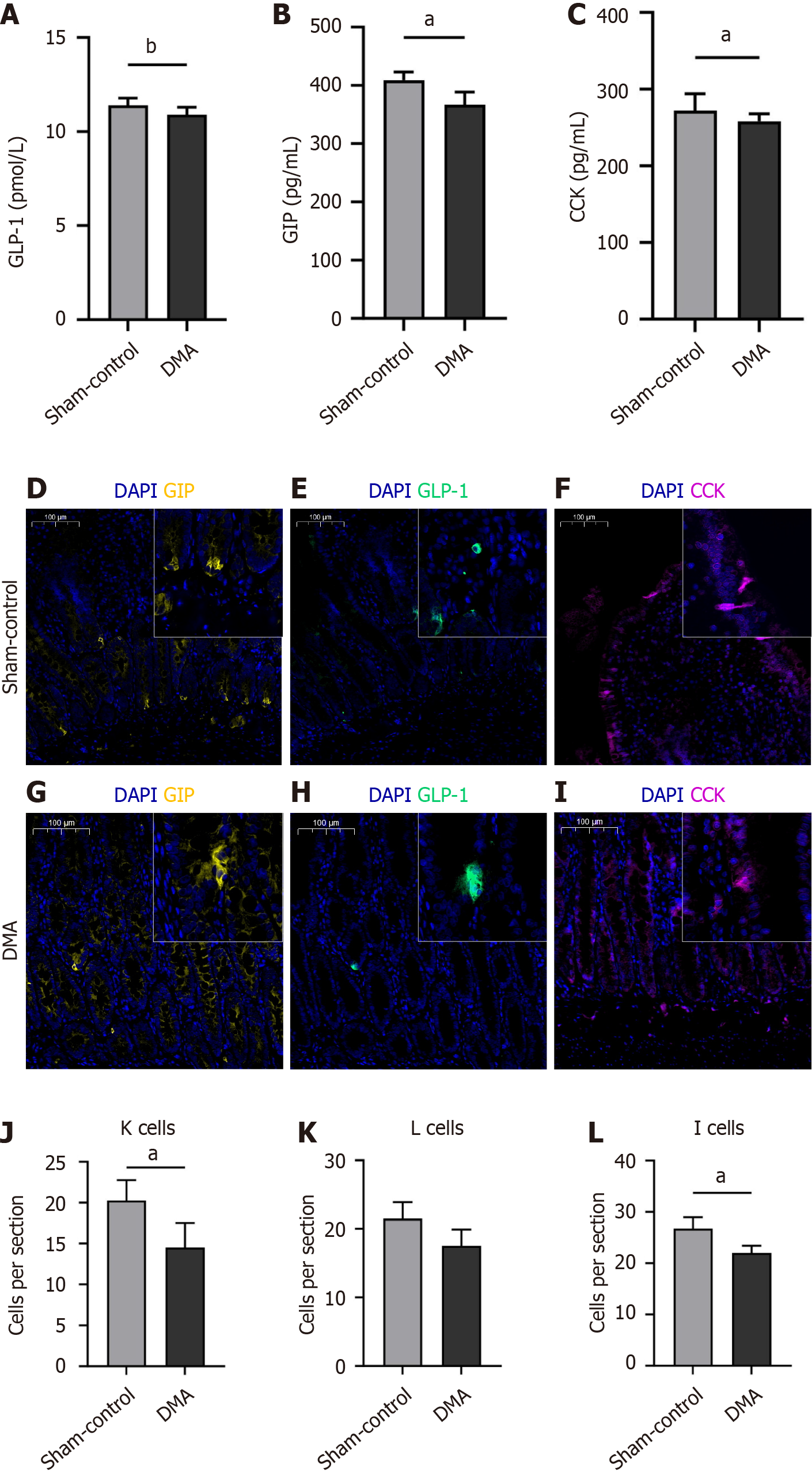
Figure 6 Enteroendocrine test and representative images of duodenal immunofluorescence staining.
A: Serum gastric inhibitory polypeptide levels; B: Glucagon-like peptide-1; C: Cholecystokinin; D-I: Immunofluorescence staining of the duodenum from sham-control and duodenal mucosal ablation groups; J-L: Number of gastric inhibitory polypeptide+ cells, glucagon-like peptide-1+, and cholecystokinin+ cells among different groups. aP < 0.05 vs sham-control group, bP < 0.01 vs sham-control group. DMA: Duodenal mucosal ablation; GIP: Gastric inhibitory polypeptide; GLP-1: Glucagon-like peptide-1; CCK: Cholecystokinin.
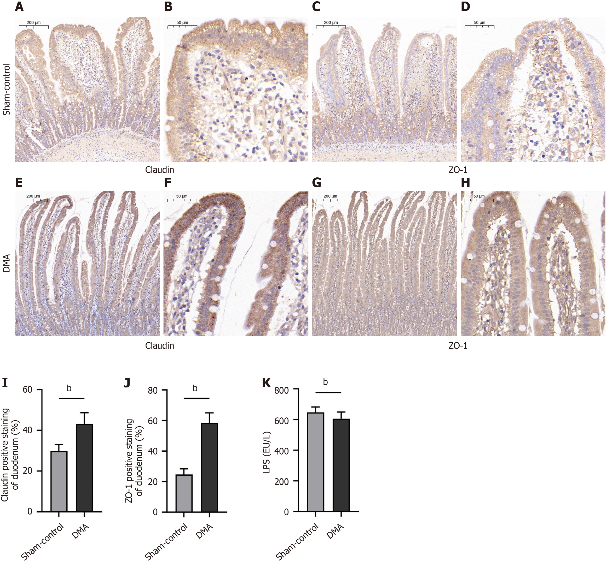
Figure 7 Immunohistochemistry staining of the duodenum among different groups.
A-H: Immunohistochemistry staining of the duodenum from the sham-control (A-D) and duodenal mucosal ablation (E-H) groups; I-K: Quantitative analysis of zonula occludens-1, claudin and lipopolysaccharide among different groups. bP < 0.01 vs sham-control group. ZO-1: Zonula occludens-1; DMA: Duodenal mucosal ablation; LPS: Lipopolysaccharide.
- Citation: Yu JW, Zhao Q, Li PX, Zhang YX, Gao BX, Xiang LB, Liu XY, Wang L, Sun YJ, Yang ZZ, Shi YJ, Chen YF, Yu MB, Zhang HK, Zhang L, Xu QH, Ren L, Li D, Lyu Y, Ren FG, Lu Q. Duodenal mucosal ablation with irreversible electroporation reduces liver lipids in rats with non-alcoholic fatty liver disease. World J Gastroenterol 2025; 31(16): 105188
- URL: https://www.wjgnet.com/1007-9327/full/v31/i16/105188.htm
- DOI: https://dx.doi.org/10.3748/wjg.v31.i16.105188


