Copyright
©The Author(s) 2025.
World J Gastroenterol. Mar 21, 2025; 31(11): 103449
Published online Mar 21, 2025. doi: 10.3748/wjg.v31.i11.103449
Published online Mar 21, 2025. doi: 10.3748/wjg.v31.i11.103449
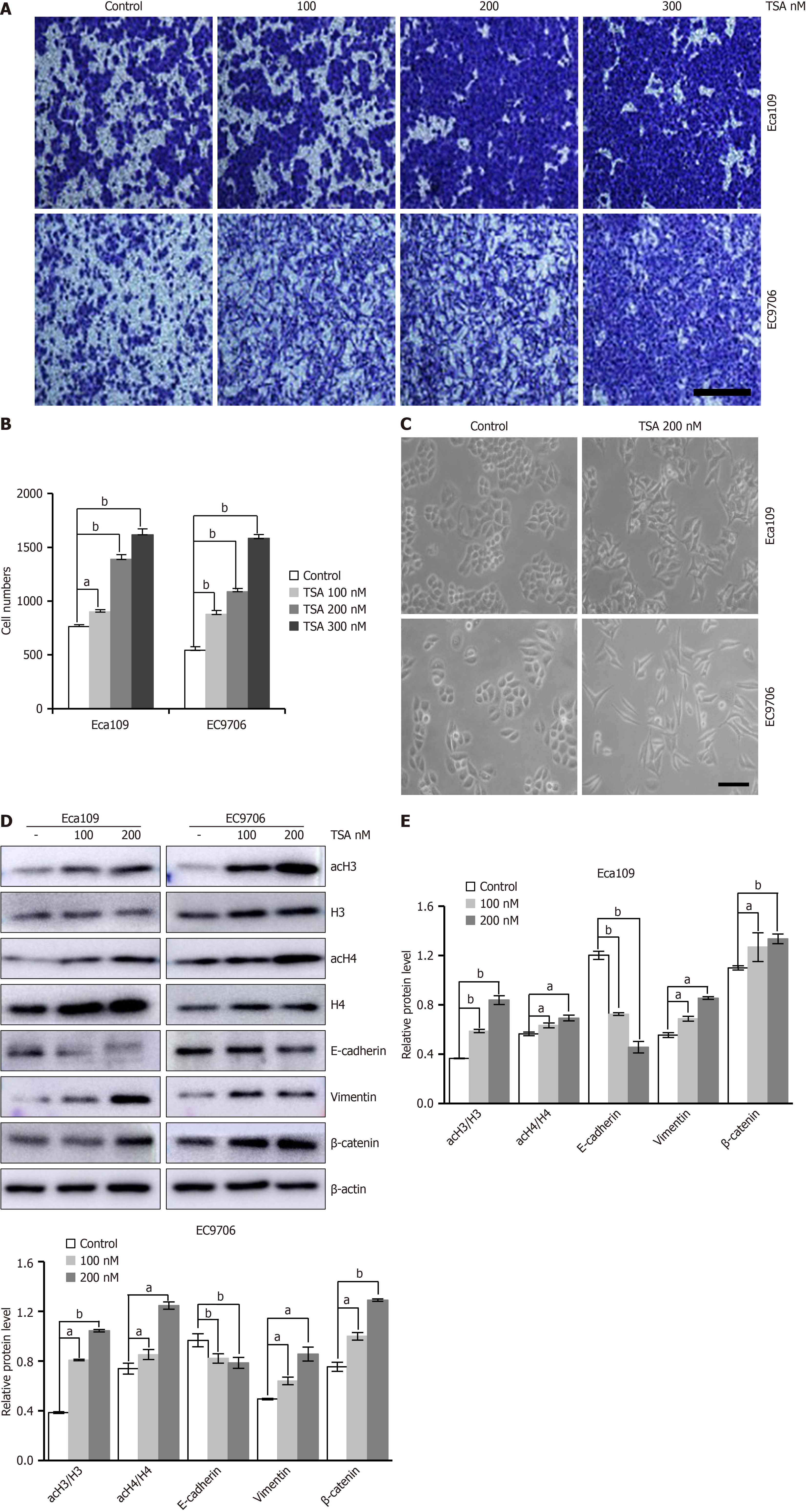
Figure 1 Trichostatin A promotes migration of esophageal squamous cell carcinoma cells by promoting epithelial-mesenchymal transition.
A: Representative images of the migration of Eca109 and EC9706 cells treated with histone deacetylase inhibitor trichostatin A (TSA) from Transwell assays; B: TSA enhanced cell migration in Transwell migration assay in dose-dependent manners; C: Changes in Eca109 and EC9706 cell morphology after TSA treatment; D and E: Western blot shows after TSA treatment that acetylated histones H3 and acetylated histones H4 levels increased and epithelial-mesenchymal transition marker E-cadherin level decreased, vimentin, β-catenin levels increased. Cell counts are for the corresponding assays of at least five random microscope fields (100 magnification). n = 5, P vs control. aP < 0.05. bP < 0.01. Panel A Bar is 50 μm, panel B Bar is 10 μm. TSA: Trichostatin A; acH3: Acetylated histones H3; acH4: Acetylated histones H4.
Figure 2 Trichostatin A promotes epithelial-mesenchymal transition of esophageal squamous cell carcinoma cells by promoting endoplasmic reticulum stress.
A: Western blot shows after trichostatin A (TSA) treatment, that endoplasmic reticulum (ER) stress marker glucose-regulated protein 78, C/EBP homologous protein and activating transcription factor 6 levels increased. These show that TSA can promote ER stress in esophageal cancer cells; B: Representative images of the migration of Eca109 and EC9706 cells treated with TSA combined with 4-phenylbutyric acid (4-PBA) from Transwell assays. The statistical results show that TSA enhanced cell migration reversed by 4-PBA in the Transwell migration assay; C: Western blot shows after TSA combined with 4-PBA, the epithelial-mesenchymal transition marker E-cadherin level increased, vimentin, β-catenin levels deceased. Cell counts are for the corresponding assays of at least five random microscope fields (100 magnification). n = 5, P vs control. aP < 0.05. bP < 0.01. Bar is 50 μm. TSA: Trichostatin A; 4-PBA: 4-phenylbutyric acid; GRP78: Glucose-regulated protein 78; CHOP: C/EBP homologous protein; ATF6: Activating transcription factor 6.
Figure 3 Trichostatin A promotes epithelial-mesenchymal transition of esophageal squamous cell carcinoma cells through cellular myelocytomatosis oncogene mediated endoplasmic reticulum stress.
A: Western blot shows after trichostatin A (TSA) treatment that cellular myelocytomatosis oncogene (c-Myc) level increased; B: Relative protein level of c-Myc in control cells and c-Myc-knockdown cells treated with TSA was examined by western blot; C: Representative images of the migration of Eca109 and EC9706 cells knockdown c-Myc protein and treated with TSA examined by Transwell assays; D: Western blot shows after TSA combined with sic-Myc that endoplasmic reticulum stress and epithelial-mesenchymal transition marker E-cadherin level increased, glucose-regulated protein 78, vimentin, β-catenin level decreased. Cell counts are for the corresponding assays of at least five random microscope fields (100 magnification). n = 5, P vs control. aP < 0.05. bP < 0.01. Bar is 50 μm. TSA: Trichostatin A; GRP78: Glucose-regulated protein 78; c-Myc: Cellular myelocytomatosis oncogene.
Figure 4 Trichostatin A promotes epithelial-mesenchymal transition of esophageal squamous cell carcinoma cells through cellular myelocytomatosis oncogene mediated endoplasmic reticulum stress may be associated with bromodomain-containing protein.
A: Western blot shows after trichostatin A (TSA) treatment that bromodomain-containing protein (BRD4) level increased; B: Relative protein level of BRD4 in control cells and BRD4-knockdown cells treated with TSA was examined by western blot; C: Representative images of the migration of Eca109 and EC9706 cells treated with TSA combined with carboxylic acid from Transwell assays; D: Representative images of the migration of Eca109 and EC9706 cells treated with TSA combined with knocking down BRD4 from Transwell assays; E: Western blot shows TSA treated of knock down BRD4 esophageal squamous cell carcinoma cells that epithelial-mesenchymal transition marker E-cadherin level increased, vimentin, β-catenin level decreased. Cell counts are for the corresponding assays of at least five random microscope fields (100 magnification). n = 5, P vs control. aP < 0.05. bP < 0.01. Bar is 50 μm. TSA: Trichostatin A; JQ-1: Carboxylic acid; GRP78: Glucose-regulated protein 78; BRD4: Bromodomain-containing protein.
Figure 5 Trichostatin A promotes epithelial-mesenchymal transition of esophageal squamous cell carcinoma cells through bromodomain-containing protein/cellular myelocytomatosis oncogene/endoplasmic reticulum-stress pathway is reversed by inhibitor of trichostatin A 1.
A: Representative images of the migration of Eca109 and EC9706 cells treated with trichostatin A (TSA) combined with inhibitor of trichostatin A 1 (ITSA1) from Transwell assays; B: Western blot shows after TSA combined with ITSA1 that acetylated histones H3, acetylated histones H4, cellular myelocytomatosis oncogene, glucose-regulated protein 78 levels decreased, and epithelial-mesenchymal transition marker E-cadherin levels increased, vimentin, β-catenin levels decreased. Cell counts are for the corresponding assays of at least five random microscope fields (100 magnification). aP < 0.05. bP < 0.01. Bar is 50 μm. TSA: Trichostatin A; ITSA1: Inhibitor of trichostatin A 1; GRP78: Glucose-regulated protein 78; acH3: Acetylated histones H3; c-Myc: Cellular myelocytomatosis oncogene.
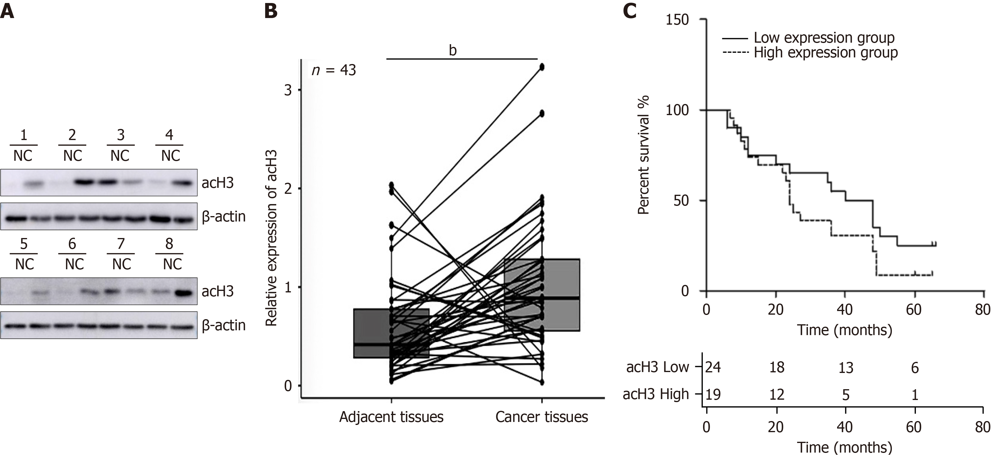
Figure 6 Expression of acetylated histones H3 is higher in esophageal squamous cell carcinoma than in paracancerous tissues, and acH3 was an independent factor affecting prognosis.
A: Representative detection of acetylated histones H3 (acH3) level in esophageal squamous cell carcinoma (ESCC) by western blot, N means adjacent tissues and C means cancer tissues; B: Statistical analysis of 43 cases of ESCC showed that the level of acH3 in ESCC tumor tissue was 1.66 times higher than that in adjacent tissue; C: Kaplan-Meier survival curve of the relationship between acH3 expression and prognosis in patients with esophageal carcinoma. P vs control. bP < 0.01. NC: Negative control; acH3: Acetylated histones H3.
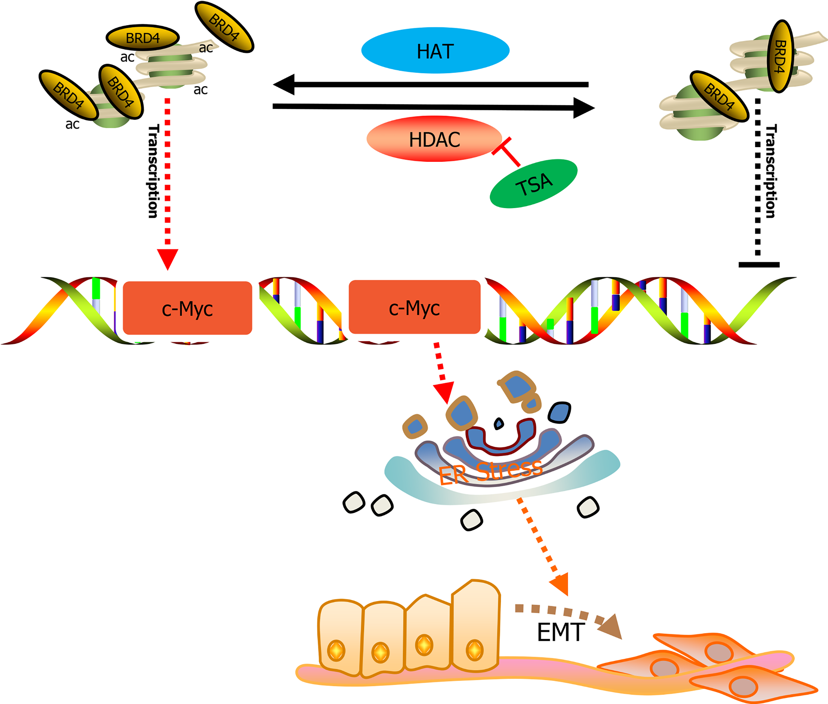
Figure 7 A proposed model illustrating the mechanism by which trichostatin A promotes esophageal squamous cell carcinoma cell epithelial-mesenchymal transition and migration.
ac: Acetylation; TSA: Trichostatin A; HAT: Histone lysine acetyltransferase; HDAC: Histone deacetylases; ER: Endoplasmic reticulum; EMT: Epithelial-mesenchymal transition; c-Myc: Cellular myelocytomatosis oncogene.
- Citation: Chen YM, Yang WQ, Fan YY, Chen Z, Liu YZ, Zhao BS. Trichostatin A augments cell migration and epithelial-mesenchymal transition in esophageal squamous cell carcinoma through BRD4/c-Myc endoplasmic reticulum-stress pathway. World J Gastroenterol 2025; 31(11): 103449
- URL: https://www.wjgnet.com/1007-9327/full/v31/i11/103449.htm
- DOI: https://dx.doi.org/10.3748/wjg.v31.i11.103449


