Copyright
©The Author(s) 2025.
World J Gastroenterol. Mar 14, 2025; 31(10): 100444
Published online Mar 14, 2025. doi: 10.3748/wjg.v31.i10.100444
Published online Mar 14, 2025. doi: 10.3748/wjg.v31.i10.100444
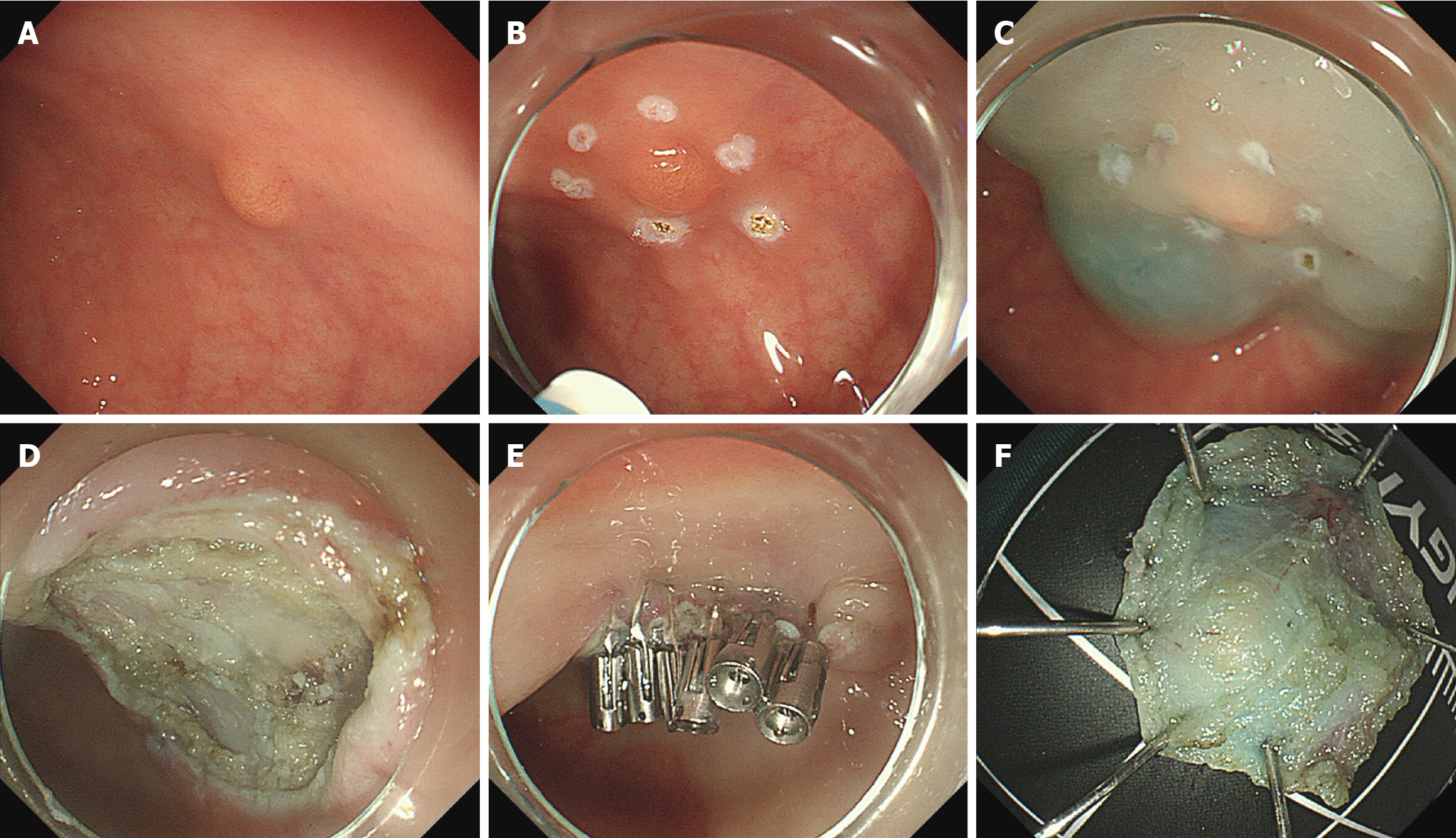
Figure 1 Endoscopic submucosal dissection.
A: A pale yellow mass with a diameter of about 4.8 mm in the rectum; B: Electrocoagulation marking in the peritumoral area by using the front end of the electrosurgical snare; C: Submucosal injection of the mass with an injection needle; D: Wound surface after the removal of the mass; E: Wound clipping with Sureclips after mass resection; F: The resected mass for pathological evaluation.
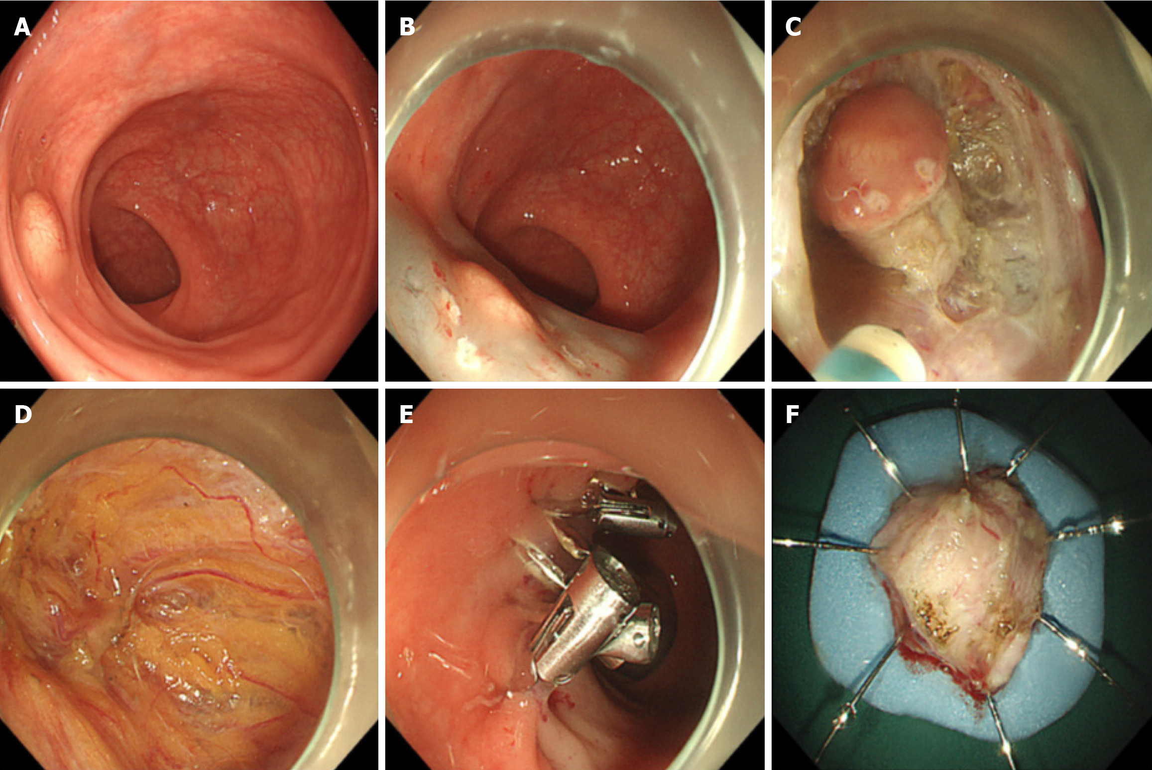
Figure 2 Endoscopic full thickness resection.
A: A pale yellow mass with a diameter of about 8 mm in the rectum; B: Electrocoagulation marking in the peritumoral area by using the front end of the electrosurgical knife and submucosal injection of the mass with an injection needle; C: Full thickness resection of the mass with electrosurgical knife; D: Wound surface after the removal of the mass; E: Wound clipping with Sureclips after mass resection; F: The resected mass for pathological evaluation.
Figure 3 Infiltration depth measurement diagram.
EFTR: Endoscopic full-thickness resection; ESD: Endoscopic submucosal dissection.
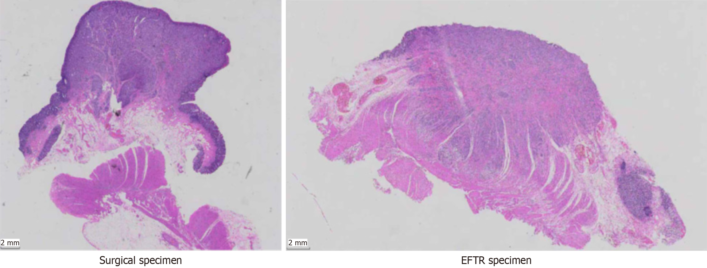
Figure 4 Pathological images of surgical and endoscopic full-thickness resection specimens.
EFTR: Endoscopic full-thickness resection.
- Citation: Zhang XL, Jiang YY, Chang YY, Sun YL, Zhou Y, Wang YH, Dou XT, Guo HM, Ling TS. Endoscopic full-thickness resection: A definitive solution for local complete resection of small rectal neuroendocrine neoplasms. World J Gastroenterol 2025; 31(10): 100444
- URL: https://www.wjgnet.com/1007-9327/full/v31/i10/100444.htm
- DOI: https://dx.doi.org/10.3748/wjg.v31.i10.100444


