Copyright
©The Author(s) 2025.
World J Gastroenterol. Jan 7, 2025; 31(1): 98479
Published online Jan 7, 2025. doi: 10.3748/wjg.v31.i1.98479
Published online Jan 7, 2025. doi: 10.3748/wjg.v31.i1.98479
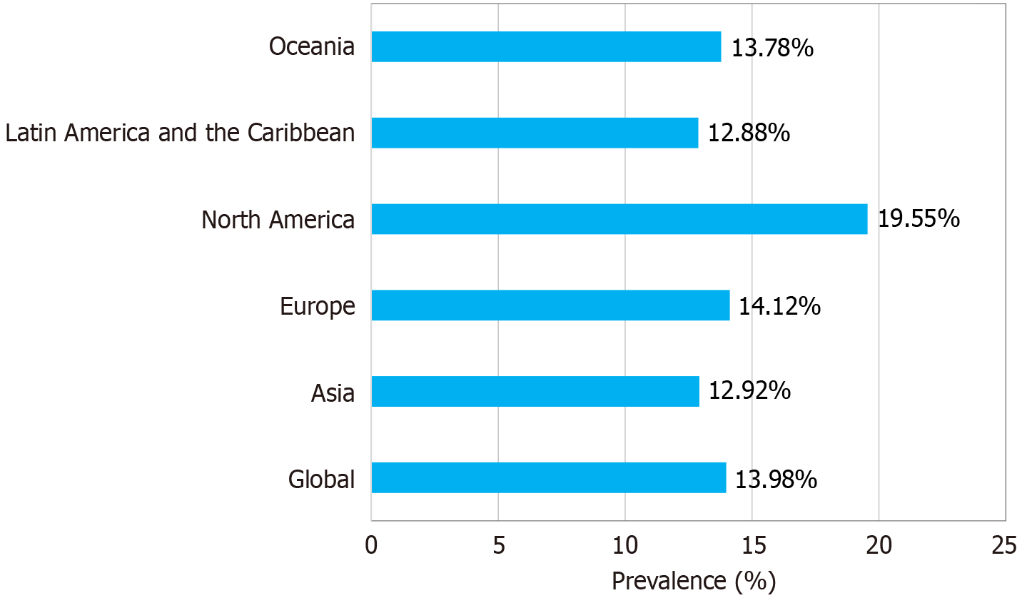
Figure 1
Pooled prevalence of gastroesophageal reflux disease according to geographical location.
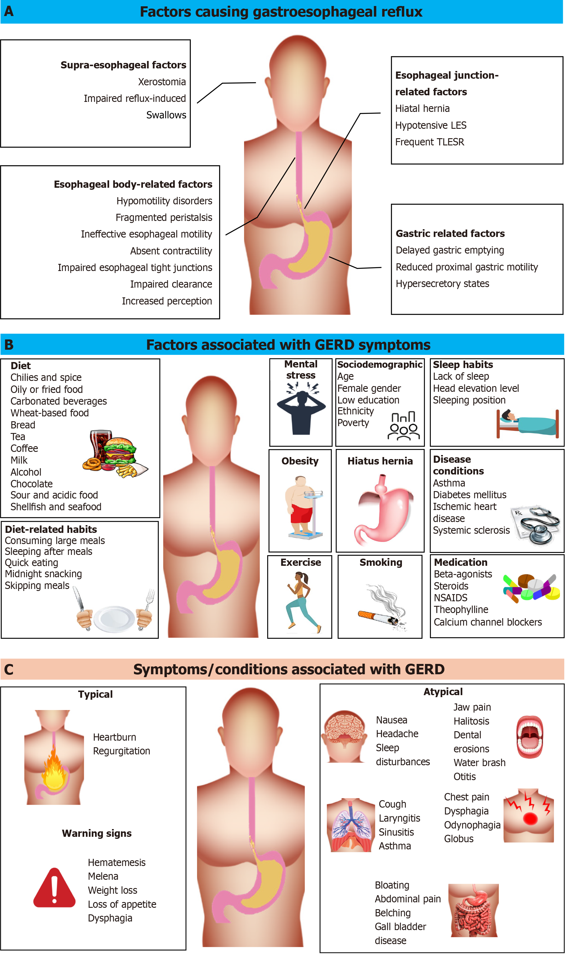
Figure 2 Gastroesophageal reflux disease.
A: Factors causing gastroesophageal reflux disease (GERD); B: Factors associated with GERD symptoms; C: Symptoms/conditions associated with GERD. LES: Lower esophageal sphincter; NSAIDS: Nonsteroidal anti-inflammatory drugs; TLESR: Transient lower esophageal sphincter relaxation.
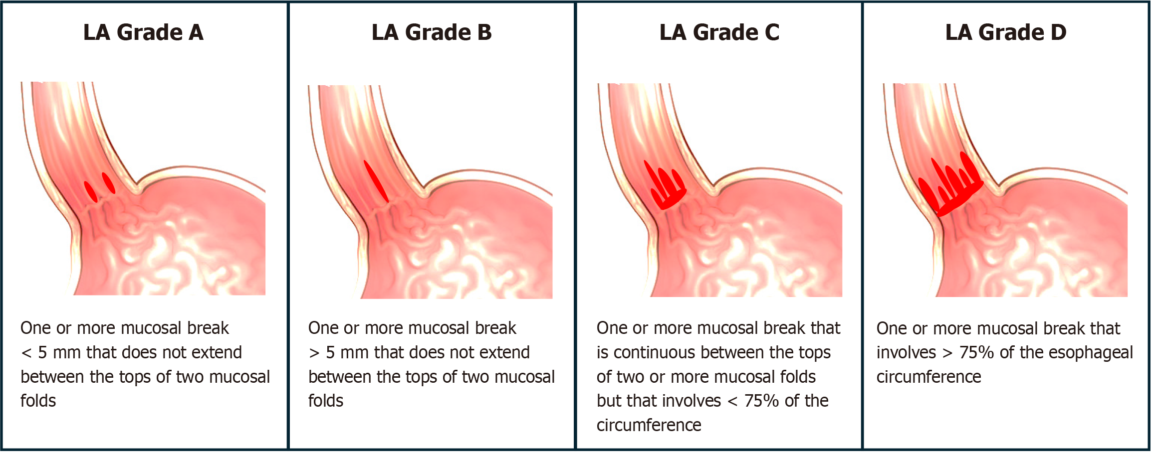
Figure 3 Los Angeles classification of esophagitis.
Grades C and D are diagnostic of gastroesophageal reflux disease. LA: Los Angeles.
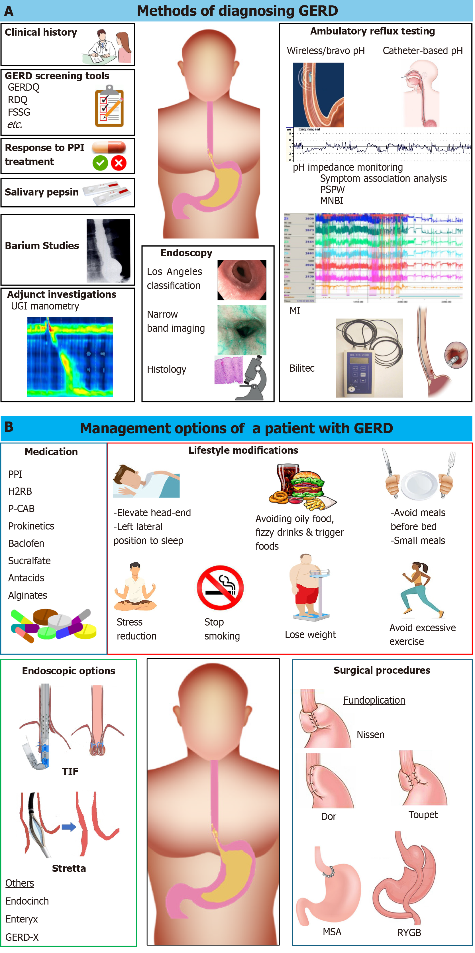
Figure 4 Diagnosis and management of gastroesophageal reflux.
A: Investigations used in gastroesophageal reflux disease (GERD) diagnosis; B: Options available for the management of GERD. FSSG: Frequency scale for the symptoms of gastroesophageal reflux disease; GERDQ: Gastroesophageal reflux disease questionnaire; GERD-X: Endoscopic full thickness plication; H2RB: Histamine 2 receptor blocker; MI: Mucosal impedance; MNBI: Mean nocturnal baseline impedance; MSA: Magnetic sphincter augmentation; P-CAB: Potassium-competitive acid blocker; PPI: Proton pump inhibitor; PSPW: Post-reflux swallow-induced peristaltic wave; RDQ: Reflux disease questionnaire; RYGB: Roux-en-Y gastric bypass; TIF: Transoral incisionless fundoplication; UGI: Upper gastrointestinal.
- Citation: Wickramasinghe N, Devanarayana NM. Unveiling the intricacies: Insight into gastroesophageal reflux disease. World J Gastroenterol 2025; 31(1): 98479
- URL: https://www.wjgnet.com/1007-9327/full/v31/i1/98479.htm
- DOI: https://dx.doi.org/10.3748/wjg.v31.i1.98479


