Copyright
©The Author(s) 2024.
World J Gastroenterol. Feb 21, 2024; 30(7): 728-741
Published online Feb 21, 2024. doi: 10.3748/wjg.v30.i7.728
Published online Feb 21, 2024. doi: 10.3748/wjg.v30.i7.728
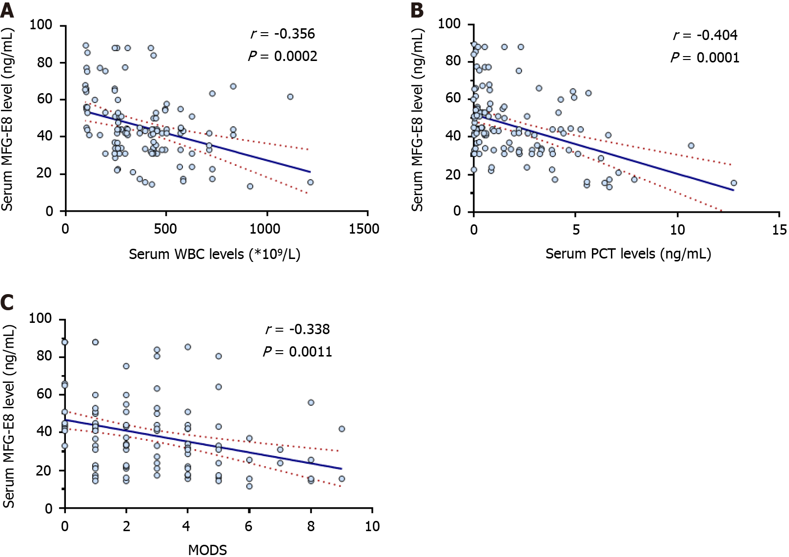
Figure 1 Serum milk fat globule epidermal growth factor 8 concentration was negatively correlated with the inflammatory severity in acute pancreatitis patients.
Blood samples from 134 acute pancreatitis patients were collected, and serum milk fat globule epidermal growth factor 8 (MFG-E8) levels were measured. A: Correlation analysis of serum MFG-E8 levels and serum white blood cell levels; B: Correlation analysis of serum MFG-E8 levels and serum procalcitonin levels; C: Correlation analysis of serum MFG-E8 levels and multiple organ failure syndrome scores. WBC: White blood cells; PCT: Procalcitonin; MODS: Multiple organ failure syndrome; MFG-E8: Milk fat globule epidermal growth factor 8.
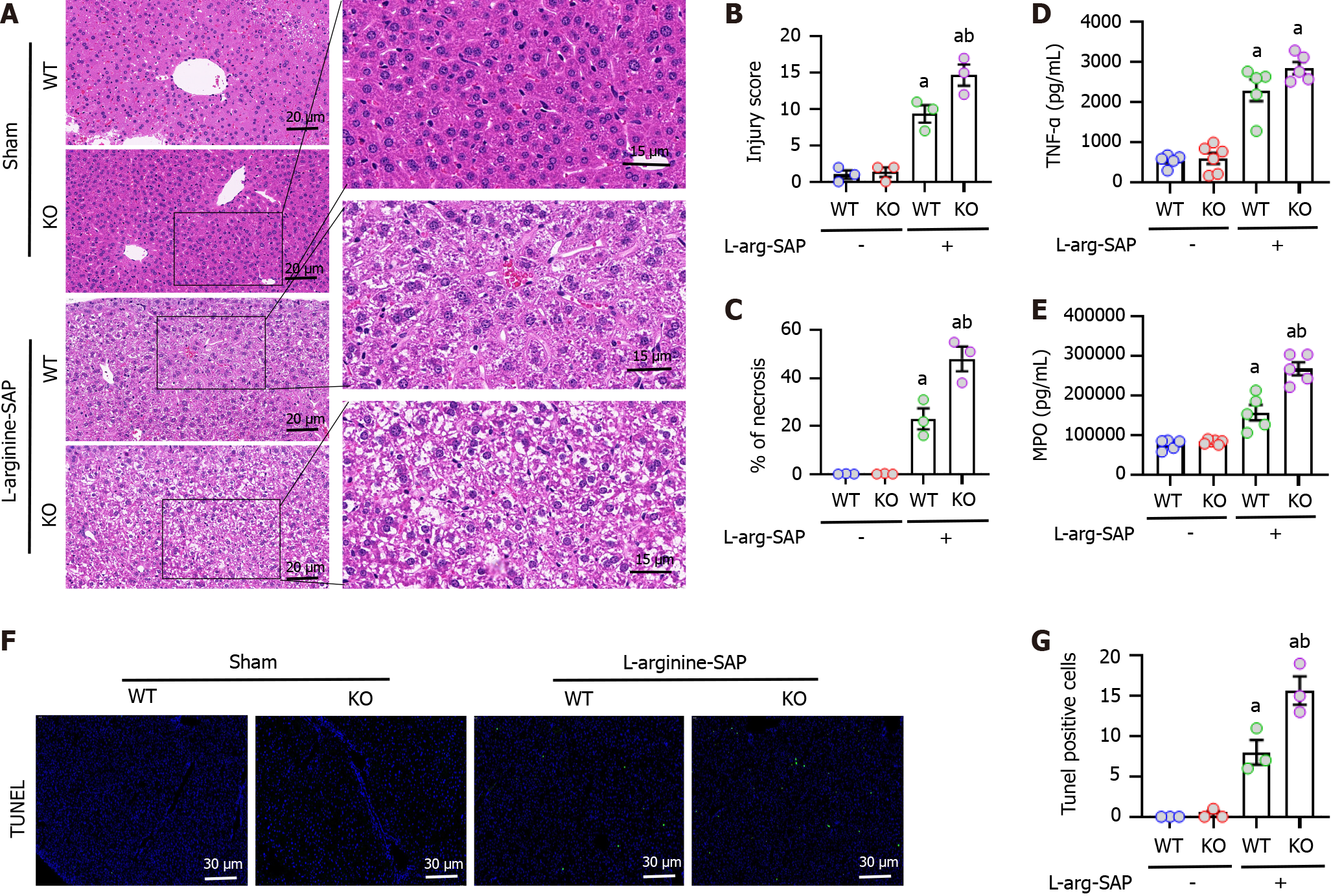
Figure 2 Milk fat globule epidermal growth factor 8 deficiency aggravated hepatic injury and inflammation in experimental severe acute pancreatitis.
In mice, acute pancreatitis (AP) was induced by 2 hly intraperitoneal injections of 4.0 g/kg L-arginine. The animals were sacrificed at 72 h after the first injection of L-arginine. Blood and tissue samples were collected. A: Representative photos of hematoxylin and eosin staining in the liver (400 × or 1000 ×); B: Hepatic injury scores; C: Percentages of necrotic areas; D: Tumor necrosis factor-α levels in the liver; E: Myeloperoxidase levels in the liver; F and G: Representative photos of TdT-mediated dUTP Nick-End Labeling (TUNEL) staining (400 ×) and quantitative of TUNEL staining. n = 3-6/group, error bars indicate the SEM; aP < 0.05 vs sham group; bP < 0.05 vs vehicle group. SAP: Severe acute pancreatitis; KO: Knockout; WT: Wild type; TNF-α: Tumor necrosis factor-α; MPO: Myeloperoxidase; TUNEL: TdT-mediated dUTP Nick-End Labeling.
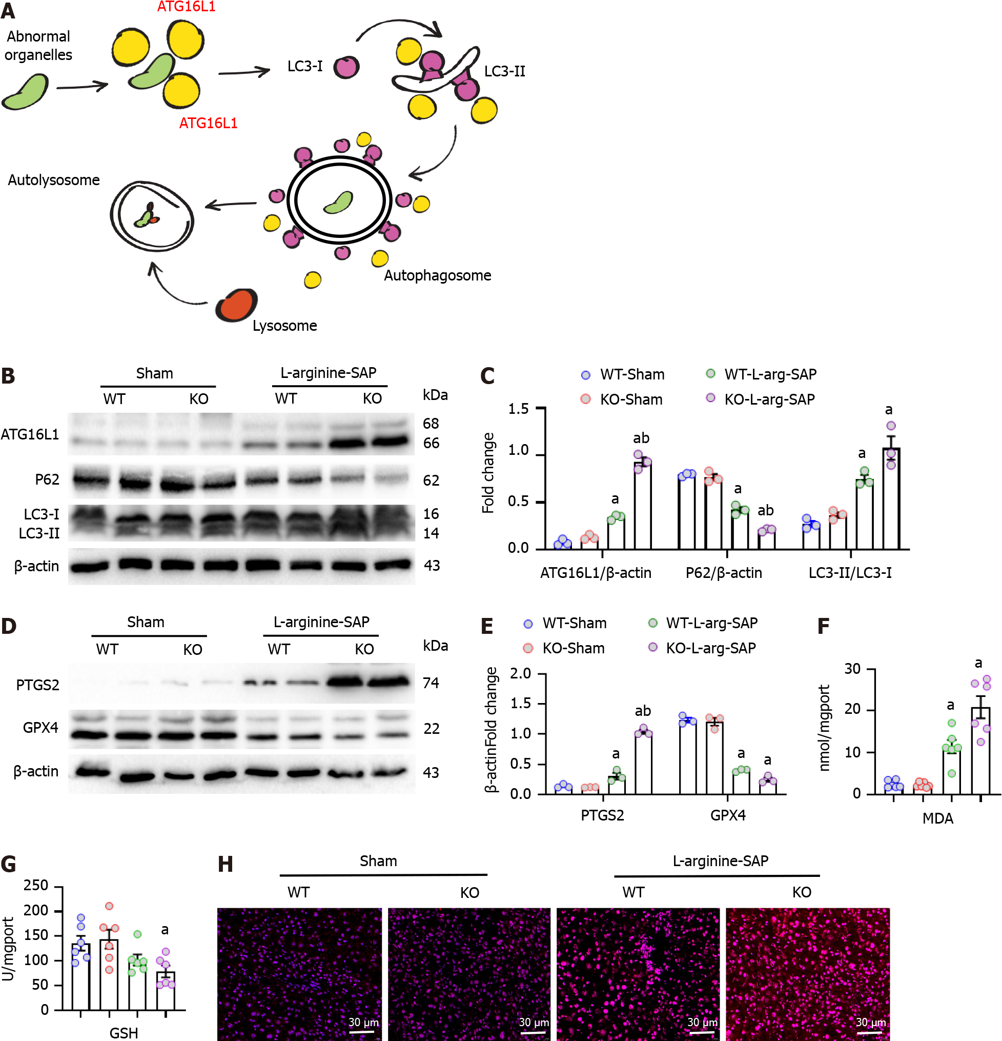
Figure 3 Milk fat globule epidermal growth factor 8 deficiency aggravated hepatic autophagy and ferroptosis in experimental severe acute pancreatitis.
In mice, severe acute pancreatitis was induced by 2 hly intraperitoneal injections of 4.0 g/kg L-arginine. The animals were sacrificed at 72 h after the first injection of L-arginine. Blood and tissue samples were collected. A: Autophagy related protein 16 like protein 1 (ATG16L1) assists LC3 in the formation of autophagosomes; B-E: Western blot analysis and quantitative of the expression of ATG16L1, P62, LC3, prostaglandin-endoperoxide synthase 2 and glutathione peroxidase 4 in the liver; F: Malondialdehyde levels in the liver; G: Glutathione levels in the liver; H: Representative images of DHE staining in the pancreas. n = 3-6/group, error bars indicate the SEM; aP < 0.05 vs sham group; bP < 0.05 vs vehicle group. SAP: Severe acute pancreatitis; KO: Knockout; WT: Wild type; ATG16L1: Autophagy related protein 16 like protein 1; PTGS2: Prostaglandin-endoperoxide synthase 2; GPX4: Glutathione peroxidase 4; MDA: Malondialdehyde; GSH: Glutathione.
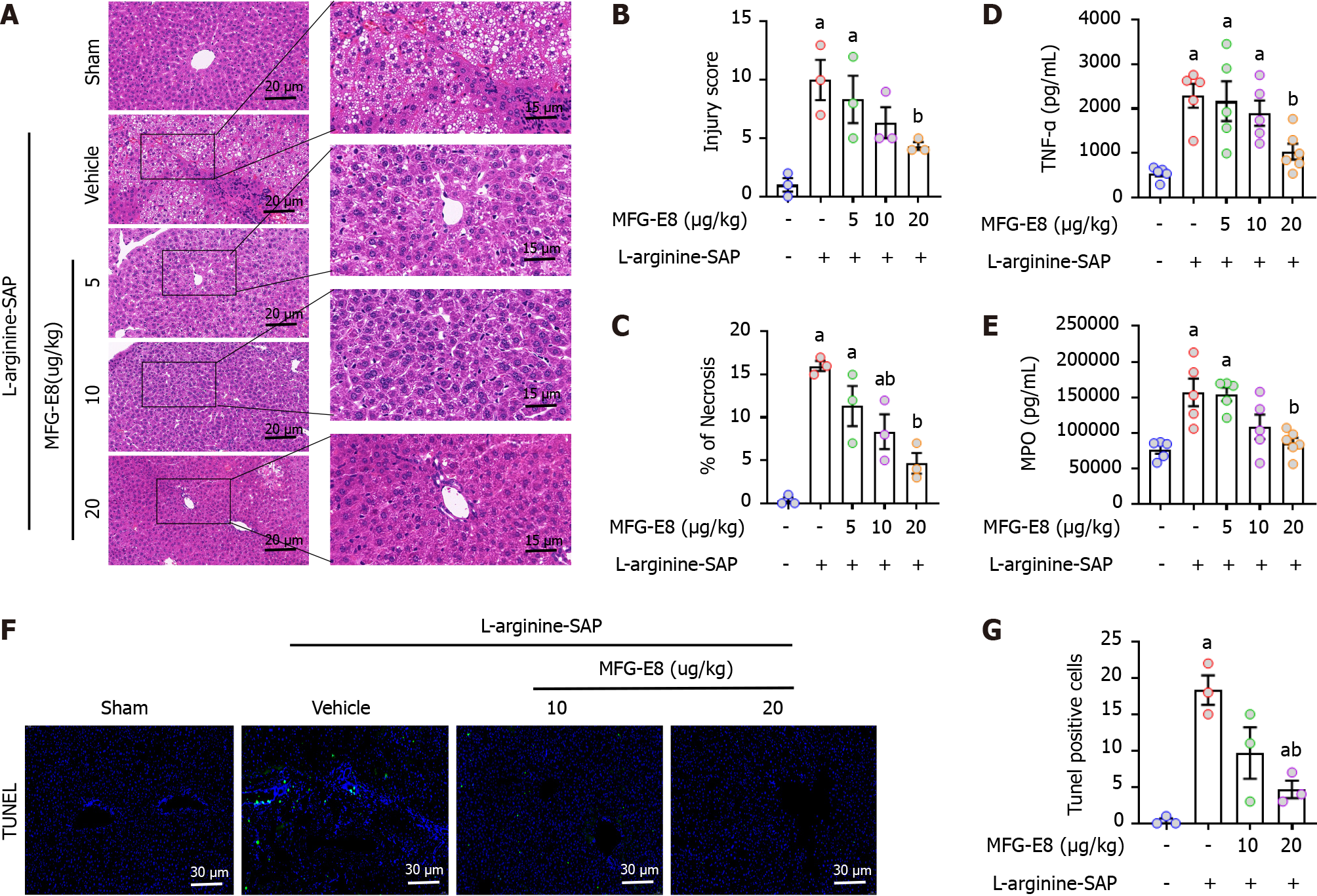
Figure 4 Milk fat globule epidermal growth factor 8 administration mitigates liver Injury in experimental severe acute pancreatitis.
In mice, severe acute pancreatitis was induced by 2 hly intraperitoneal injections of 4.0 g/kg L-arginine. At 2 h after the last injection of L-arginine, normal saline (vehicle) or 5, 10, or 20 μg/kg milk fat globule epidermal growth factor 8 (MFG-E8) was administered through intraperitoneal injection. The animals were sacrificed at 69 h after MFG-E8 treatment (i.e., 72 h after the first injection of L-arginine). Blood and tissue samples were collected. A: Representative photos of hematoxylin and eosin staining in the liver (400 × or 1000 ×); B: Hepatic injury scores; C: Percentages of necrotic areas; D: Tumor necrosis factor-α levels in the liver; E: Myeloperoxidase levels in the liver; F and G: Representative photos of TdT-mediated dUTP Nick-End Labeling (TUNEL) staining (400 ×) and quantitative of TUNEL staining. n = 3-6/group, error bars indicate the SEM; aP < 0.05 vs sham group; bP < 0.05 vs vehicle group. SAP: Severe acute pancreatitis; TNF-α: Tumor necrosis factor-α; MPO: Myeloperoxidase; TUNEL: TdT-mediated dUTP Nick-End Labeling; MFG-E8: Milk fat globule epidermal growth factor 8.
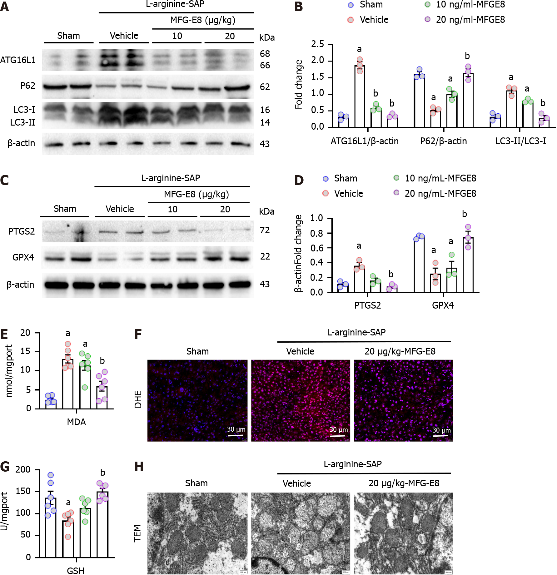
Figure 5 Milk fat globule epidermal growth factor 8 administration mitigates hepatic autophagy and ferroptosis in experimental severe acute pancreatitis.
In mice, arginine-severe acute pancreatitis was induced by 2 hly intraperitoneal injections of 4.0 g/kg L-arginine. At 2 h after the last injection of L-arginine, normal saline (vehicle) or 10, or 20 μg/kg milk fat globule epidermal growth factor 8 (MFG-E8) was administered through intraperitoneal injection. The animals were sacrificed at 69 h after MFG-E8 treatment (i.e., 72 h after the first injection of L-arginine). Blood and tissue samples were collected. A-D: Western blot analysis and quantitative of the expression of autophagy related protein 16 like protein 1, P62, LC3, prostaglandin-endoperoxide synthase 2 and glutathione peroxidase 4 in the liver; E: Malondialdehyde levels in the liver; F: Representative images of DHE staining in the pancreas; G: Glutathione levels in the liver; H: Ultrastructural changes of mitochondria in hepatocytes (electron microscopy). n = 3-6/group, error bars indicate the SEM; aP < 0.05 vs sham group; bP < 0.05 vs vehicle group. SAP: Severe acute pancreatitis; ATG16L1: Autophagy related protein 16 like protein 1; PTGS2: Prostaglandin-endoperoxide synthase 2; GPX4: Glutathione peroxidase 4; MDA: Malondialdehyde; GSH: Glutathione; MFG-E8: Milk fat globule epidermal growth factor 8.
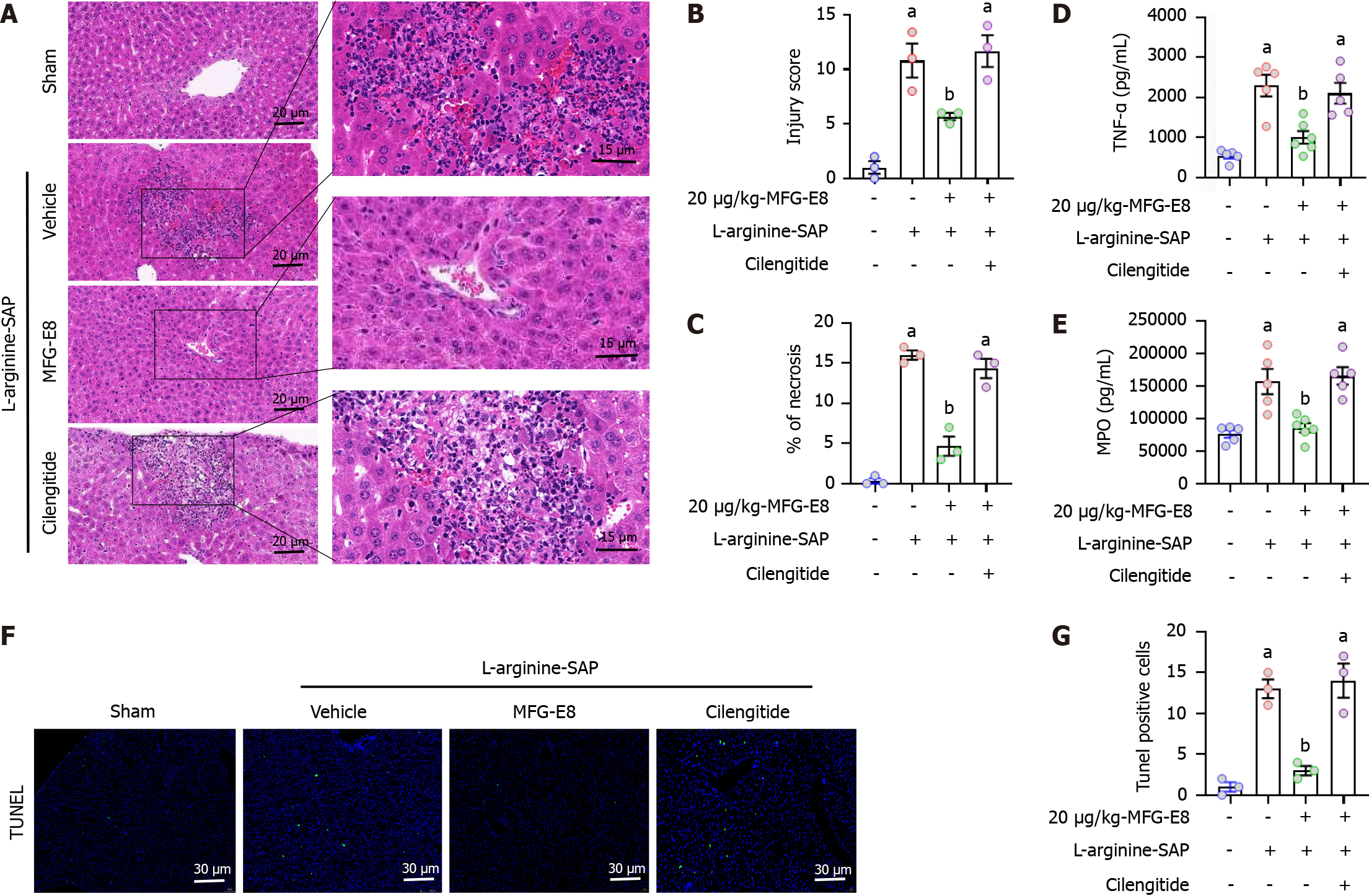
Figure 6 Milk fat globule epidermal growth factor 8 protects the liver from damage through the integrin αVβ3/5.
In mice, severe acute pancreatitis was induced by 2 hly intraperitoneal injections of 4.0 g/kg L-arginine. At 2 h after the last injection of L-arginine, normal saline (vehicle) or 20 μg/kg milk fat globule epidermal growth factor 8 (MFG-E8) was administered through intraperitoneal injection. To determine whether the protective effect of MFG-E8 in liver is mediated through integrin αVβ3/5, 20 mg/kg cilengitide, a specific integrin αVβ3/5 antagonist, was administered intraperitoneally at 1 h before the injection of MFG-E8 (i.e., 1 h after the last injection of L-arginine). The animals were sacrificed at 69 h after MFG-E8 treatment (i.e., 72 h after the first injection of L-arginine). Blood and tissue samples were collected. A: Representative photos of hematoxylin and eosin staining in the liver (400 × or 1000 ×); B: Hepatic injury scores; C: Percentages of necrotic areas; D: Tumor necrosis factor-α levels in the liver; E: Myeloperoxidase levels in the liver; F and G: Representative photos of TdT-mediated dUTP Nick-End Labeling (TUNEL) staining (400 ×) and quantitative of TUNEL staining. n = 3-6/group, error bars indicate the SEM; aP < 0.05 vs sham group; bP < 0.05 vs vehicle group. SAP: Severe acute pancreatitis; TNF-α: Tumor necrosis factor-α; MPO: Myeloperoxidase; TUNEL: TdT-mediated dUTP Nick-End Labeling; MFG-E8: Milk fat globule epidermal growth factor 8.
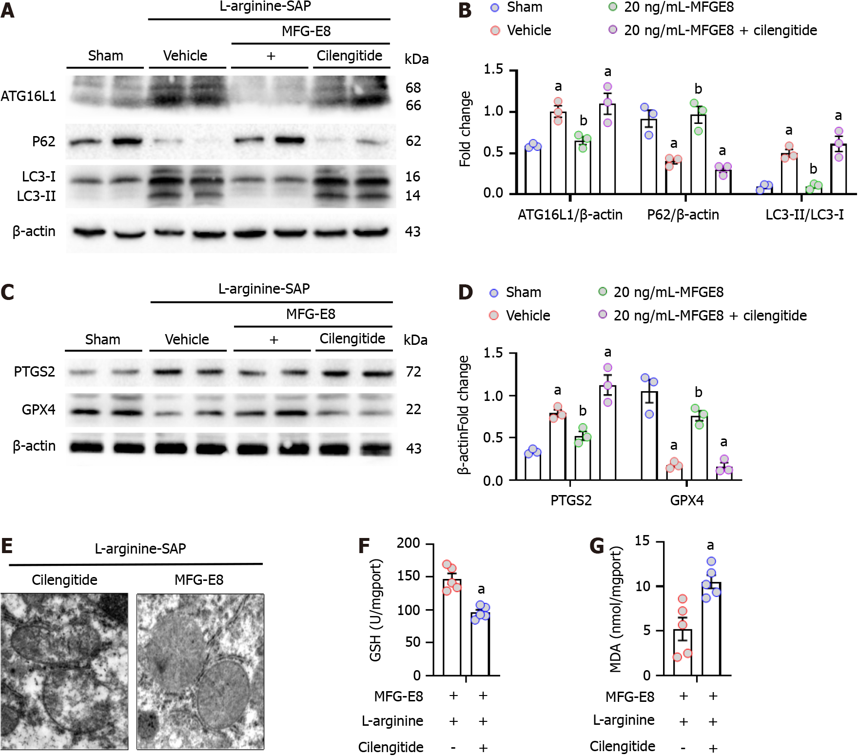
Figure 7 Milk fat globule epidermal growth factor 8 mitigates hepatic autophagy and ferroptosis through the integrin αVβ3/5.
In mice, severe acute pancreatitis was induced by 2 hly intraperitoneal injections of 4.0 g/kg L-arginine. At 2 h after the last injection of L-arginine, normal saline (vehicle) or 20 μg/kg milk fat globule epidermal growth factor 8 (MFG-E8) was administered through intraperitoneal injection. To determine whether the protective effect of MFG-E8 in liver is mediated through integrin αVβ3/5, 20 mg/kg cilengitide, a specific integrin αVβ3/5 antagonist, was administered intraperitoneally at 1 h before the injection of MFG-E8 (i.e., 1 h after the last injection of L-arginine). The animals were sacrificed at 69 h after MFG-E8 treatment (i.e., 72 h after the first injection of L-arginine). Blood and tissue samples were collected. A-D: Western blot analysis and quantitative of the expression of autophagy related protein 16 like protein 1, P62, LC3, prostaglandin-endoperoxide synthase 2 and glutathione peroxidase 4 in the liver; E: Ultrastructural changes of mitochondria in hepatocytes (electron microscopy); F: Glutathione levels in the liver; G: Malondialdehyde levels in the liver. n = 3-6/group, error bars indicate the SEM; aP < 0.05 vs sham group; bP < 0.05 vs vehicle group. SAP: Severe acute pancreatitis; ATG16L1: Autophagy related protein 16 like protein 1; PTGS2: Prostaglandin-endoperoxide synthase 2; GPX4: Glutathione peroxidase 4; MDA: Malondialdehyde; GSH: Glutathione; MFG-E8: Milk fat globule epidermal growth factor 8.
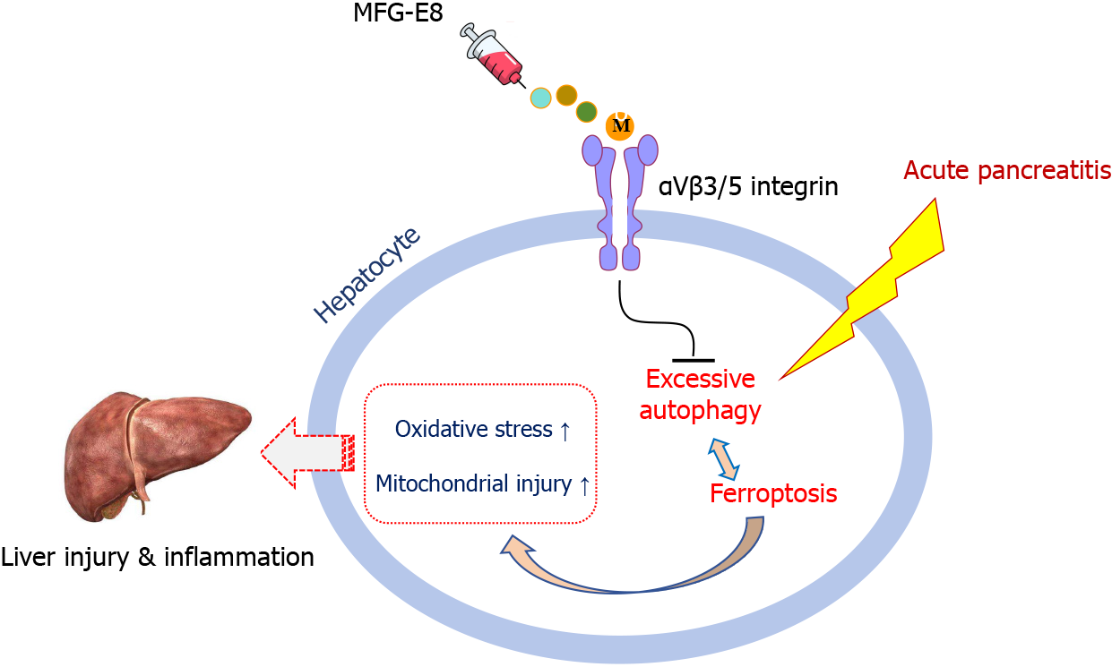
Figure 8 Graphical abstract.
Exogenous milk fat globule epidermal growth factor 8 restored impaired autophagy, reduced ferroptosis and alleviated liver injury by acting on integrin αVβ3/5. MFG-E8: Milk fat globule epidermal growth factor 8.
- Citation: Cui Q, Liu HC, Liu WM, Ma F, Lv Y, Ma JC, Wu RQ, Ren YF. Milk fat globule epidermal growth factor 8 alleviates liver injury in severe acute pancreatitis by restoring autophagy flux and inhibiting ferroptosis in hepatocytes. World J Gastroenterol 2024; 30(7): 728-741
- URL: https://www.wjgnet.com/1007-9327/full/v30/i7/728.htm
- DOI: https://dx.doi.org/10.3748/wjg.v30.i7.728


