Copyright
©The Author(s) 2024.
World J Gastroenterol. Apr 28, 2024; 30(16): 2220-2232
Published online Apr 28, 2024. doi: 10.3748/wjg.v30.i16.2220
Published online Apr 28, 2024. doi: 10.3748/wjg.v30.i16.2220
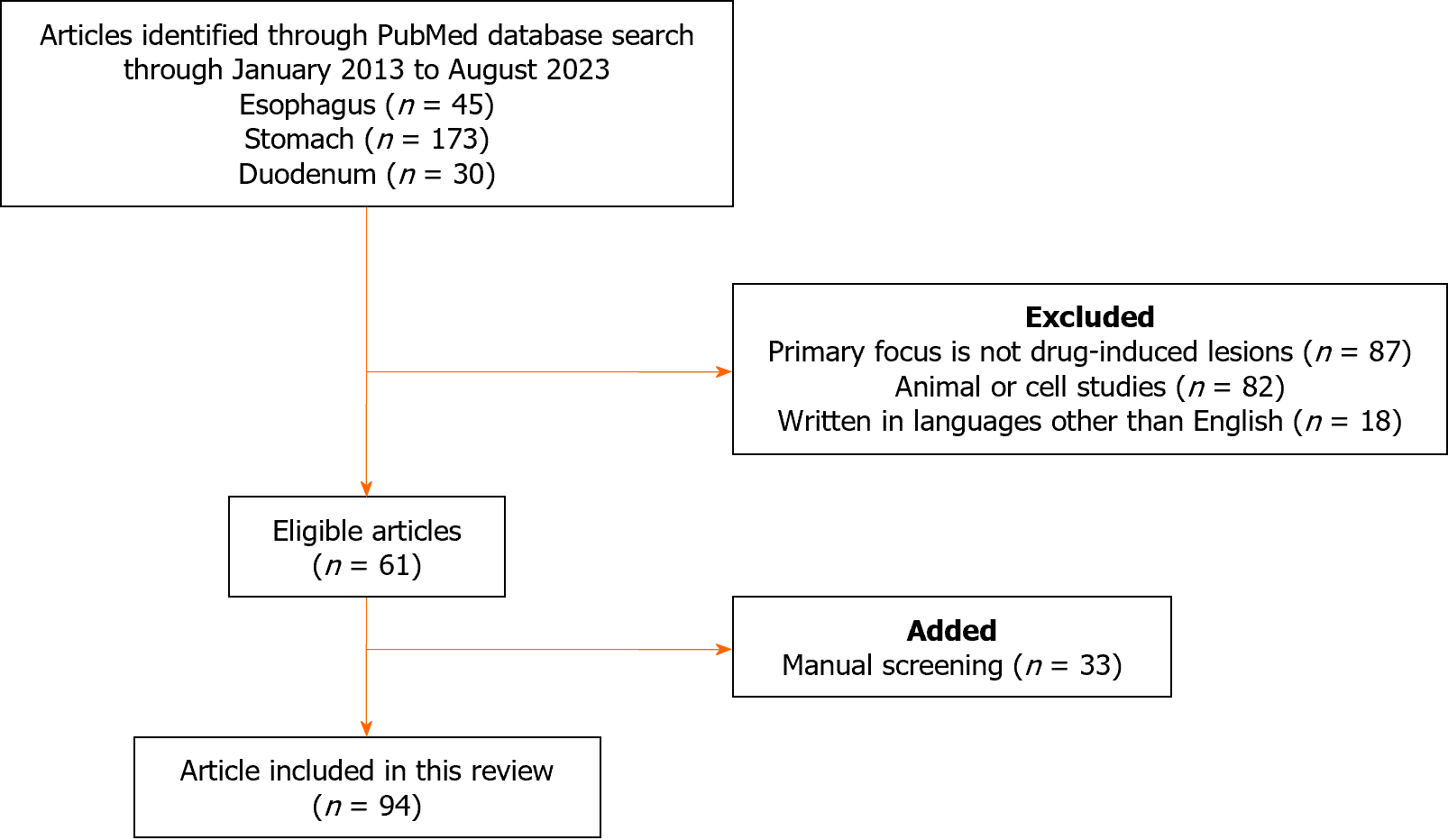
Figure 1 Flow diagram summarizing the identification, screening, eligibility, and exclusion processes of the literature search.
Figure 2 Dabigatran-induced desquamative esophagitis.
A-C: White membranous material is observed in the middle to lower esophagus of a 73-year-old woman taking dabigatran.
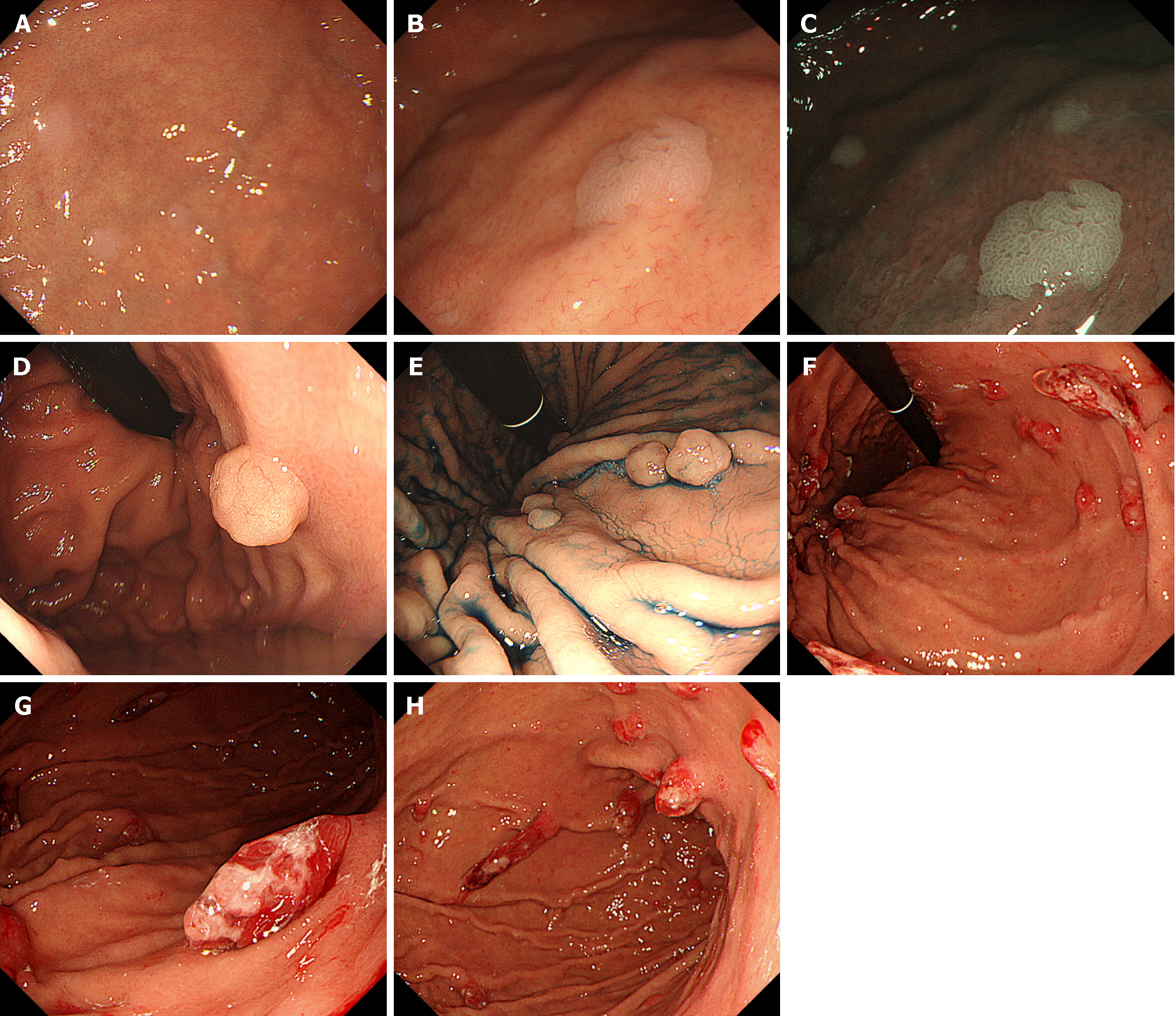
Figure 3 Proton pump inhibitor-induced gastric mucosal changes.
A-C: Multiple white and flat elevated lesions. Whitish slight elevations are observed in the gastric fornix of a patient taking proton pump inhibitor (PPI). Lesions are easily identified on narrow-band imaging observation (C); D and E: Fundic gland polyps in a PPI user. After indigo carmine dye spraying (E); F-H: Hyperplastic polyps in the stomach. Multiple reddish, friable, long polyps are seen.
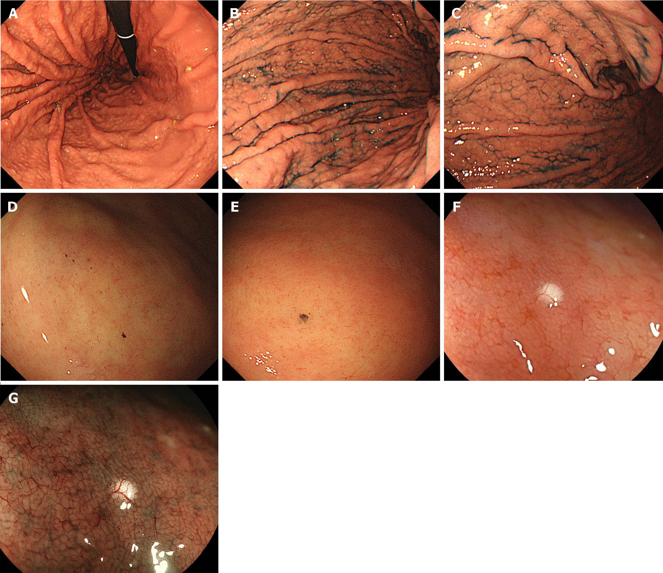
Figure 4 Proton pump inhibitor-induced gastric mucosal changes.
A-C: Cobblestone-like mucosa. Numerous, approximately 3-5 mm-sized, elevated mucosal lesions are seen in the gastric body of a proton pump inhibitor user. After indigo carmine dye spraying (B and C); D and E: Black spots. Dark, dot-like spots are observed in the gastric body; F and G: White globe appearance. A small, round, white deposit observed during esophagogastroduodenoscopy. Magnifying endoscopic observation with blue laser imaging emphasized the lesion (G).
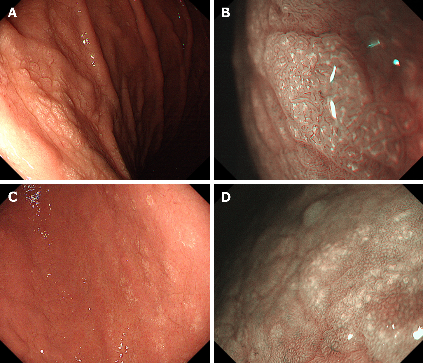
Figure 5 Lanthanum deposition in the stomach.
A and B: Lanthanum deposition shows diffuse white lesions in non-atrophic mucosa. Magnifying observation with narrow-band imaging reveals tiny whitish depositions within the gastric mucosa (B); C and D: Multiple circular white lesions are seen in the gastric antrum with atrophic change. Magnifying observation with narrow-band imaging of the circular white lesions (D).
Figure 6 Zinc acetate hydrate tablet-induced gastric lesions.
A 73-year-old Japanese woman had been taking zinc acetate dihydrate tablets for eight months to treat dysgeusia and hypozincemia. A: A round erosion with adhesion of the white coat is observed; B: Linear erosions are also seen in the gastric body (arrow); C: Esophagogastroduodenoscopy performed two months after cessation of zinc acetate hydrate tablet shows a resolution of erosions.
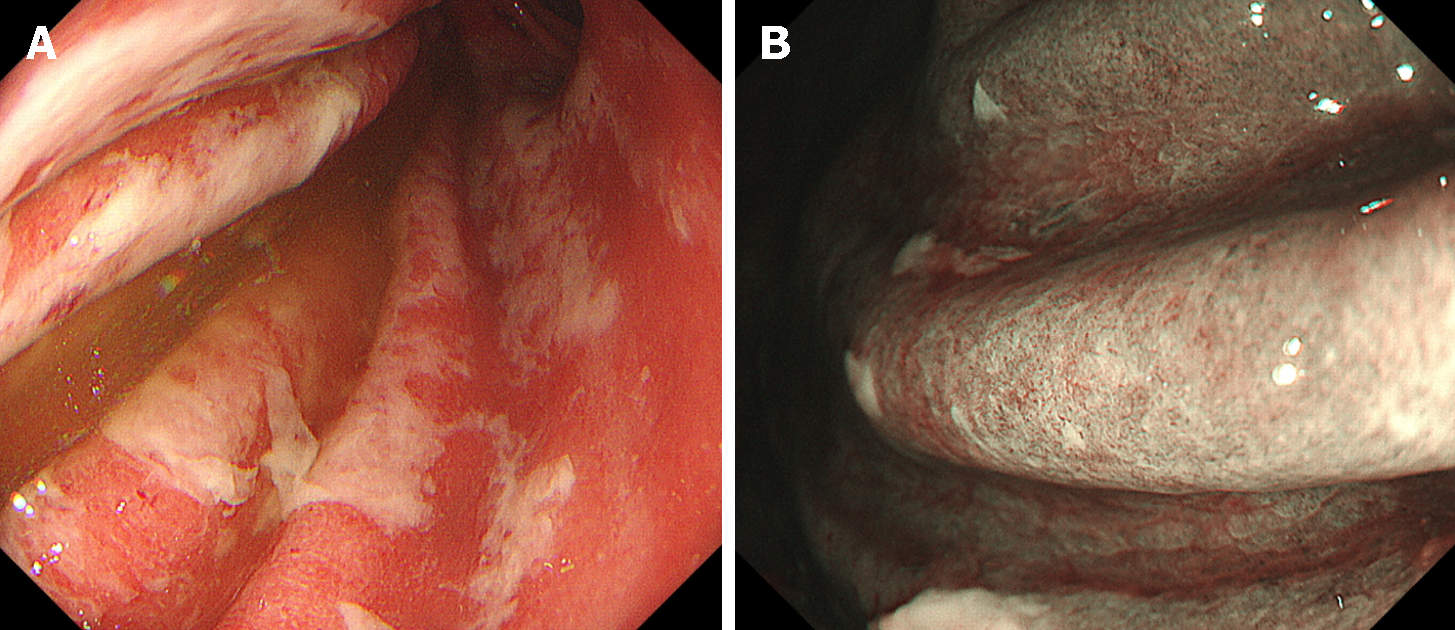
Figure 7 Immune-related adverse events gastritis.
Esophagogastroduodenoscopy images after 16 wk of pembrolizumab administration in a 57-year-old female. A: White exudate and coarse mucosa are observed in the gastric body; B: Magnifying observation with narrow-band imaging shows that the glandular structures are absent.
Figure 8 Pseudomelanosis of the duodenum.
A-C: A dark brown pigmentation is observed in the duodenal bulb. Narrow-band imaging (C).
Figure 9 Lanthanum deposition in the duodenum.
A: The duodenal mucosa is whitish; B: Magnifying observation reveals numerous dot-like white deposits in the duodenal villi; C: Magnifying observation with blue laser imaging emphasized the white deposits.
- Citation: Iwamuro M, Kawano S, Otsuka M. Drug-induced mucosal alterations observed during esophagogastroduodenoscopy. World J Gastroenterol 2024; 30(16): 2220-2232
- URL: https://www.wjgnet.com/1007-9327/full/v30/i16/2220.htm
- DOI: https://dx.doi.org/10.3748/wjg.v30.i16.2220


