Copyright
©The Author(s) 2024.
World J Gastroenterol. Mar 28, 2024; 30(12): 1706-1713
Published online Mar 28, 2024. doi: 10.3748/wjg.v30.i12.1706
Published online Mar 28, 2024. doi: 10.3748/wjg.v30.i12.1706
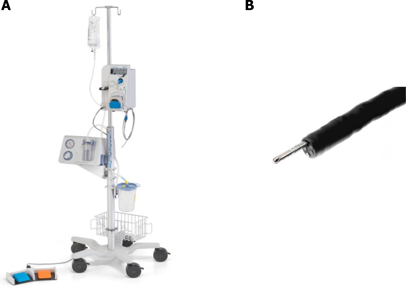
Figure 1 EndoRotor endoscopic powered resection System.
A: EndoRotor endoscopic powered resection system; B: Beveled tip single-use catheter.
Figure 2 Session one: Examination of two recurrent polyps on the previous resection site with white light narrow-band imaging and Indigo.
A: White light; B: Narrow-band imaging; C: Indigo.
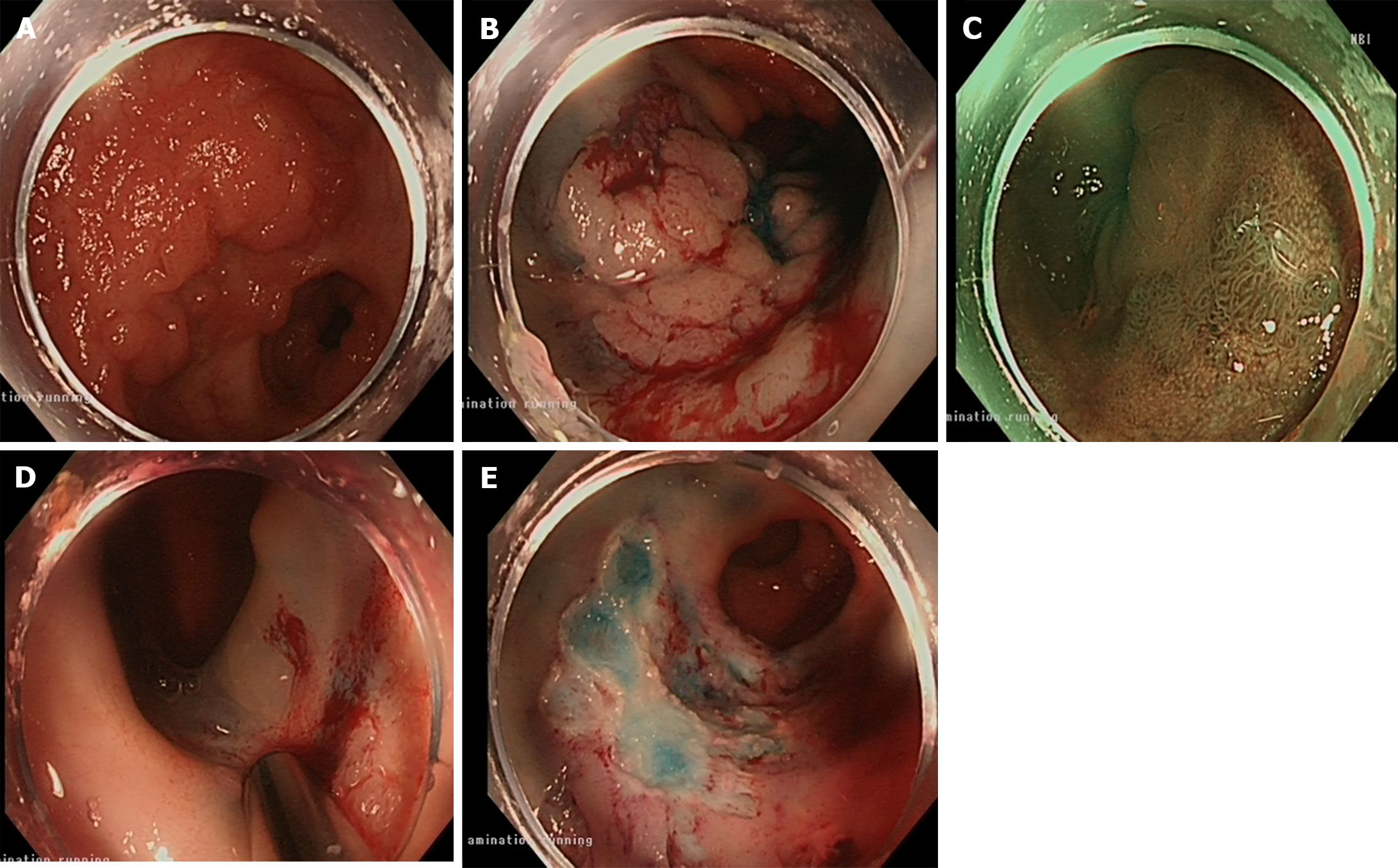
Figure 3 Session two: Examination of a residual 15 mm flat elevated lesion with white light and narrow band imaging; application of EndoRotor; resection site after EndoRotor and endoscopic mucosal resection.
A and B: White light; C: Narrow band imaging; D: EndoRotor; E: Resection site after EndoRotor and endoscopic mucosal resection.
Figure 4 Session three: Examination of the residual polyp with white light and narrow band imaging and resection site after EndoRotor and argon plasma coagulation.
A: White light; B: Narrow band imaging; C: Resection site after EndoRotor and argon plasma coagulation.
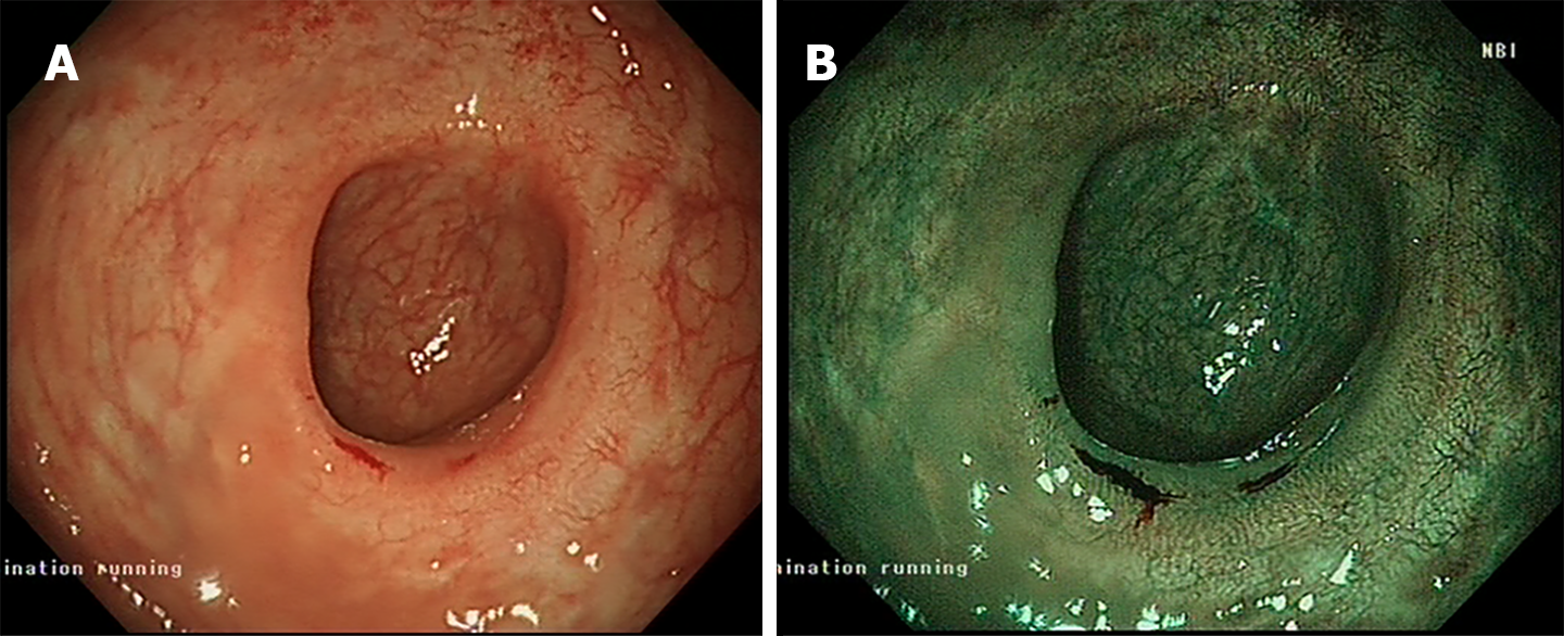
Figure 5 Session four: Examination of the scar tissue via the white light and narrow band imaging methods.
A: White light; B: Narrow band imaging.
- Citation: Zaghloul M, Rehman H, Sansone S, Argyriou K, Parra-Blanco A. Endoscopic treatment of scarred polyps with a non-thermal device (Endorotor): A review of the literature. World J Gastroenterol 2024; 30(12): 1706-1713
- URL: https://www.wjgnet.com/1007-9327/full/v30/i12/1706.htm
- DOI: https://dx.doi.org/10.3748/wjg.v30.i12.1706


