Copyright
©The Author(s) 1997.
World J Gastroenterol. Dec 15, 1997; 3(4): 242-245
Published online Dec 15, 1997. doi: 10.3748/wjg.v3.i4.242
Published online Dec 15, 1997. doi: 10.3748/wjg.v3.i4.242
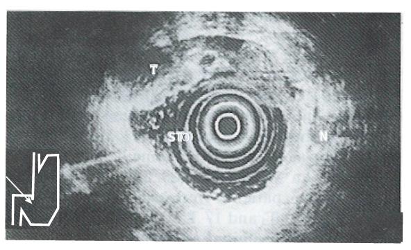
Figure 1 Endoscopic ultrasonography-T2 cancer.
Endoscopic ultrasonography shows a hypoechoic tumor (T) adjacent to an ulcerative lesion (U) invading the muscularis propria (Mp).
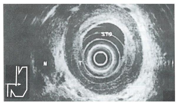
Figure 2 Endoscopic ultrasonography-T3 cancer.
Endoscopic ultrasonography findings in linitis plastica. Hypoechoic diffuse wall thickening is shown, with preservation of layers as distinctive structures. Note invasion of all layers but absence of invasion of adjacent organs.
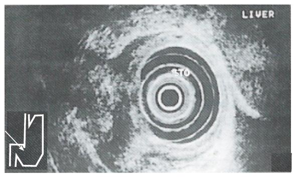
Figure 3 Endoscopic ultrasonography-T4 cancer.
Endoscopic ultrasonography shows a transmural hypoechoic tumor with penetration into adjacent structures.
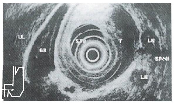
Figure 4 Peritumorous lymph node metastasis.
- Citation: Guo W, Zhang YL, Li GX, Zhou DY, Zhang WD. Comparison of preoperative TN staging of gastric carcinoma by endoscopic ultrasonography with CT examination. World J Gastroenterol 1997; 3(4): 242-245
- URL: https://www.wjgnet.com/1007-9327/full/v3/i4/242.htm
- DOI: https://dx.doi.org/10.3748/wjg.v3.i4.242


