Copyright
©The Author(s) 1997.
World J Gastroenterol. Dec 15, 1997; 3(4): 228-230
Published online Dec 15, 1997. doi: 10.3748/wjg.v3.i4.228
Published online Dec 15, 1997. doi: 10.3748/wjg.v3.i4.228
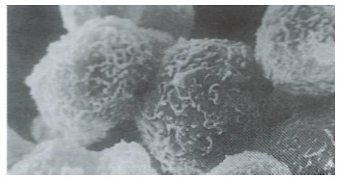
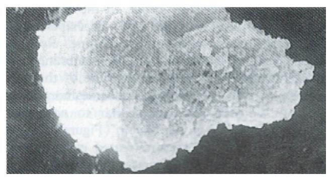
Figure 1 Normal human fetal hepatocytes.
The cellular membrane is intact and microvilli can be clearly seen. × 7500
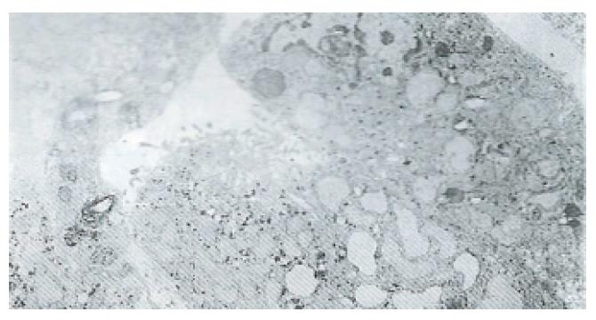
Figure 2 Normal human fetal hepatocytes.
× 4500
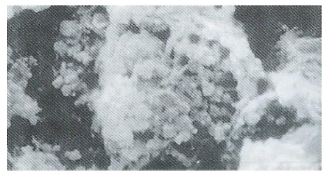
Figure 3 Hepatocytes in the control group following exposure to CCl4.
The cellular membrane appears mesh-like. × 10500
Figure 4 Hepatocytes pretreated with silybin and exposed to CCl4.
The changes of the cellular membrane are similar to Figure 5. × 8000.
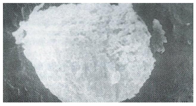
Figure 5 Hepatocytes pretreated with PSP and exposed to CCl4.
The cellular membrane is intact and microvilli swelling is present. × 7500
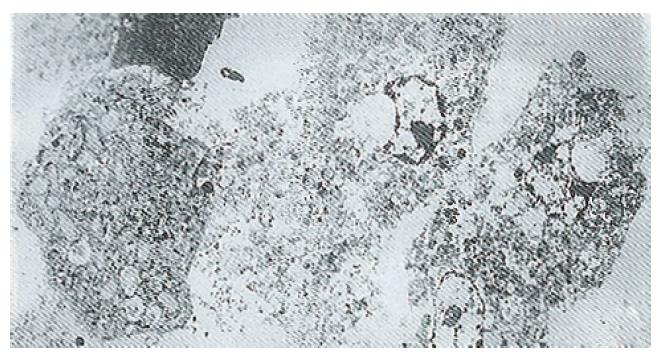
Figure 6 Hepatocytes in the non-pretreated control group after exposure to CCl4.
× 4500
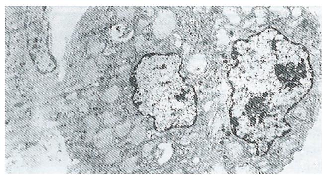
Figure 7 Hepatocytes in the PSP pretreatment group.
The ultrastructure is well preserved. × 4500
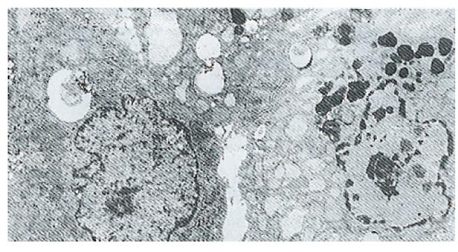
Figure 8 Hepatocytes in the silybin pretreatment group.
The changes are similar to those in Figure 7. × 4500.
- Citation: Wang MR, Le MZ, Xu JZ, He CL. Establishment and application of an experimental model of human fetal hepatocytes for investigation of the protective effects of silybin and polyporus umbellalus polysaccharides. World J Gastroenterol 1997; 3(4): 228-230
- URL: https://www.wjgnet.com/1007-9327/full/v3/i4/228.htm
- DOI: https://dx.doi.org/10.3748/wjg.v3.i4.228


