Copyright
©The Author(s) 1997.
World J Gastroenterol. Dec 15, 1997; 3(4): 213-217
Published online Dec 15, 1997. doi: 10.3748/wjg.v3.i4.213
Published online Dec 15, 1997. doi: 10.3748/wjg.v3.i4.213
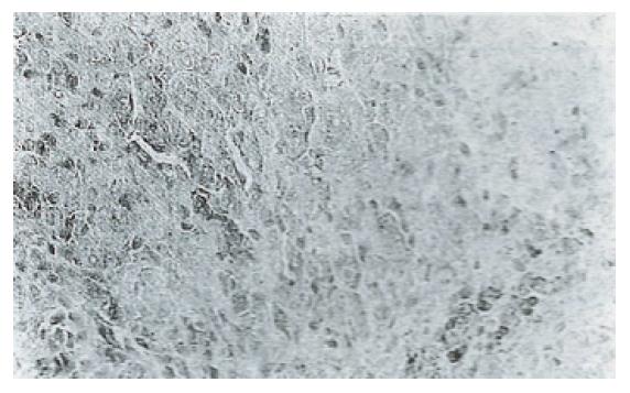
Figure 1 Immunostaining of a rat malignant nodule with anti-p21 monoclonal IgG2b antibody detected by the biotin-streptavidin method.
Magnification × 10.
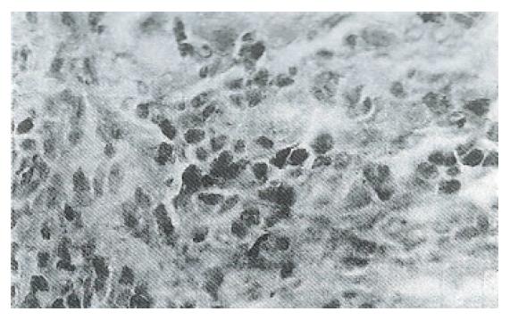
Figure 2 Immunostaining of a rat malignant foci with anti-p53 polyclonal antibody detected by the biotin-streptavidin method.
Magnification × 40.
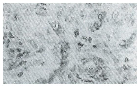
Figure 3 Immunostaining of a rat malignant foci with anti-bcl-2 polyclonal antibody detected by the biotin-streptavidin method.
Magnification × 40.
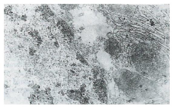
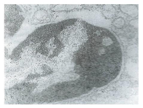
Figure 4 Immunostaining of ultrastructure of a rat normal hepatocyte with anti-p21 IgG/colloidal gold (φ 10 nm) antibody.
Magnification × 2000
Figure 5 Immunostaining of ultrastructure of a rat malignant hepatocyte with anti-p21 IgG/colloidal gold (φ 10 nm) antibody.
Magnification × 2000.
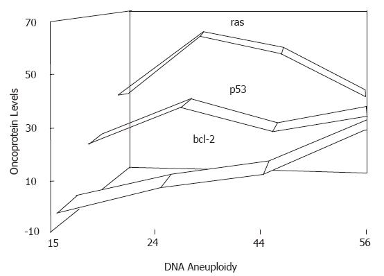
Figure 6 Relationship between hepatocarcinoma malignancy and expression of oncogene proteins detected by immunofluorencence flow cytometry.
- Citation: Yang JM, Han DW, Liang QC, Zhao JL, Hao SY, Ma XH, Zhao YC. Effects of endotoxin on expression of ras, p53 and bcl-2 oncoprotein in hepatocarcinogenesis induced by thioacetamide in rats. World J Gastroenterol 1997; 3(4): 213-217
- URL: https://www.wjgnet.com/1007-9327/full/v3/i4/213.htm
- DOI: https://dx.doi.org/10.3748/wjg.v3.i4.213


