Copyright
©The Author(s) 1997.
World J Gastroenterol. Sep 15, 1997; 3(3): 147-149
Published online Sep 15, 1997. doi: 10.3748/wjg.v3.i3.147
Published online Sep 15, 1997. doi: 10.3748/wjg.v3.i3.147
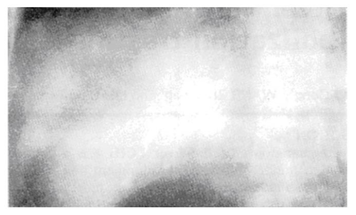
Figure 1 The tumor appeared as a filling defect.
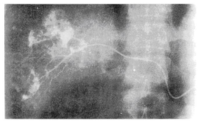
Figure 2 Radiograph by immediate visualization of tumor body after injection intubation of hepatic artery branch.
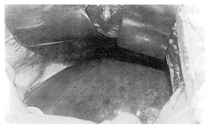
Figure 3 Methylene blue staining of the liver except the tumor area.
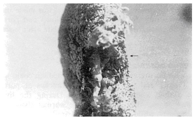
Figure 4 Specimen cast filled with methyl methacrylate via portal vein and hepatic vein, the tumor area appeared as a round vacant cavity.
- Citation: Li GW, Zhao ZR, Li BS, Liu XG, Wang ZL, Liu QF. Source of blood supply and embolization treatment in cavernous hemangioma and sclerosis of the liver. World J Gastroenterol 1997; 3(3): 147-149
- URL: https://www.wjgnet.com/1007-9327/full/v3/i3/147.htm
- DOI: https://dx.doi.org/10.3748/wjg.v3.i3.147


