Copyright
©The Author(s) 1997.
World J Gastroenterol. Sep 15, 1997; 3(3): 143-146
Published online Sep 15, 1997. doi: 10.3748/wjg.v3.i3.143
Published online Sep 15, 1997. doi: 10.3748/wjg.v3.i3.143
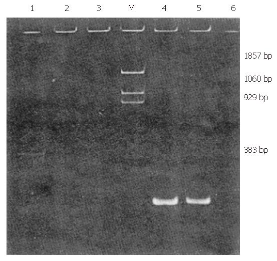
Figure 1 Electrophoretic pattern of RT-nested PCR for hepatitis G virus (HGV) RNA detection.
1, 4 were products of PCR from original sera, 2 and 5 products from 10-6 diluted sera, 3 and 6 were negative controls. 1, 2 and 3 were the frist PCR products, 4, 5 and 6 were the second PCR products. M was DNA marker, PBR 322/BstN I.
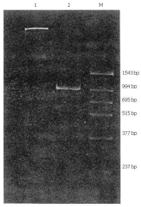
Figure 2 RT-nested PCR for amplification of long fragment cDNA of hepatitis G virus (HGV) NS5 region.
1 was negative control, 2 was positive PCR product, M was DNA and PCR marker.
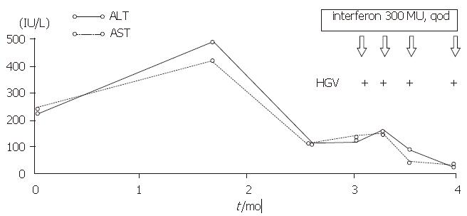
Figure 3 Clinical manifestation of the patient with chronic non-A-E hepatitis
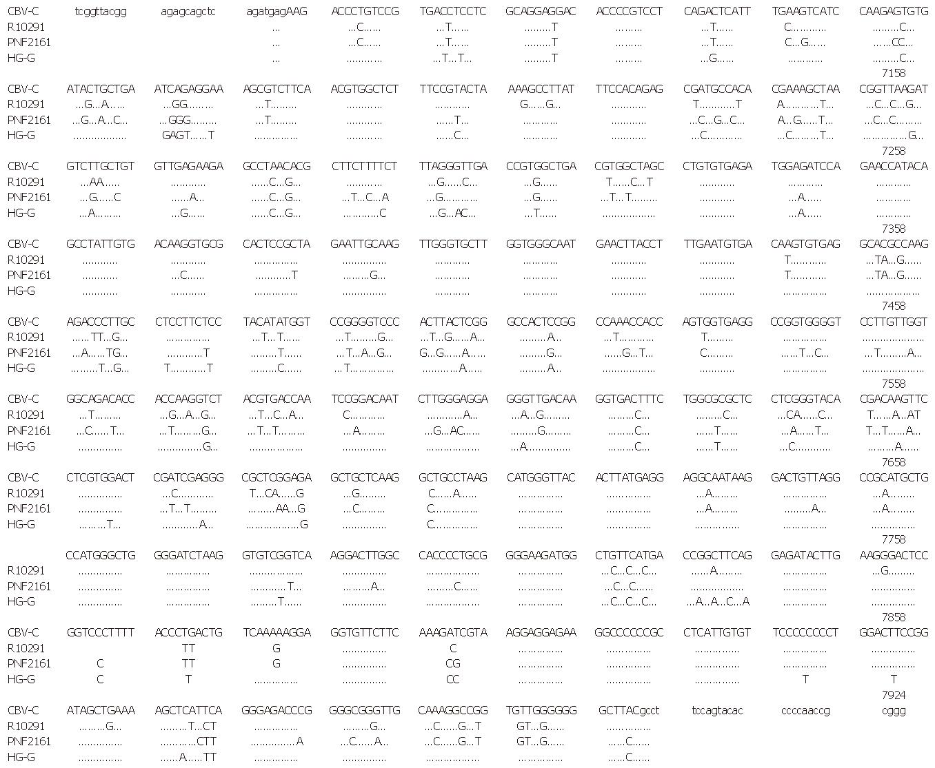
Figure 4 Nucleotide sequence of partial hepatitis G virus (HGV) NS5 gene in the serum of a patient with chronic non-A-E hepatitis and its comparison with West African and American isolates.
GBV-C is West African isolate; R10291 and PNF2161 are American isolates; HG-G is a patient with chronic non-A-E hepatitis. —denotes the same as GBV-C sequence. Capital letter is nucleotide detected, small letter is primer sequence.
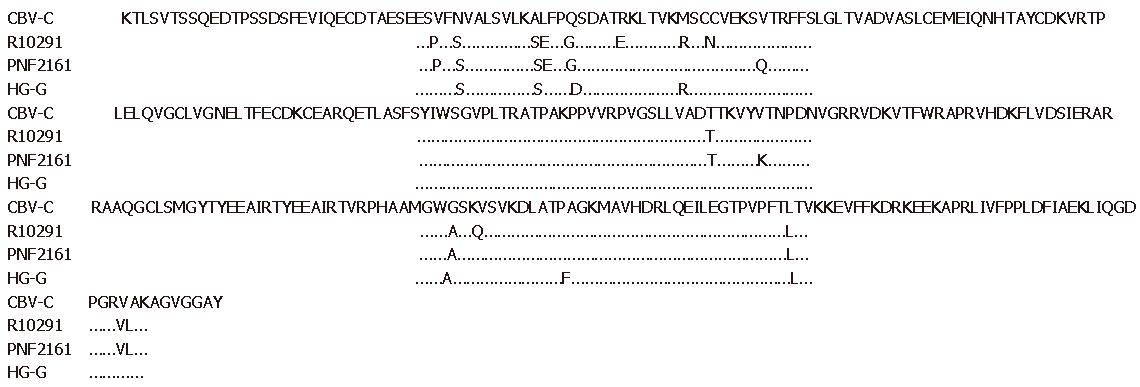
Figure 5 Amino acid sequence of hepatitis G virus (HGV) NS5 region in the serum of the patient with chronic non-A-E hepatitis and its comparison with West African and American isolates.
- Citation: Chang JH, Wei L, Du SC, Wang H, Sun Y, Tao QM. Hepatitis G virus infection in patients with chronic non-A–E hepatitis. World J Gastroenterol 1997; 3(3): 143-146
- URL: https://www.wjgnet.com/1007-9327/full/v3/i3/143.htm
- DOI: https://dx.doi.org/10.3748/wjg.v3.i3.143


