Copyright
©The Author(s) 2023.
World J Gastroenterol. Mar 7, 2023; 29(9): 1475-1491
Published online Mar 7, 2023. doi: 10.3748/wjg.v29.i9.1475
Published online Mar 7, 2023. doi: 10.3748/wjg.v29.i9.1475
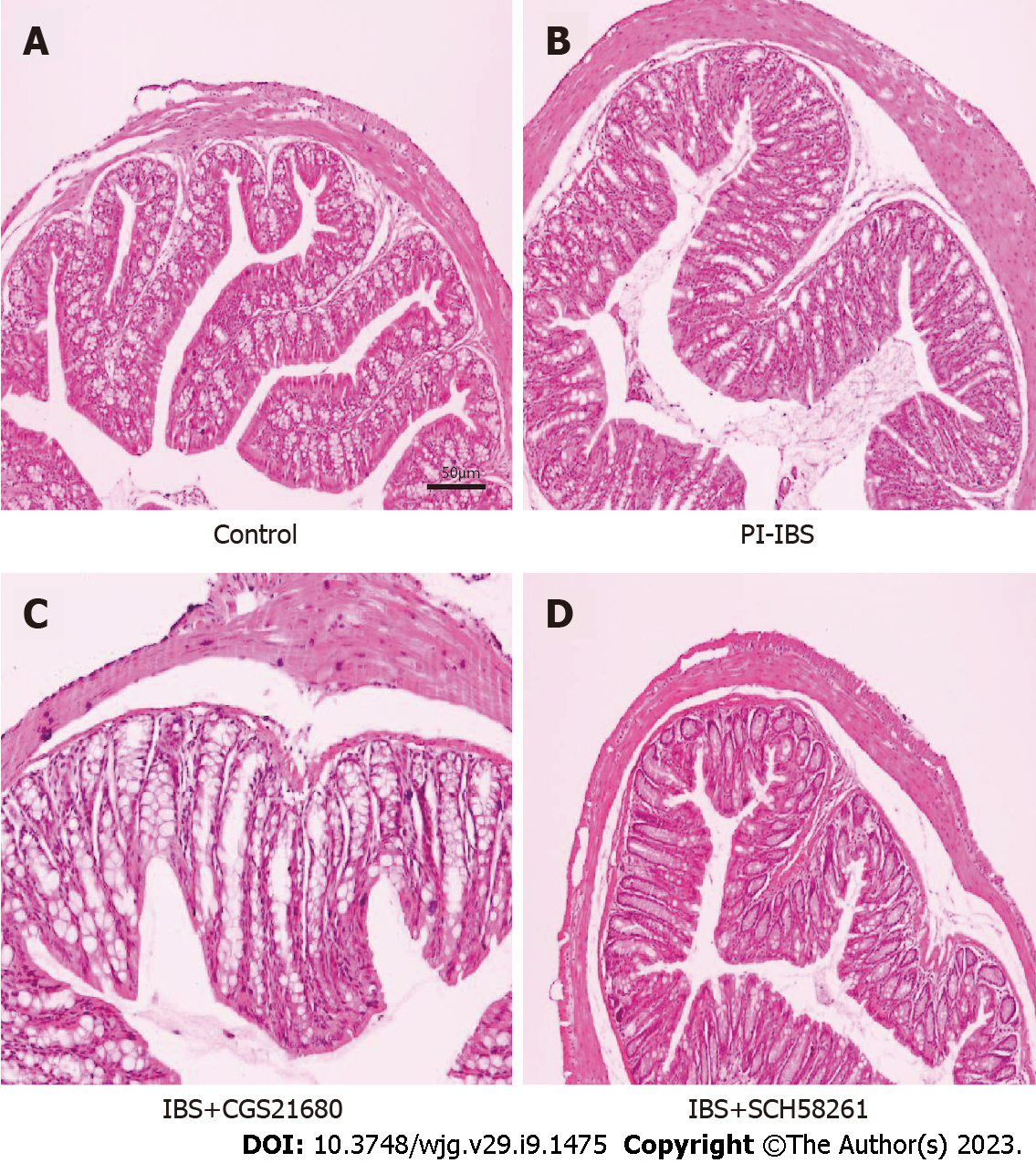
Figure 1 The effect of adenosine 2A receptor on the histopathological changes in post-infectious irritable bowel syndrome mouse.
A and B: The Post-infectious irritable bowel syndrome (PI-IBS) model (B) exhibits no substantial inflammatory alterations in its colon tissue compared to the control animal (A); C: Adenosine 2A receptor (A2AR) agonist CGS21680 leads to some inflammatory alterations in the colon tissue of the PI-IBS model mice, including the infiltration of certain inflammatory cells; D: A2AR antagonist SCH58261 relieves inflammation in the colon tissue of the PI-IBS model mice. A2AR: Adenosine 2A receptor; PI-IBS: Post-infectious irritable bowel syndrome. Scale bars, 50 μm.
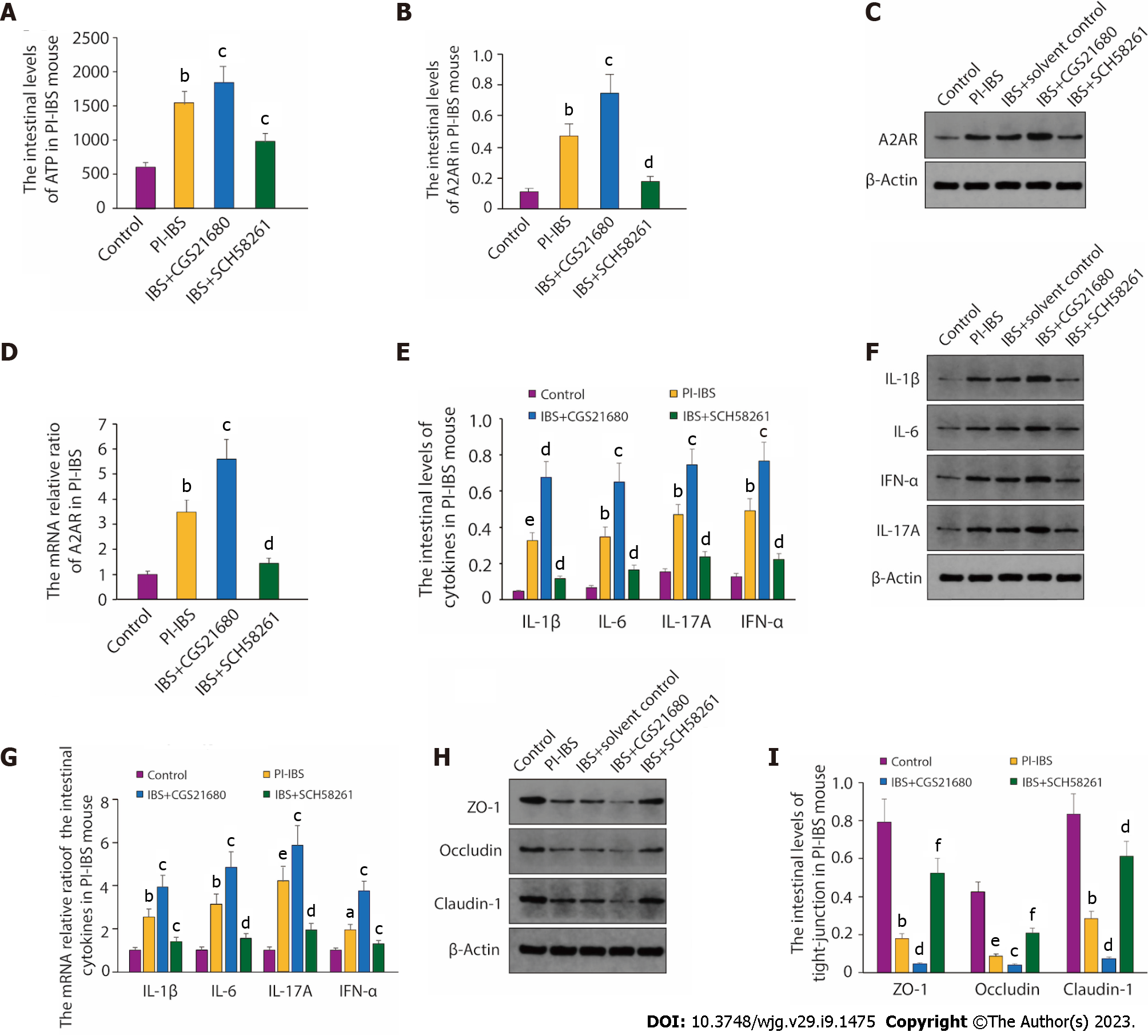
Figure 2 The intestinal ATP and adenosine 2A receptor expression in post-infectious irritable bowel syndrome mouse.
A: The level of ATP in post-infectious irritable bowel syndrome (PI-IBS) mouse. The results were independently repeated three times; B and C: The intestinal levels of adenosine 2A receptor (A2AR) in PI-IBS mouse; D: The mRNA relative ratio of A2AR in PI-IBS mouse. The results were independently repeated three times; E and F: The intestinal levels of the inflammatory cytokines in PI-IBS mice were measured by ELISA and western blot, respectively. The ELISA results were obtained from six mice; G: The mRNA relative ratio of the intestinal cytokines in PI-IBS mouse. The samples were from mice mentioned in E and F. The results were independently repeated three times; H and I: The intestinal levels of tight junction proteins in PI-IBS mice were tested through western blot and quantified by image J software. β-Actin is used as the loading control for both western blot and reverse transcription polymerase chain reaction. aP < 0.05, bP < 0.01, eP < 0.001 vs the Control group; cP < 0.05, dP < 0.01, fP < 0.001 vs the PI-IBS group.
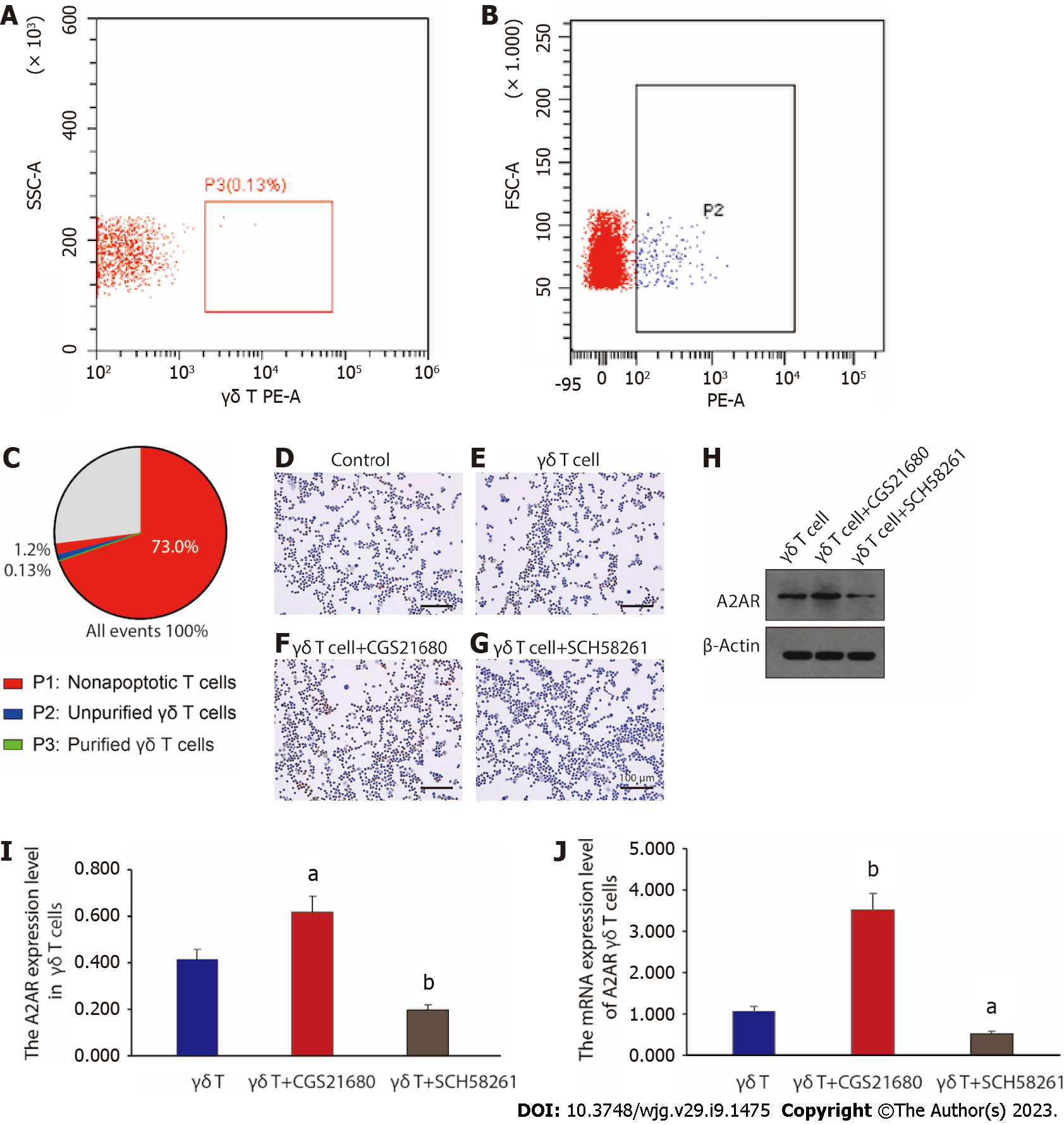
Figure 3 γδ T cells’ isolation and functional evaluation.
A-C: Based on the results of fluorescence-activated cell sorting, γδ T cells were effectively extracted and purified. P2, unpurified γδ T cells; P3, purified γδ T cells; D-G: Immunohistochemistry labeling of adenosine 2A receptor (A2AR) expression in γδT cells. Scale bars, 100 μm; H: Western blot analysis of A2AR expression levels in γδ T cells. β-Actin is used as the loading control; I: The quantitive result of A2AR expression level in γδ T cells was analyzed from data shown in H. Results from three times of independently repeated experiments were analysed; J: Reverse transcription polymerase chain reaction analysis of the expression level of A2AR mRNA in γδ T cells. The results were independently repeated three times. A2AR: Adenosine 2A receptor. aP < 0.05, bP < 0.01 vs the γδ T cell group.
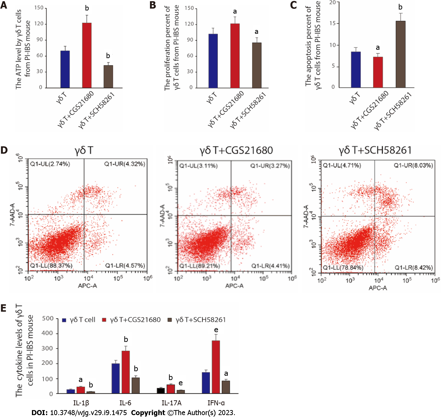
Figure 4 Functional evaluation of γδ T cells in vitro.
A: The ATP level changed by differently treated γδ T cells from post-infectious irritable bowel syndrome (PI-IBS) mice. The data was obtained from six mice in each group; B: The proliferation percent of γδ T cells from PI-IBS mouse was analysed through CCK8 assay. The samples were taken from six mice in each group; C: The apoptosis percent of γδ T cells from PI-IBS mouse measured by fluorescence-activated cell sorting. The γδ T cells were taken from six mice in each group; D: Apoptosis rates were detected according to Annexin V and PI double-staining method; E: The cytokines level of IL-1β, IL-6, IL-17A and IFN-α from PI-IBS mice after reinfusing control γδ T cells and γδ T cells treated with CGS21680 or SCH58261, respectively. The data was obtained from six mice in each group. PI-IBS: Post-infectious irritable bowel syndrome. aP < 0.05, bP < 0.01, eP < 0.001 vs the γδ T cell group.
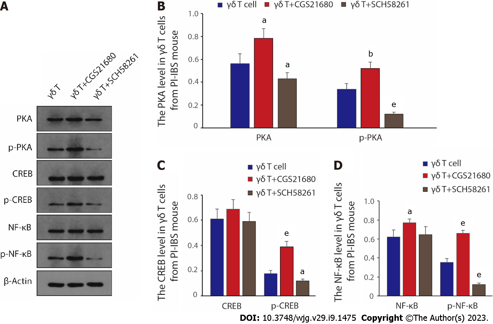
Figure 5 Adenosine 2A receptor mediated signaling pathway that could regulate the function of γδ T cells.
A: The PKA/p-PKA, CREB/p-CREB, NF-κB/p-NF-κB protein levels in γδ T cells of post-infectious irritable bowel syndrome (PI-IBS) mice. β-Actin is used as the loading control; B: The relative PKA level in γδ T cells from mice with PI-IBS; C: The relative CREB level in γδ T cells from PI-IBS mice; D: The relative NF-κB level in γδ T cells from mice with PI-IBS. The results from three times of independently repeated experiments were gathered and analyzed. PI-IBS: Post-infectious irritable bowel syndrome. aP < 0.05, bP < 0.01, eP < 0.001 vs the γδ T cell group.
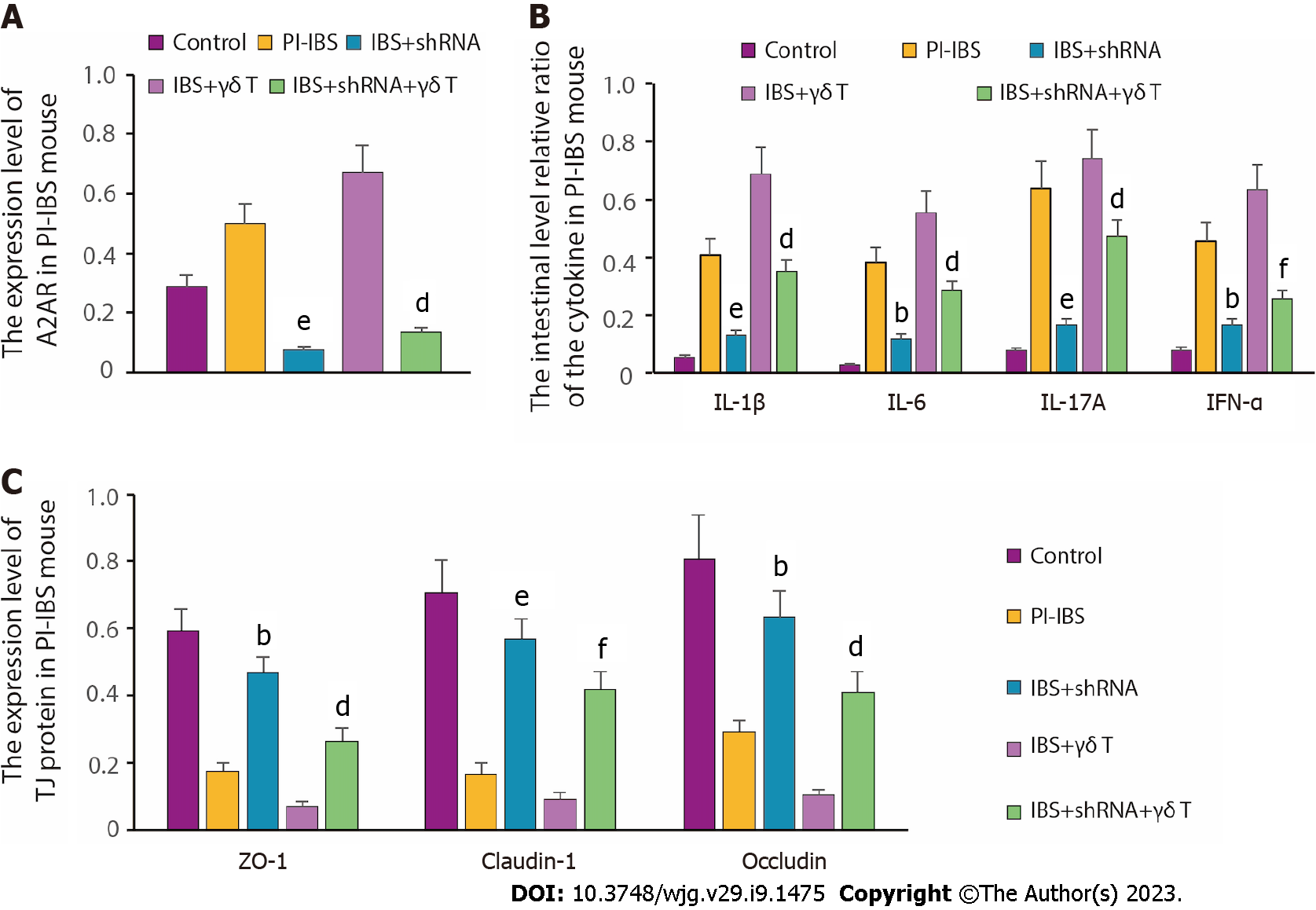
Figure 6 The inflammatory response alteration after adenosine 2A receptor knockdown.
A: The altered level of adenosine 2A receptor (A2AR) expression after A2AR-shRNA transfection or γδ T cell reinfusion in post-infectious irritable bowel syndrome (PI-IBS) mice was assessed. The results were obtained from three times of independently repeated experiments; B: The relative ratio of the intestinal cytokine level in PI-IBS mice (n = 6, per group); C: The degree of TJ (tight junction protein) protein expression in PI-IBS mice (n = 6, per group). aP < 0.05, bP < 0.01, eP < 0.001 vs the PI-IBS group; cP < 0.05, dP < 0.01, fP < 0.001 vs the IBS + γδ T group.
- Citation: Dong LW, Chen YY, Chen CC, Ma ZC, Fu J, Huang BL, Liu FJ, Liang DC, Sun DM, Lan C. Adenosine 2A receptor contributes to the facilitation of post-infectious irritable bowel syndrome by γδ T cells via the PKA/CREB/NF-κB signaling pathway. World J Gastroenterol 2023; 29(9): 1475-1491
- URL: https://www.wjgnet.com/1007-9327/full/v29/i9/1475.htm
- DOI: https://dx.doi.org/10.3748/wjg.v29.i9.1475


