Copyright
©The Author(s) 2023.
World J Gastroenterol. Mar 7, 2023; 29(9): 1460-1474
Published online Mar 7, 2023. doi: 10.3748/wjg.v29.i9.1460
Published online Mar 7, 2023. doi: 10.3748/wjg.v29.i9.1460
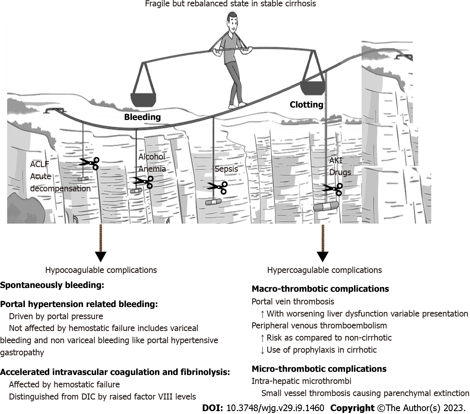
Figure 1 Rebalanced hemostasis in cirrhosis.
ACLF: Acute-on-chronic liver failure; AKI: Acute kidney injury; DIC: Disseminated intravascular coagulation.
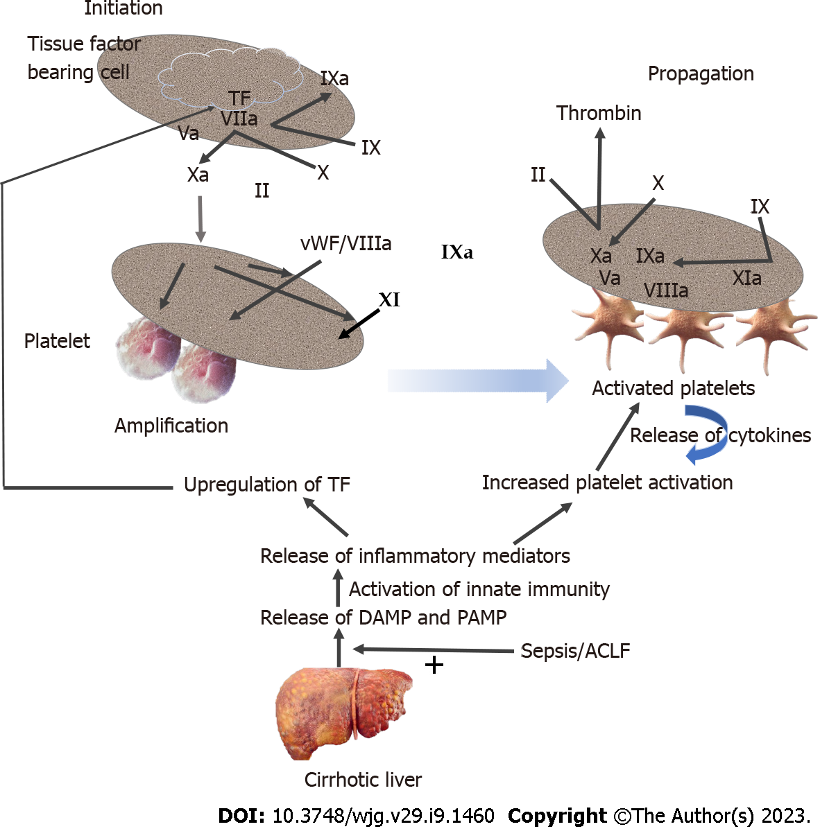
Figure 2 Dynamic coagulation profile in cirrhosis.
ACLF: Acute-on-chronic liver failure; DAMP: Damage-associated molecular patterns; PAMP: Pathogen-associated molecular patterns; TF: Tissue factor.
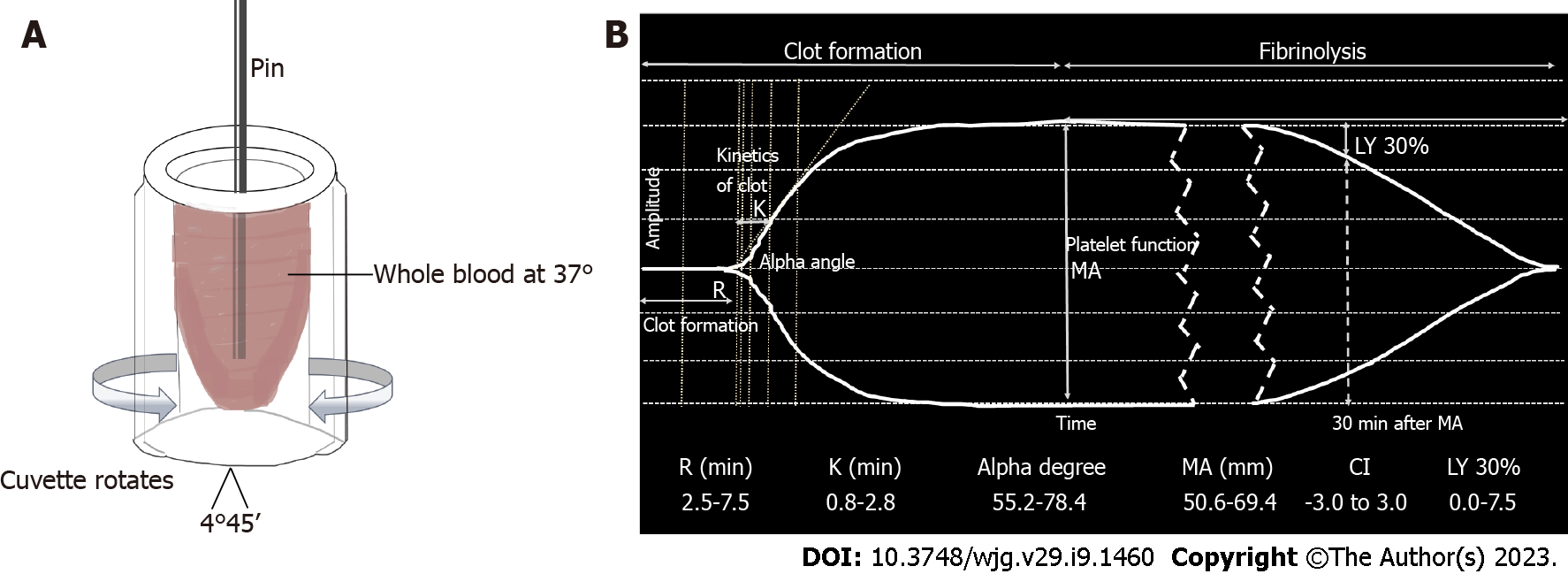
Figure 3 Basis and results of the thromboelastography.
A: Basis of the thromboelastography (TEG) test; B: TEG tracing and relevant parameters (kaolin-activated). MA: Maximum amplitude.
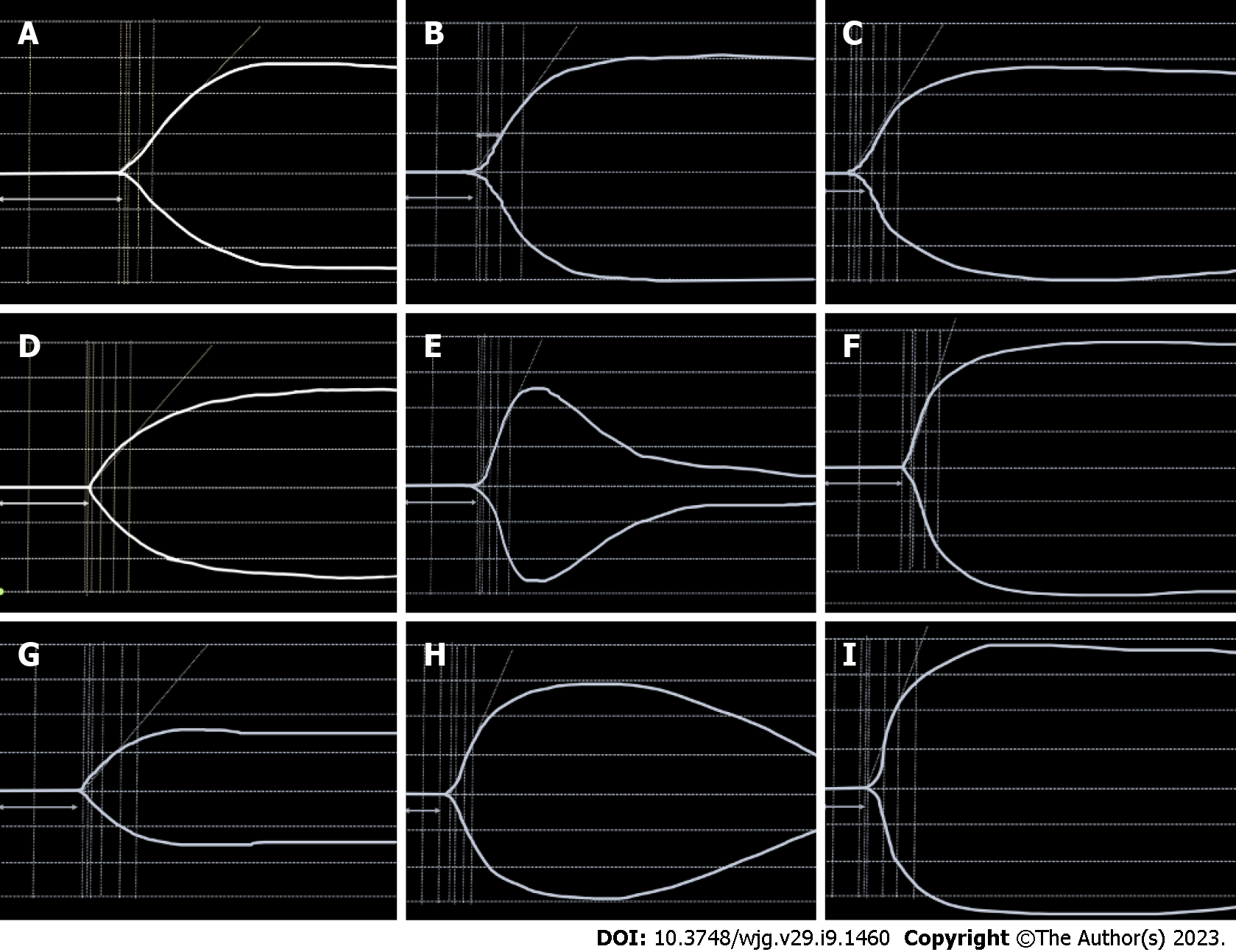
Figure 4 Tracing of thromboelastography in various clinical conditions.
A: Low clotting factors; B: Normal trace; C: Enzymatic hypercoagulability; D: Low fibrinogen levels; E: Primary fibrinolysis; F: Platelet hypercoagulability; G: Low platelet function; H: Secondary fibrinolysis; I: Enzymatic and platelet hypercoagulability.
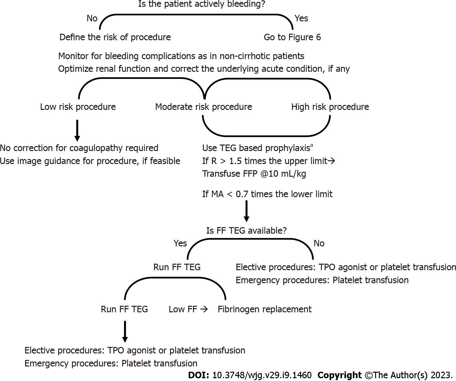
Figure 5 Algorithm for coagulation factor administration in the cirrhotic patient with coagulopathy undergoing an invasive procedure.
FF: Functional fibrinogen; FFP: Fresh frozen plasma; MA: Maximum amplitude; TEG: Thromboelastography; TPO: Thrombopoietin.
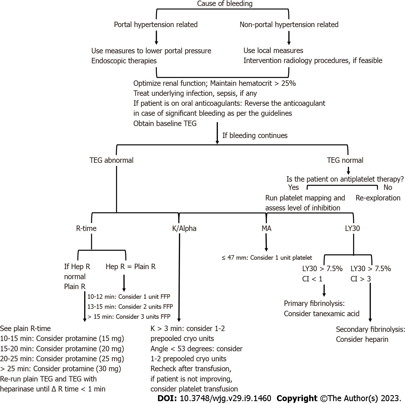
Figure 6 Algorithm for guiding blood product transfusion by thromboelastography.
Cryo: Cryoprecipitate; FFP: Fresh frozen plasma; Hep R: Heparinase R-Time; MA: Maximum amplitude; TEG: Thromboelastography.
- Citation: Kataria S, Juneja D, Singh O. Approach to thromboelastography-based transfusion in cirrhosis: An alternative perspective on coagulation disorders. World J Gastroenterol 2023; 29(9): 1460-1474
- URL: https://www.wjgnet.com/1007-9327/full/v29/i9/1460.htm
- DOI: https://dx.doi.org/10.3748/wjg.v29.i9.1460


