Copyright
©The Author(s) 2023.
World J Gastroenterol. Feb 28, 2023; 29(8): 1261-1288
Published online Feb 28, 2023. doi: 10.3748/wjg.v29.i8.1261
Published online Feb 28, 2023. doi: 10.3748/wjg.v29.i8.1261
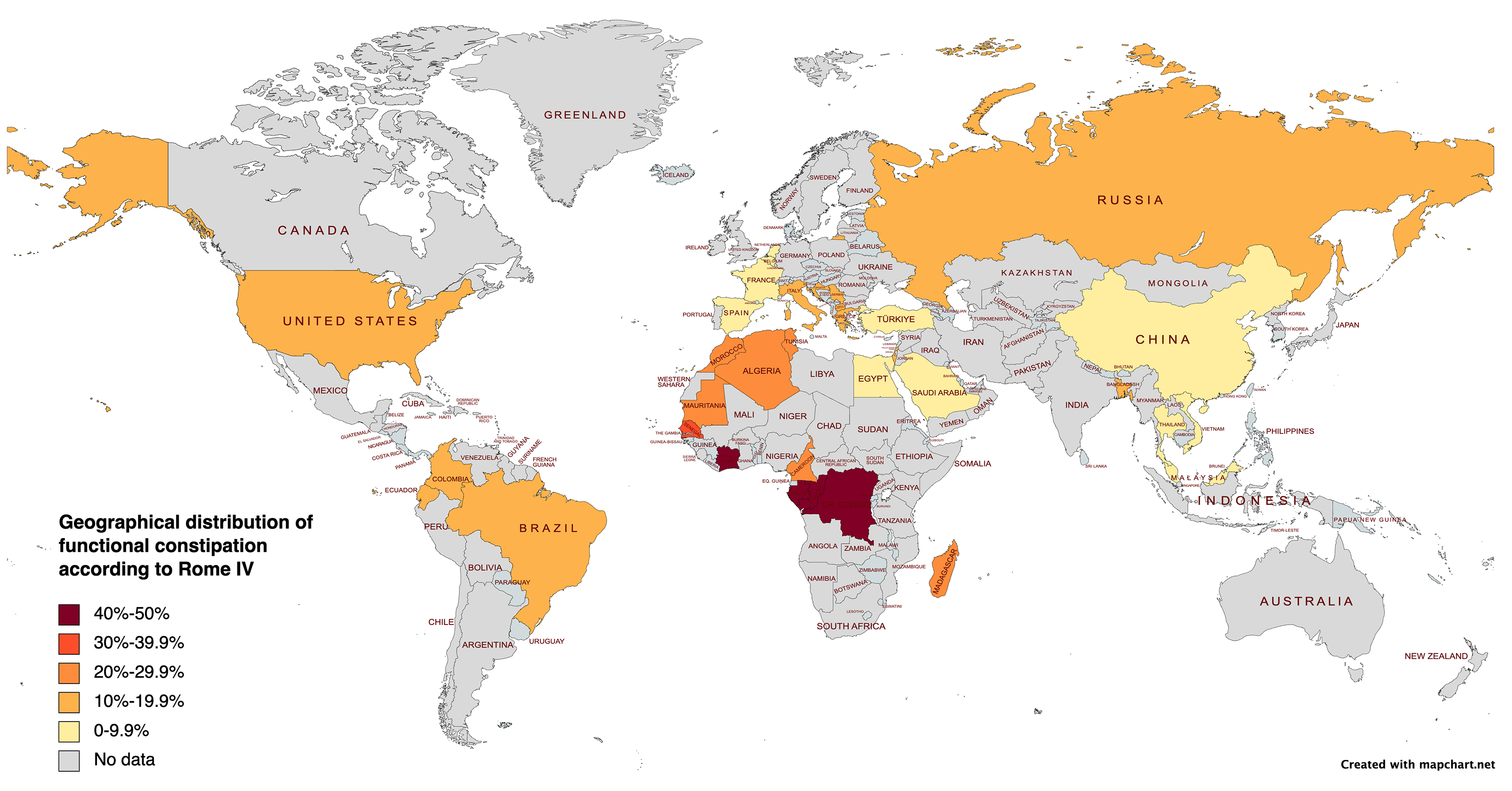
Figure 1 Geographical distribution of functional constipation in children worldwide, according to Rome IV.
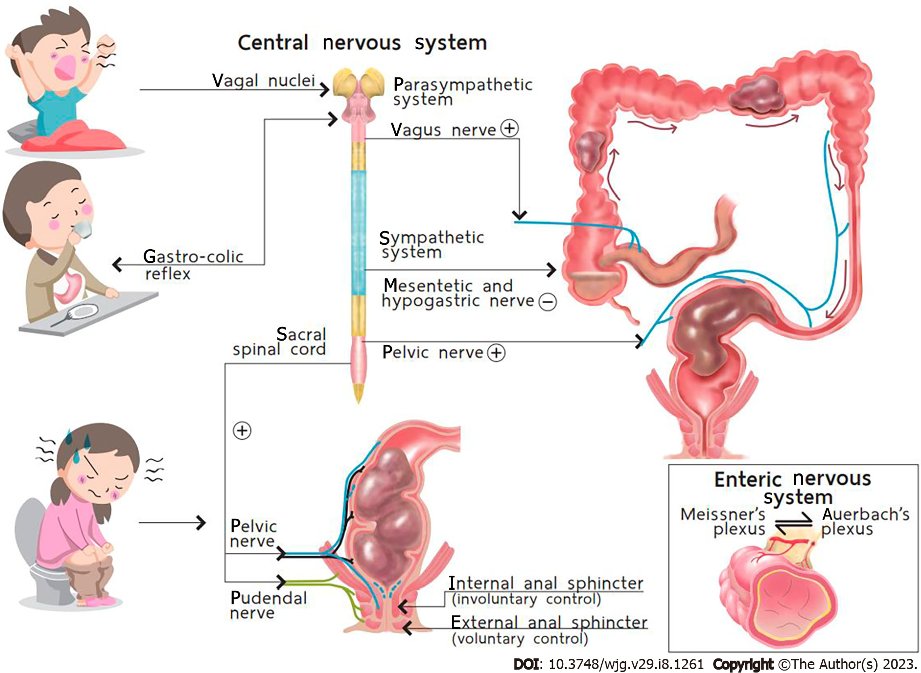
Figure 2 Normal defecation dynamics.
Citation: Palittiya Sintusek. Pediatric lower gastrointestinal motility disorders. In: Werawatganon D, editor. Practical Neurogastroenterology and motility: Basic, testing, and treatment. Bangkok: Printable Inc, 2021: 417. Copyright ©The Authors 2021. Published by Thai Neurogastroenterology and Motility Society. The authors have obtained the permission for figure using from Thai Neurogastroenterology and Motility Society (Supplementary material).
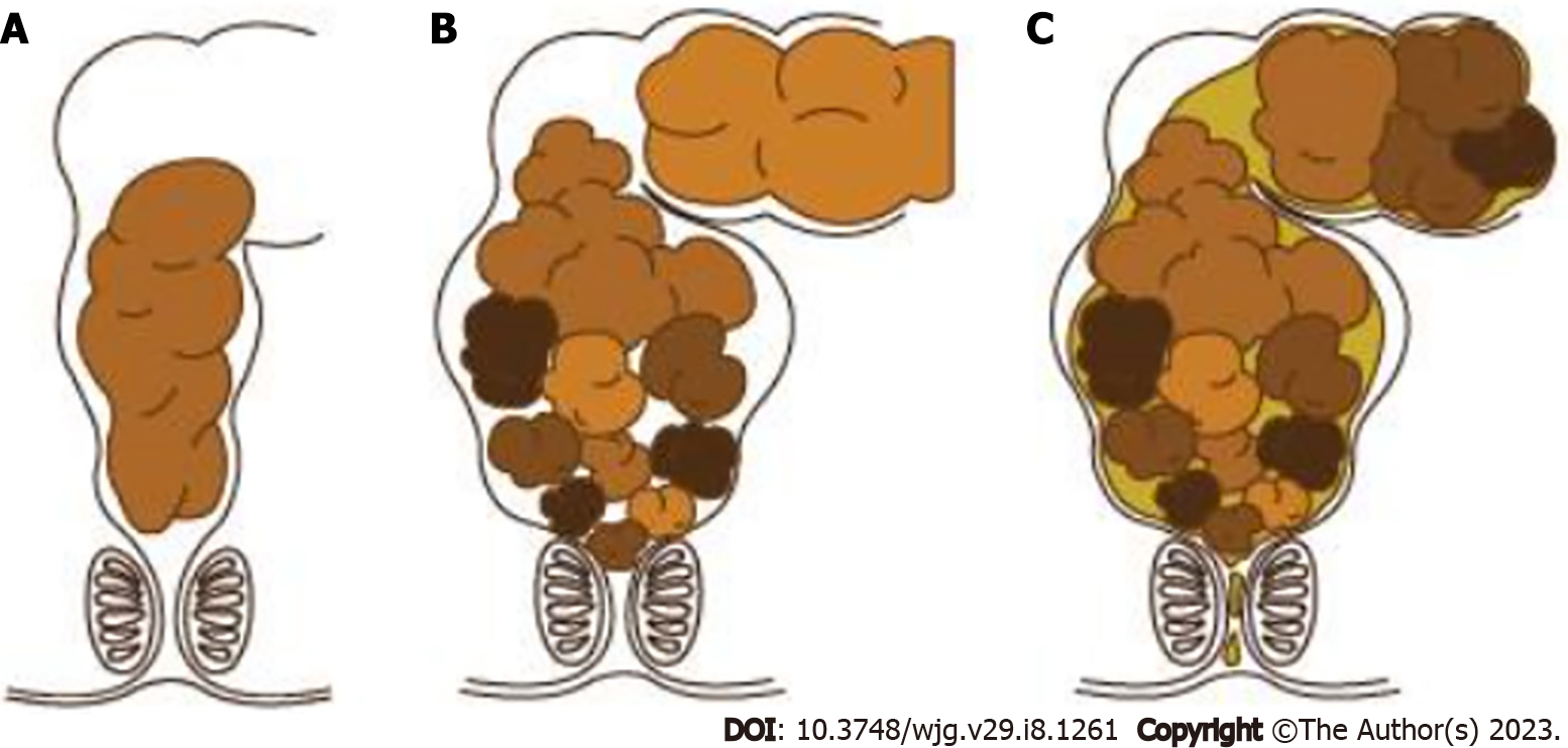
Figure 3 Accumulation of fecal mass in the rectum causes rectal wall distension and fecal soiling.
A: Normal stool; B: Massive stool are retained in the rectum from withholding behavior; C: Liquid stool can leak out the anus or fecal soiling. A-C: Citation: Palittiya Sintusek. Chronic constipation. In: Supapitiporn K, editor. Pediatric practice: simple&applicable. Bangkok: Chulalongkorn University, 2021: 96. Copyright ©The Authors 2017. Published by Department of Pediatrics, Chulalongkorn University. The authors have obtained the permission for figure using from Department of Pediatrics, Chulalongkorn University (Supplementary material).
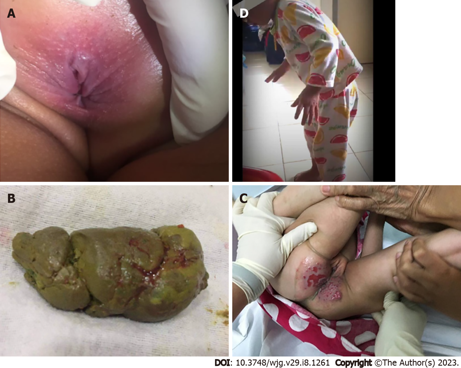
Figure 4 Clinical manifestations in constipated children.
A: Anal fissure usually at the 6 o’clock or posterior parts; B: Blood coated, hard, and lumpy stool; C: Rectal abrasion from fecal incontinence that was misdiagnosed as chronic diarrhea; D: Retentive posture. A-D: Citation: Palittiya Sintusek. Chronic constipation. In: Supapitiporn K, editor. Pediatric practice: simple&applicable. Bangkok: Chulalongkorn University, 2021: 96. Copyright ©The Authors 2017. Published by Department of Pediatrics, Chulalongkorn University. The authors have obtained the permission for figure using from Department of Pediatrics, Chulalongkorn University (Supplementary material).
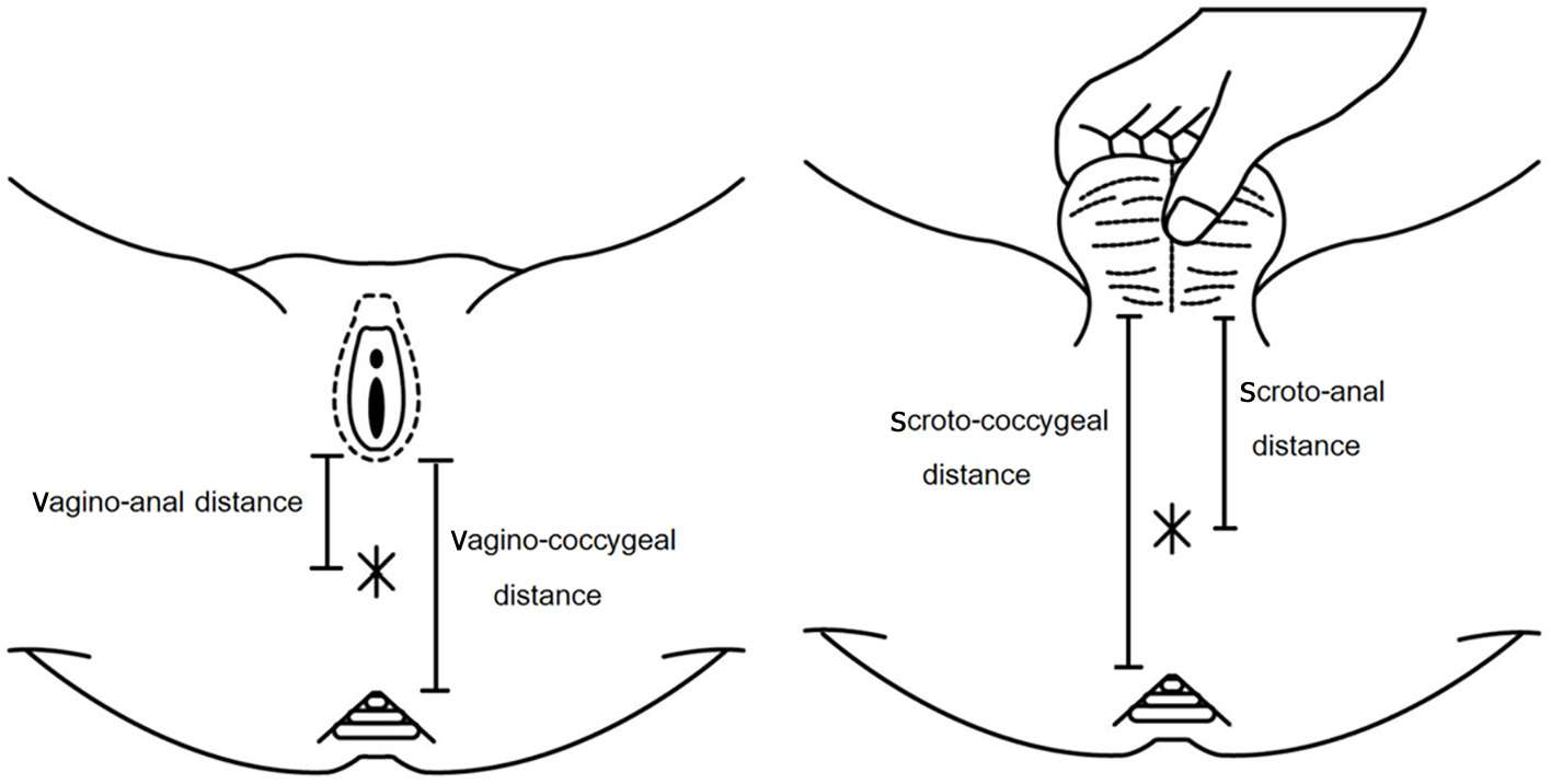
Figure 5 Landmarks for the measurement of the anogenital index.
Citation: Palittiya Sintusek. Chronic constipation. In: Supapitiporn K, editor. Pediatric practice: simple&applicable. Bangkok: Chulalongkorn University, 2021: 96. Copyright ©The Authors 2017. Published by Department of Pediatrics, Chulalongkorn University. The authors have obtained the permission for figure using from Department of Pediatrics, Chulalongkorn University (Supplementary material).
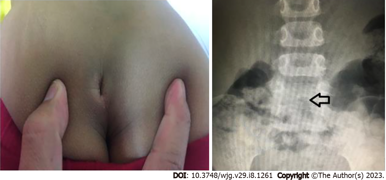
Figure 6 Sacral dimple and spinal dysraphism on spinal radiography (arrow) in a 5-year-old boy with intractable constipation.
A: Sacral dimple; B: Spinal dysraphism.
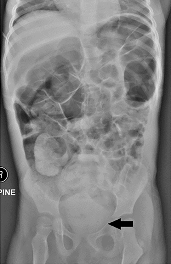
Figure 7 Abdominal radiography of a 3-year-old girl with a chief complaint of chronic diarrhea for 4 mo shows fecal impaction in the rectum.
Citation: Palittiya Sintusek. Chronic constipation. In: Supapitiporn K, editor. Pediatric practice: simple&applicable. Bangkok: Chulalongkorn University, 2021: 96. Copyright ©The Authors 2017. Published by Department of Pediatrics, Chulalongkorn University. The authors have obtained the permission for figure using from Department of Pediatrics, Chulalongkorn University (Supplementary material).
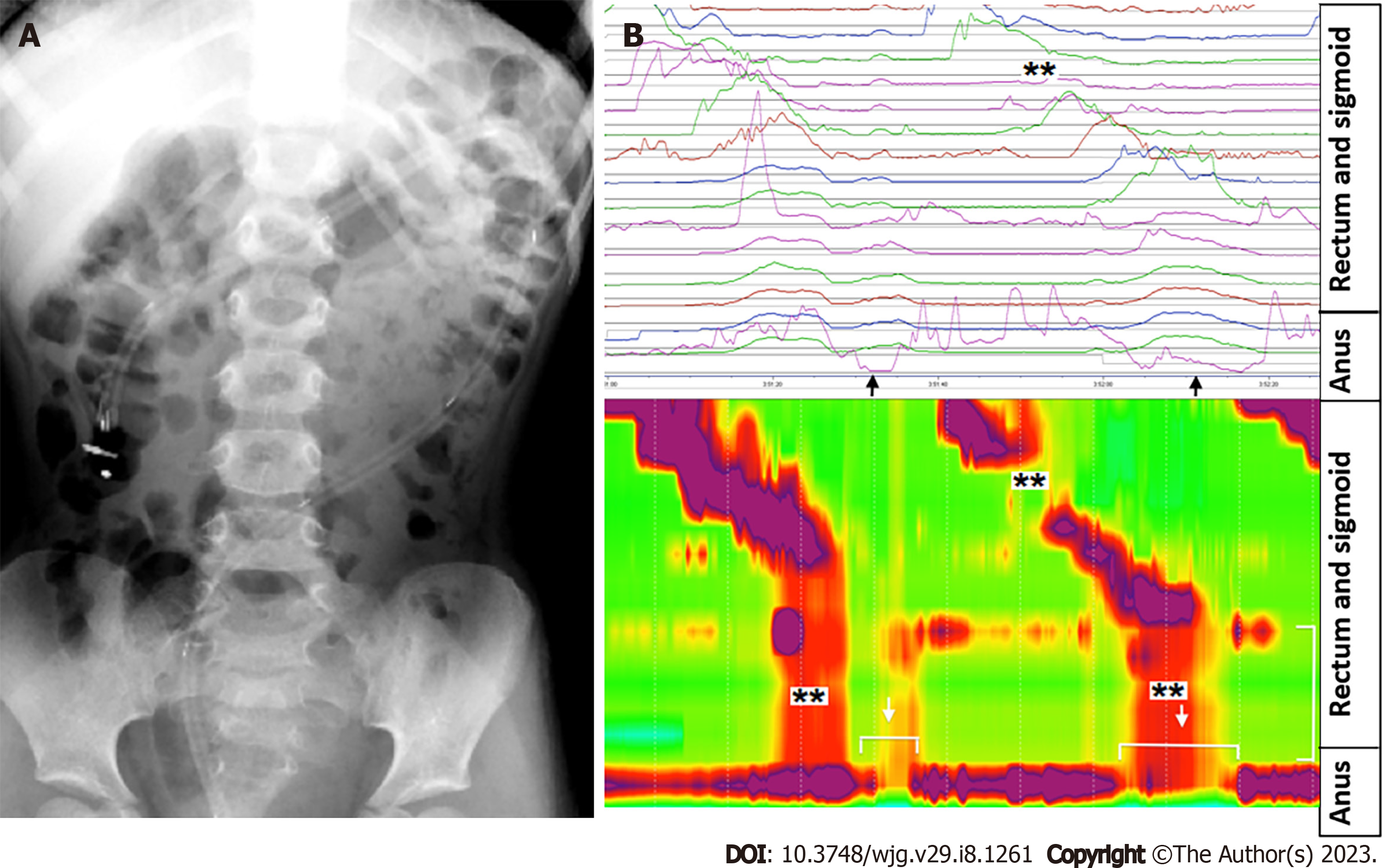
Figure 8 High amplitude propagating contractions.
A: Proper position of the colonic catheter; B: graphical demonstration of abnormal high amplitude propagating contractions (HAPCs) with absent HAPCs at the sigmoid region (**) but preserved colo-anal reflex (arrow). A and B: Citation: Palittiya Sintusek. Chronic constipation. In: Supapitiporn K, editor. Pediatric practice: simple&applicable. Bangkok: Chulalongkorn University, 2021: 96. Copyright ©The Authors 2017. Published by Department of Pediatrics, Chulalongkorn University. The authors have obtained the permission for figure using from Department of Pediatrics, Chulalongkorn University (Supplementary material).
Figure 9 Investigations for organic constipation.
A: Transition zone after barium enema in a child diagnosed with Hirschsprung’s disease; B: Histopathology revealing no ganglion cells in the nerve plexuses (myenteric plexus) of the muscular propria layer. Arrow shows thick nerve trunk but without ganglion cells; C: Anorectal manometry showing rectoanal inhibitory reflex. A-C: Citation: Palittiya Sintusek. Pediatric lower gastrointestinal motility disorders. In: Werawatganon D, editor. Practical Neurogastroenterology and motility: Basic, testing, and treatment. Bangkok: Printable Inc, 2021: 417. Copyright ©The Authors 2021. Published by Thai Neurogastroenterology and Motility Society. The authors have obtained the permission for figure using from Thai Neurogastroenterology and Motility Society (Supplementary material).
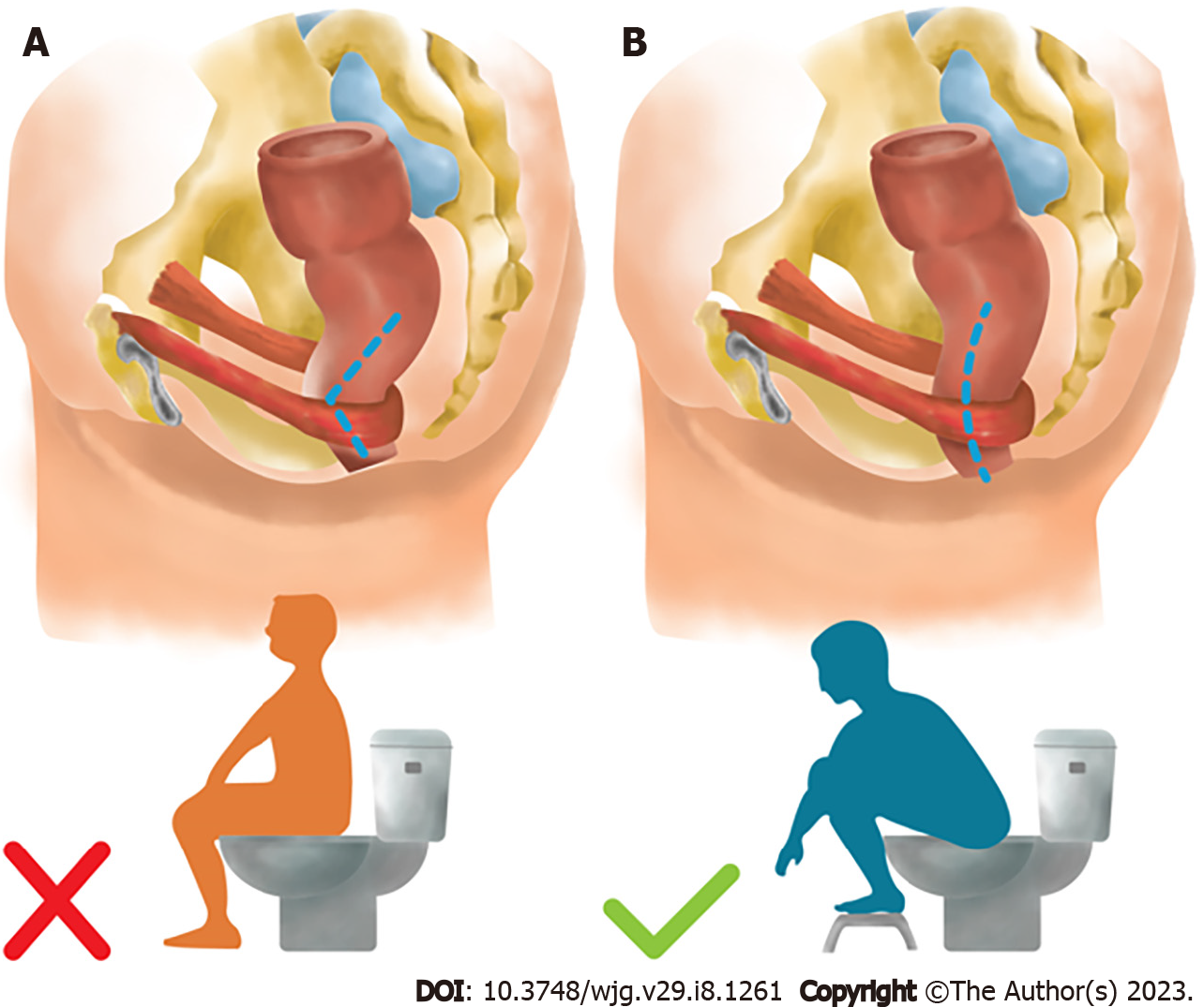
Figure 10 Position during defecation.
A: Improper position during defecation; B: Proper position to open the rectoanal angle and facilitate stool expulsion during defecation.
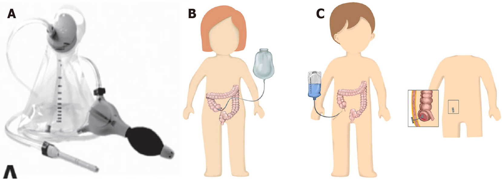
Figure 11 Rectal irrigation method.
A: Transanal irrigation; B and C: Antegrade continence enema. A-C: Citation: Palittiya Sintusek. Pediatric lower gastrointestinal motility disorders. In: Werawatganon D, editor. Practical Neurogastroenterology and motility: Basic, testing, and treatment. Bangkok: Printable Inc, 2021: 423-424. Copyright ©The Authors 2021. Published by Thai Neurogastroenterology and Motility Society. The authors have obtained the permission for figure using from Thai Neurogastroenterology and Motility Society (Supplementary material).
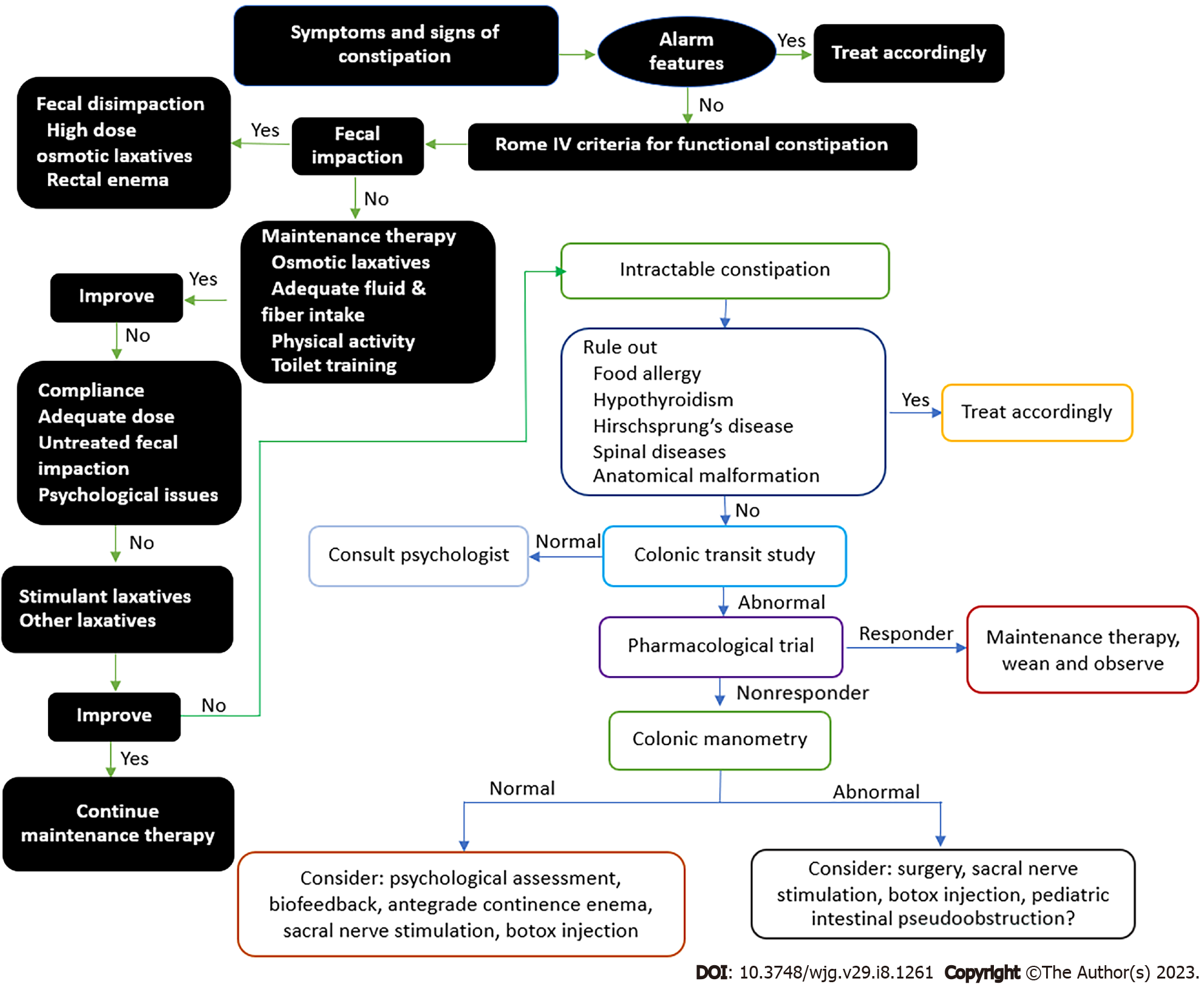
Figure 12 Algorithm for the management of children presenting with signs and symptoms of constipation.
- Citation: Tran DL, Sintusek P. Functional constipation in children: What physicians should know. World J Gastroenterol 2023; 29(8): 1261-1288
- URL: https://www.wjgnet.com/1007-9327/full/v29/i8/1261.htm
- DOI: https://dx.doi.org/10.3748/wjg.v29.i8.1261


