Copyright
©The Author(s) 2023.
World J Gastroenterol. Feb 7, 2023; 29(5): 867-878
Published online Feb 7, 2023. doi: 10.3748/wjg.v29.i5.867
Published online Feb 7, 2023. doi: 10.3748/wjg.v29.i5.867
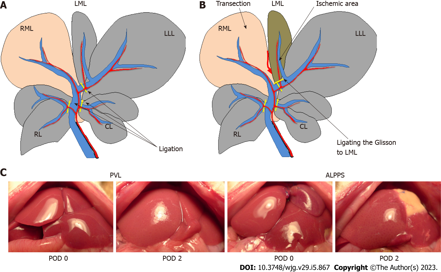
Figure 1 Schema of experimental models.
A: Portal vein ligation (PVL) group. Portal vein branches were ligated, other than the right median lobe; B: Associating liver partition and PVL for staged hepatectomy group. In addition to ligating the portal vein as performed in the PVL group, the median lobe was transected, and the left Glisson was ligated; C: Macroscopic findings after operations in each group. ALPPS: Associating liver partition and portal vein ligation for staged hepatectomy; PVL: Portal vein ligation; RML: Right median lobe; LML: Left median lobe; LLL: Left lateral lobe; RL: Right lobe; CL: Caudate lobe; POD: Postoperative day.
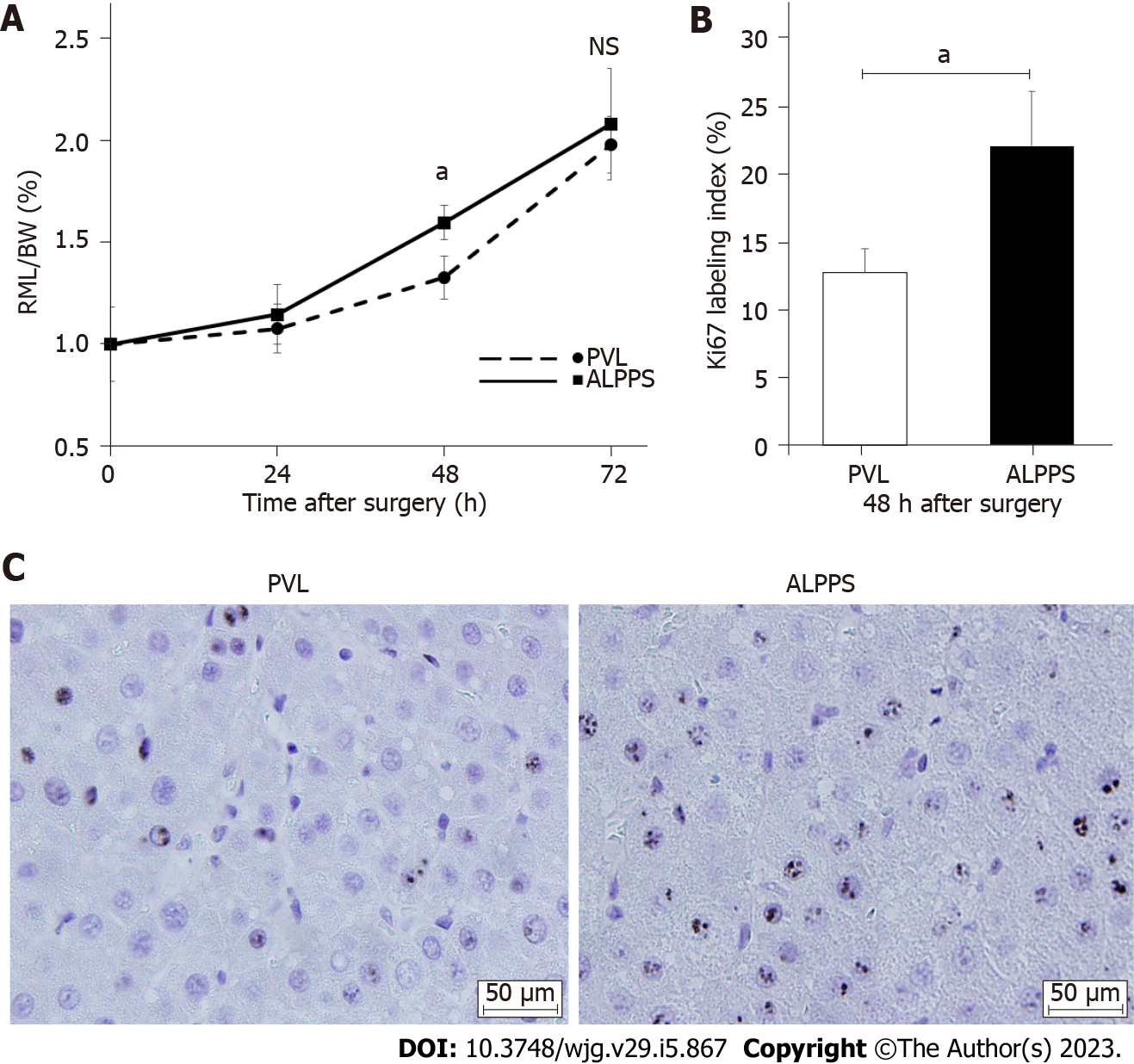
Figure 2 Changes in the right side of the median lobe weight to body weight ratio and Ki-67 index after surgery.
A: Future liver remnant/body weight ratio up to 72 h after surgery; B: Immunohistochemistry of Ki-67 at 48 h after the operation; C: Ki-67 labeling index at 48 h after the surgery. Values are expressed as the mean ± SD; n = 5 for each group; aP < 0.05; NS: Not significant; RML/BW: Right side of the median lobe weight/body weight; PVL: Portal vein ligation; ALPPS: Associating liver partition and portal vein ligation for staged hepatectomy.
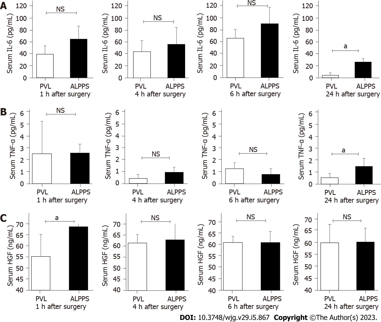
Figure 3 Expression of inflammatory cytokines and hepatocyte growth factor in serum and right side of the median lobe tissue.
A: Serum interleukin-6 concentrations at 1, 4, 6, and 24 h after surgery; B: Tumor necrosis factor-α concentrations at 1, 4, 6, and 24 h after surgery; C: Hepatocyte growth factor concentration at 1, 4, 6, and 24 h after surgery. Values are expressed as the mean ± SD; n = 5 for each group; aP < 0.05; NS: Not significant; PVL: Portal vein ligation; ALPPS: Associating liver partition and portal vein ligation for staged hepatectomy; IL: Interleukin; TNF: Tumor necrosis factor; HGF: Hepatocyte growth factor.
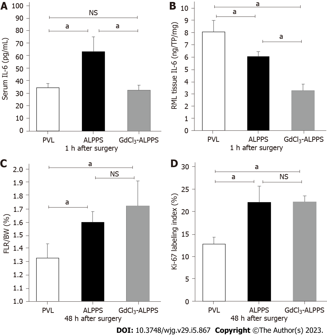
Figure 4 Interleukin-6 expression and liver regeneration in the gadolinium chloride model.
A: The serum concentration of interleukin (IL)-6 at 1 h after surgery; B: IL-6 concentration in right side of the median lobe tissue at 1 h after surgery; C: Future liver remnant/body weight ratio at 48 h after surgery; D: Ki-67 labeling index. Values are expressed as mean ± SD; n = 3 or 5 for each group; aP < 0.05; NS: Not significant; PVL: Portal vein ligation; ALPPS: Associating liver partition and portal vein ligation for staged hepatectomy; IL: Interleukin; RML: Right median lobe; FLR/BW: Future liver remnant/body weight; CdCl3: Gadolinium chloride.
Figure 5 Western blotting of Akt-endothelial nitric oxide synthase pathway-related proteins.
A: Western blotting was used to evaluate the expression of phosphorylated Akt and endothelial nitric oxide synthase (eNOS) in right side of the median lobe at 1, 4, and 6 h after surgery in portal vein ligation (PVL) and associating liver partition and portal vein ligation for staged hepatectomy (ALPPS) groups; B: Comparison of the expression of P-Akt Ser473 and P-eNOS Ser1177 in PVL and ALPPS groups (quantification of western blots, n = 5 for each group). Values are expressed as the mean ± SD; n = 5 for each group; aP < 0.05; NS: Not significant; eNOS: Endothelial nitric oxide synthase; PVL: Portal vein ligation; ALPPS: Associating liver partition and portal vein ligation for staged hepatectomy.
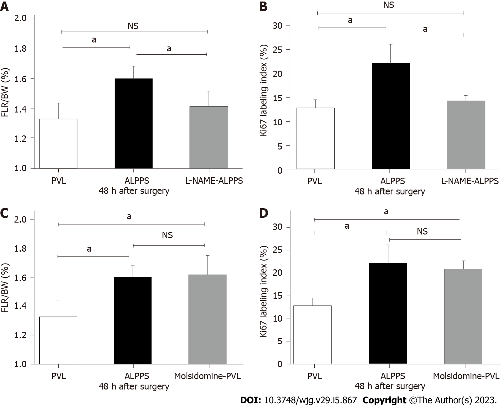
Figure 6 Changes in liver regeneration and cell proliferation due to drug administration.
A: Future liver remnant/body weight (FLR/BW) ratio in the N-nitro-arginine methyl ester (L-NAME)-administered associating liver partition and portal vein ligation for staged hepatectomy (ALPPS) group; B: Ki-67 labeling index in the L-NAME-administered ALPPS group; C: FLR/BW ratio in the molsidomine-administered portal vein ligation (PVL) group; D: Ki-67 labeling index in the molsidomine-administered PVL group; n = 5 for each group; aP < 0.05; NS: Not significant; PVL: Portal vein ligation; ALPPS: Associating liver partition and portal vein ligation for staged hepatectomy; FLR/BW: Future liver remnant/body weight; L-NAME: N-nitro-arginine methyl ester.
Figure 7 Evaluation of hepatic artery and portal vein volumetric blood flow in the future liver remnant after surgery.
A: Portal vein (PV) flow in control, PV ligation (PVL), and associating liver partition and portal vein ligation for staged hepatectomy (ALPPS) groups; B: Hepatic artery flow in control, PVL, and ALPPS groups; C: Total blood flow in control, PVL, and ALPPS groups. Values are expressed as the mean ± SD; n = 4 for each group; aP < 0.05; NS: Not significant; PVL: Portal vein ligation; ALPPS: Associating liver partition and portal vein ligation for staged hepatectomy; HA: Hepatic artery.
- Citation: Masuo H, Shimizu A, Motoyama H, Kubota K, Notake T, Yoshizawa T, Hosoda K, Yasukawa K, Kobayashi A, Soejima Y. Impact of endothelial nitric oxide synthase activation on accelerated liver regeneration in a rat ALPPS model. World J Gastroenterol 2023; 29(5): 867-878
- URL: https://www.wjgnet.com/1007-9327/full/v29/i5/867.htm
- DOI: https://dx.doi.org/10.3748/wjg.v29.i5.867


