Copyright
©The Author(s) 2023.
World J Gastroenterol. Dec 21, 2023; 29(47): 6138-6147
Published online Dec 21, 2023. doi: 10.3748/wjg.v29.i47.6138
Published online Dec 21, 2023. doi: 10.3748/wjg.v29.i47.6138
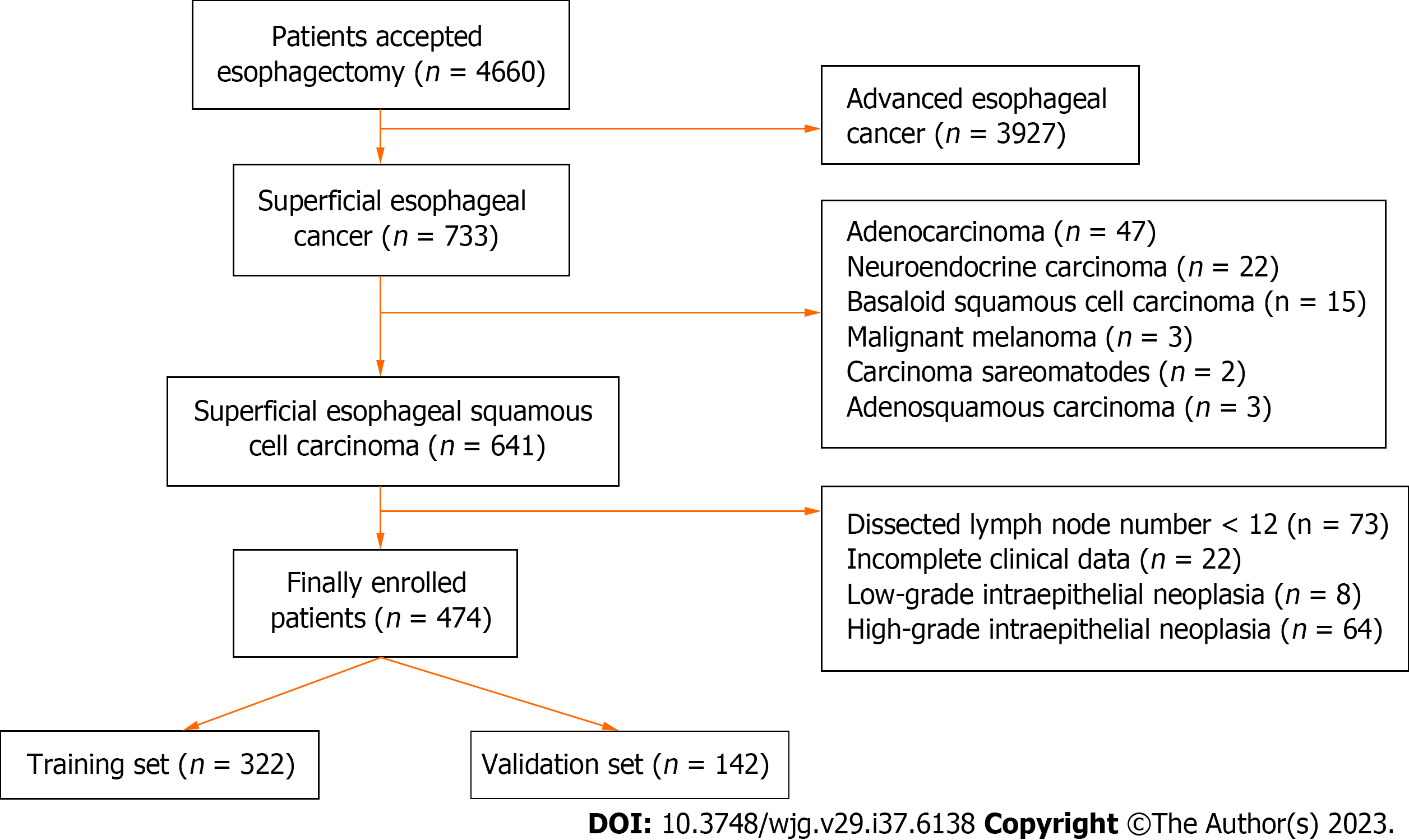
Figure 1
Flowchart of enrollment process.
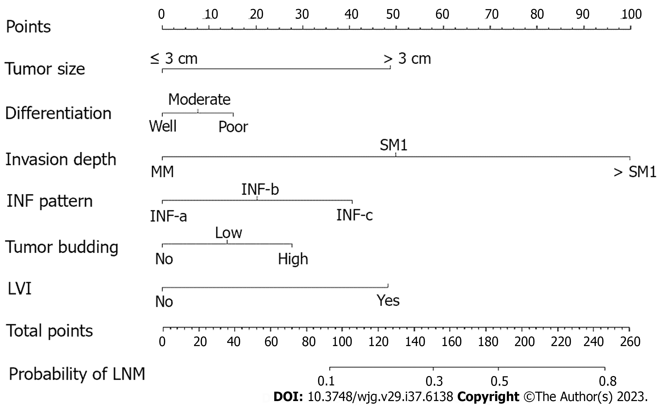
Figure 2 Nomogram for predicting the probability of lymph node metastasis in superficial esophageal squamous cell carcinoma patients.
Calculate the total points of different characteristics, and drop a vertical line from the total points row to obtain the probability of lymph node metastasis. MM: Muscularis mucosae; SM1: Upper third of submucosa; SM2: Middle third of submucosa; SM3: Lower third of submucosa; INF pattern: Infiltrative growth pattern; LVI: Lymphovascular invasion; LNM: Lymph node metastasis.
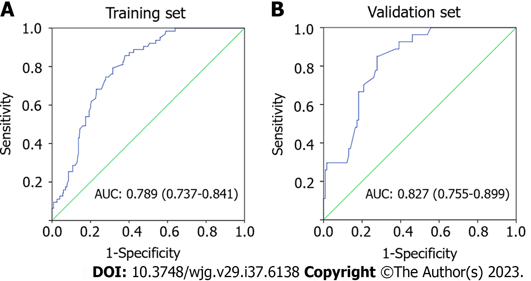
Figure 3 Receiver operating characteristics curve of the nomogram for predicting lymph node metastasis.
A: Receiver operating characteristics (ROC) curve in the training set; B: ROC curve in the validation set. AUC: Area under the curve.
- Citation: Wang J, Zhang X, Gan T, Rao NN, Deng K, Yang JL. Risk factors and a predictive nomogram for lymph node metastasis in superficial esophageal squamous cell carcinoma. World J Gastroenterol 2023; 29(47): 6138-6147
- URL: https://www.wjgnet.com/1007-9327/full/v29/i47/6138.htm
- DOI: https://dx.doi.org/10.3748/wjg.v29.i47.6138


