Copyright
©The Author(s) 2023.
World J Gastroenterol. Nov 21, 2023; 29(43): 5834-5847
Published online Nov 21, 2023. doi: 10.3748/wjg.v29.i43.5834
Published online Nov 21, 2023. doi: 10.3748/wjg.v29.i43.5834
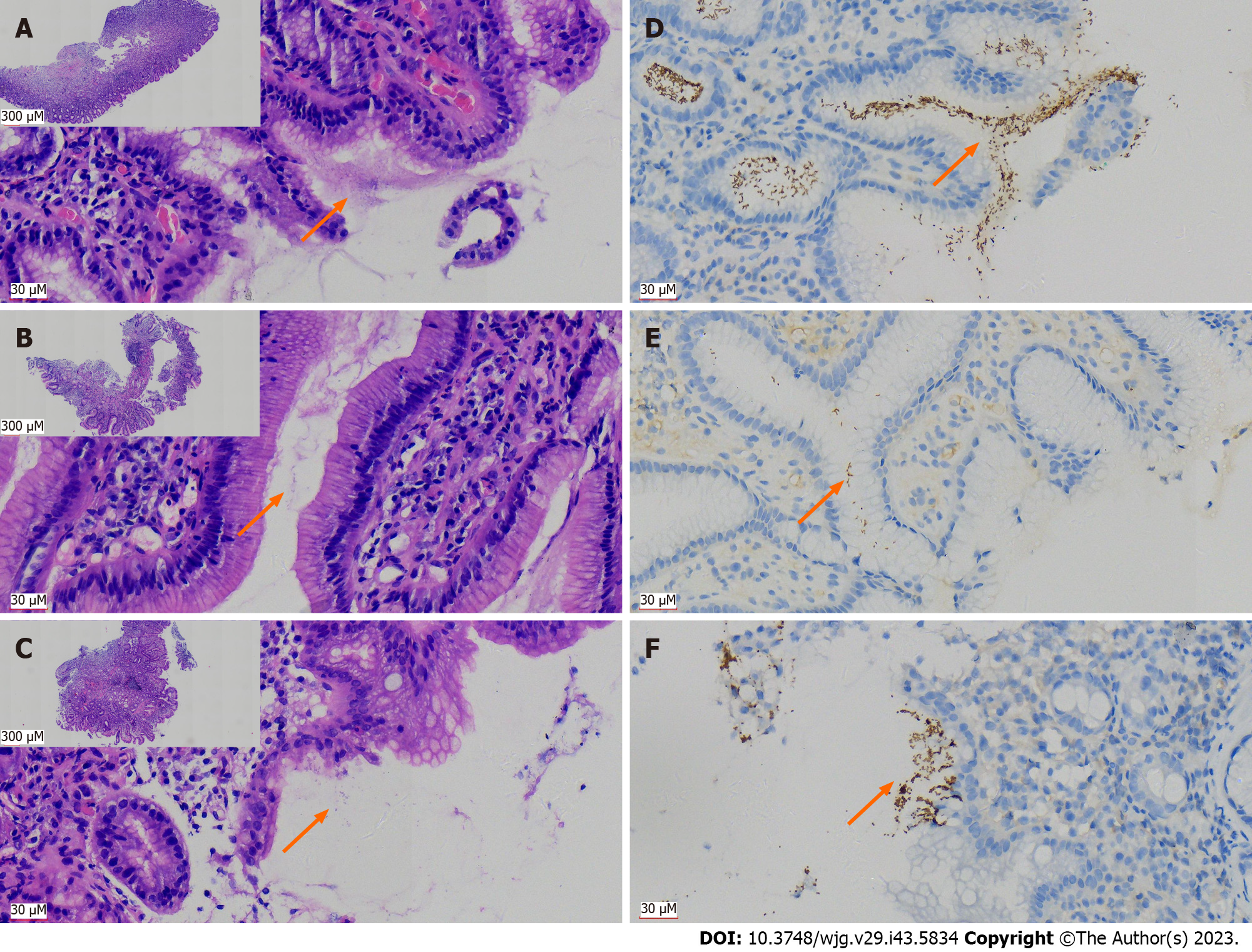
Figure 1 Immunohistochemical staining using antibodies against Helicobacter pylori has significant advantages over hematoxylin and eosin staining in the pathological tissue for gastroscopy biopsy.
A: Distribution of curved rod-shaped bacteria [hematoxylin and eosin (H&E, 400 ×)], on the surface of glands and in the mucus of typical Helicobacter pylori (H. pylori)-positive patients in H&E section; B: A case with low bacterial count of H. pylori (H&E, 400 ×); C: A case with atypical H. pylori morphology (mainly coccoid rather than rod-shaped), which was difficult to identify (H&E, 400 ×), lower magnification figures of A-C are in the upper left corner (H&E, 40 ×); D: Immunohistochemistry (IHC) with antibodies against H. pylori indicated a large number of bacteria, exhibiting brown yellow deep staining (DAB, high magnification, one case the same as in A); E: IHC displayed H. pylori bacilli more clearly than H&E due to a lower bacterial count (DAB staining, high magnification, one case the same as in B); F: Atypical coccoid H. pylori were clearly present and confirmed by IHC (DAB staining, high magnification, one case the same as in C); orange arrows in A-F indicate distribution of H. pylori.
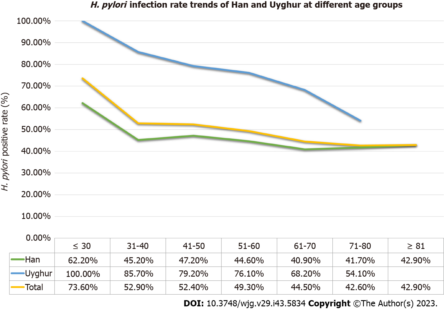
Figure 2 Helicobacter pylori positive rate changed with age in Xinjiang Uyghur Autonomous Region.
Regardless of the Uyghur, Han or the overall series, the Helicobacter pylori (H. pylori) infection rates calculated by age group showed a decreasing trend as age increased. In Han group, the positive rate of H. pylori infection increased in two age groups after the age of 70 years. H. pylori: Helicobacter pylori.
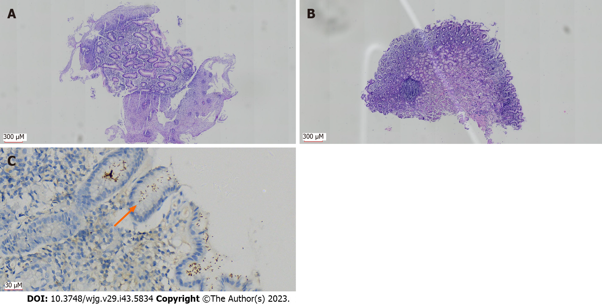
Figure 3 Patients with Barrett’s esophagus concurrent with Helicobacter pylori infection.
A: Changes with Barrett’s esophagus: Many hyperplastic glands in the esophageal wall, partially replacing the squamous epithelial mucosa [hematoxylin and eosin (H&E, 40 ×)]; B: Mild inflammation (H&E, 40 ×) in the gastric wall tissue biopsied simultaneously; C: Immunohistochemistry showing Helicobacter pylori (H. pylori) infection (DAB, 400 ×); orange arrow in C indicates distribution of H. pylori.
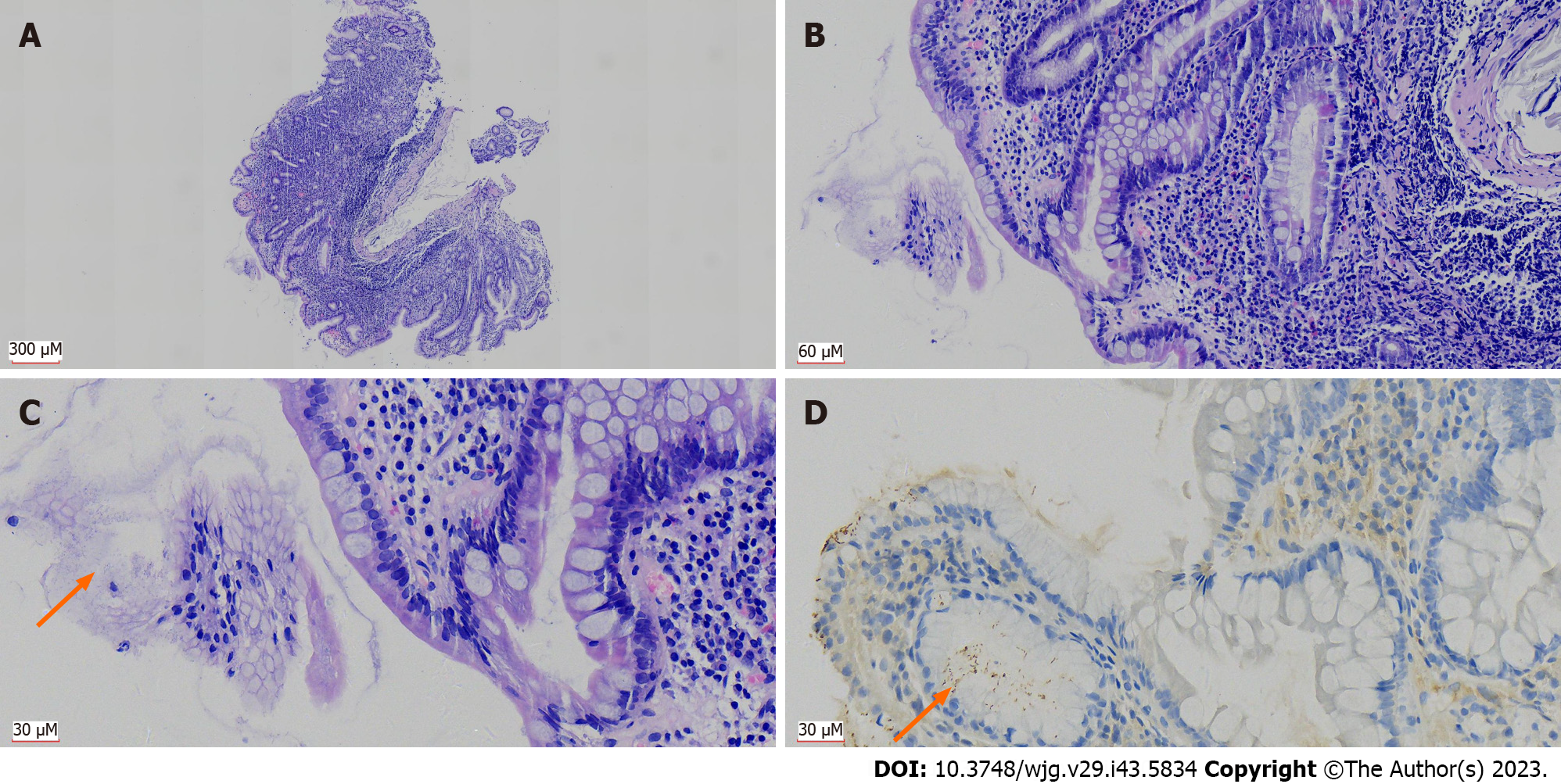
Figure 4 Helicobacter pylori infection associated with elevated grade of chronic/active gastritis and intestinal metaplasia.
A: Patient with severe chronic gastritis (grade 3) in the gastric wall biopsy tissue; inflammatory cell infiltration can be seen throughout the lamina propria of the mucosa [hematoxylin and eosin (H&E, 40 ×)]; B: Infiltrating inflammatory cells were mainly lymphocytes, with scattered neutrophils, indicating chronic gastritis combined with active gastritis (H&E, 200 ×); C: Gastric glands were infected by Helicobacter pylori (H. pylori) and were accompanied by severe intestinal metaplasia (H&E, 400 ×); D: H. pylori infection was confirmed by immunohistochemistry (DAB, 400 ×); all images were from the same patient, orange arrows in C and D indicates distribution of H. pylori.
- Citation: Peng YH, Feng X, Zhou Z, Yang L, Shi YF. Helicobacter pylori infection in Xinjiang Uyghur Autonomous Region: Prevalence and analysis of related factors. World J Gastroenterol 2023; 29(43): 5834-5847
- URL: https://www.wjgnet.com/1007-9327/full/v29/i43/5834.htm
- DOI: https://dx.doi.org/10.3748/wjg.v29.i43.5834


