Copyright
©The Author(s) 2023.
World J Gastroenterol. Jul 21, 2023; 29(27): 4289-4316
Published online Jul 21, 2023. doi: 10.3748/wjg.v29.i27.4289
Published online Jul 21, 2023. doi: 10.3748/wjg.v29.i27.4289
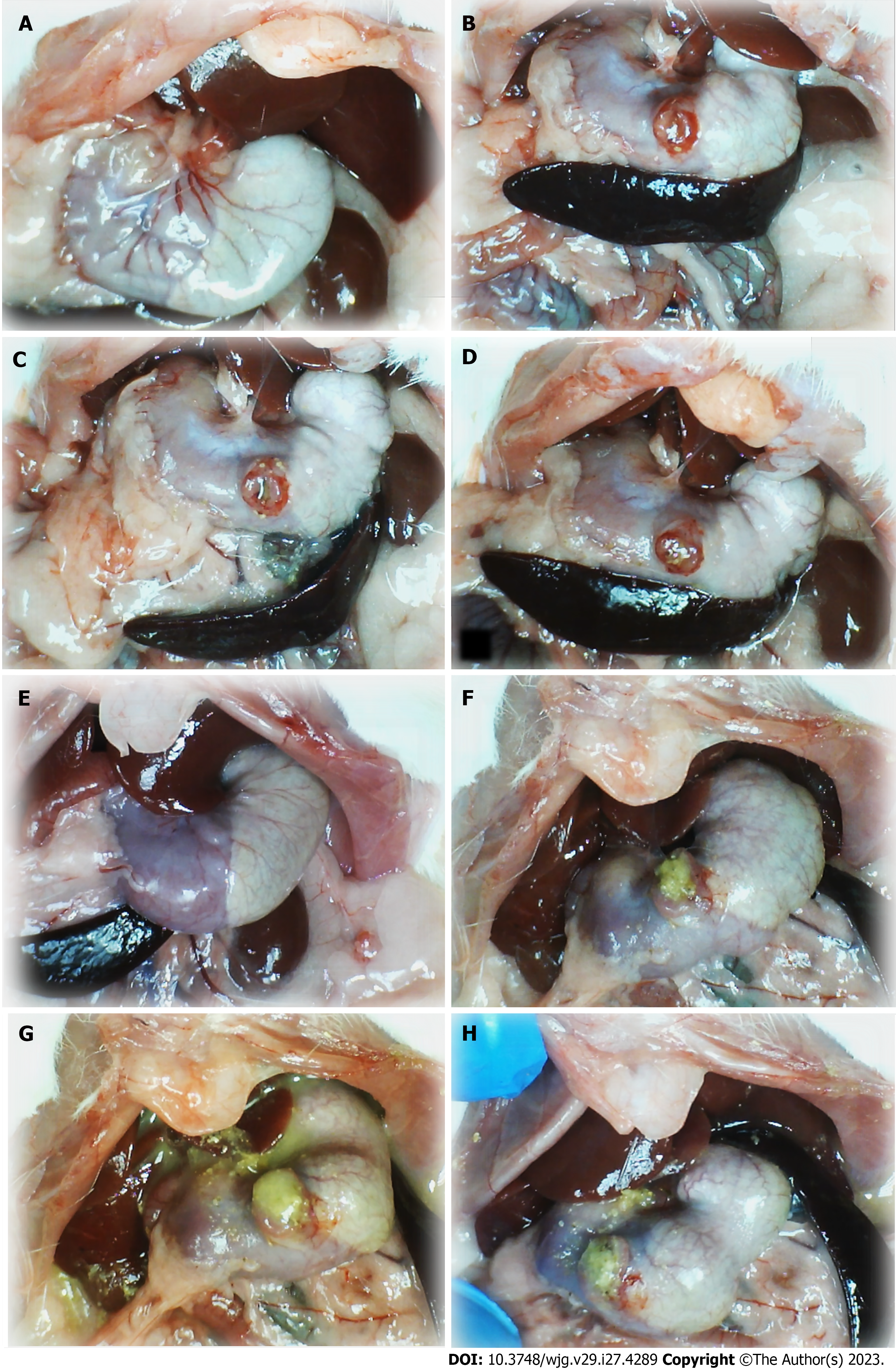
Figure 1 Stomach illustrative presentation.
A and E: Before procedure; B and F: After perforation before therapy; C and D: Course after given therapy into the perforated defect, saline; G and H: BPC 157. Control rats: (1) Before procedure (normal rats, A); (2) after perforation before therapy application (B); (3) after saline application (1 mL/rat into the perforated defect in the stomach) (C and D), immediately thereafter (C); and (4) later (at 5 min following medication, D). BPC 157 rats: (1) Stomach before perforation (normal rats, E); (2) after perforation, but before BPC 157 therapy application (F), and (3) specifically after BPC 157 application (10 µg or 10 ng/kg, 1 mL/rat into the perforated defect in the stomach, G and H), immediately thereafter (G), and later (at 5 min following medication, H).
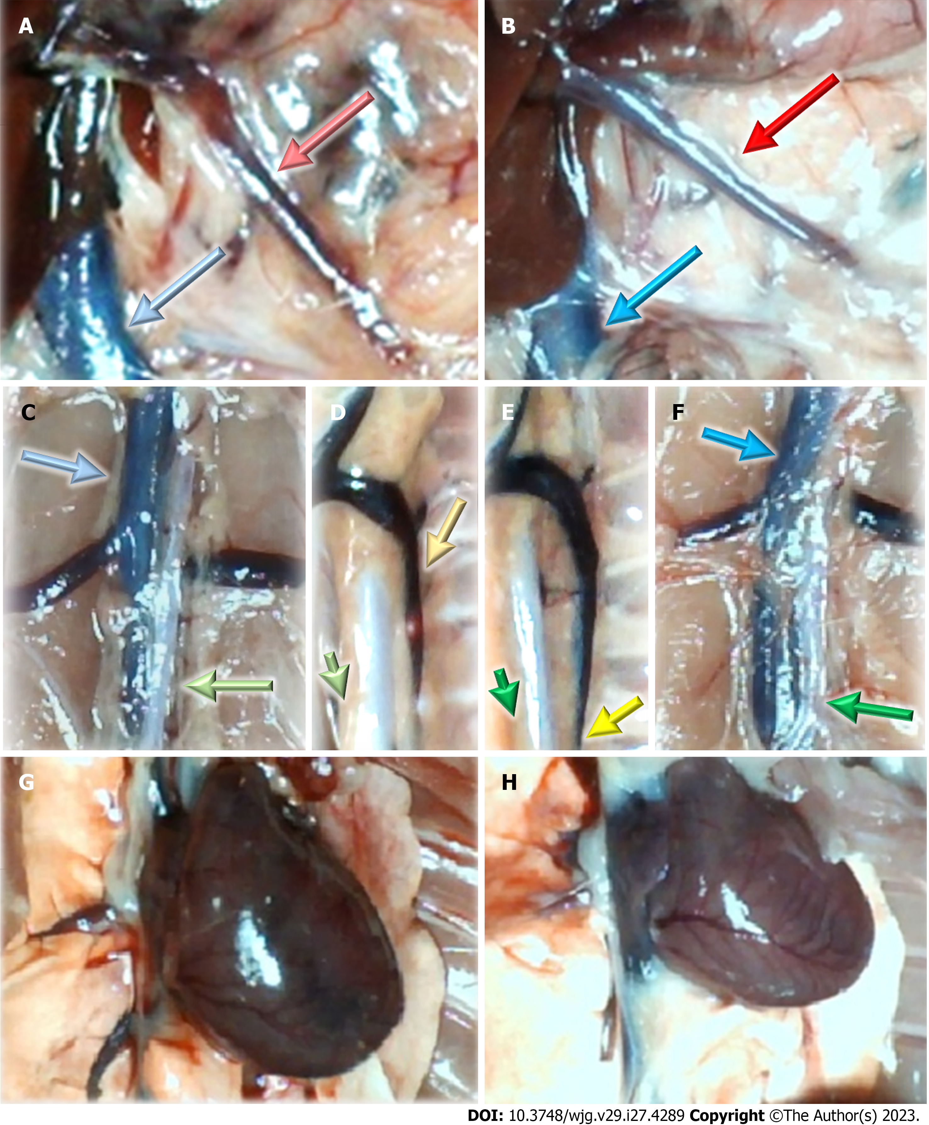
Figure 2 Vessels and heart illustrative presentation.
A, C, D, G: In the control rats (light color arrows); B, E, F, H: In the BPC 157 rats (dark color arrows); superior mesenteric vein (red arrows), inferior caval vein (blue arrows), aorta (green arrows), azygos vein (yellow arrows), and heart (G and H).
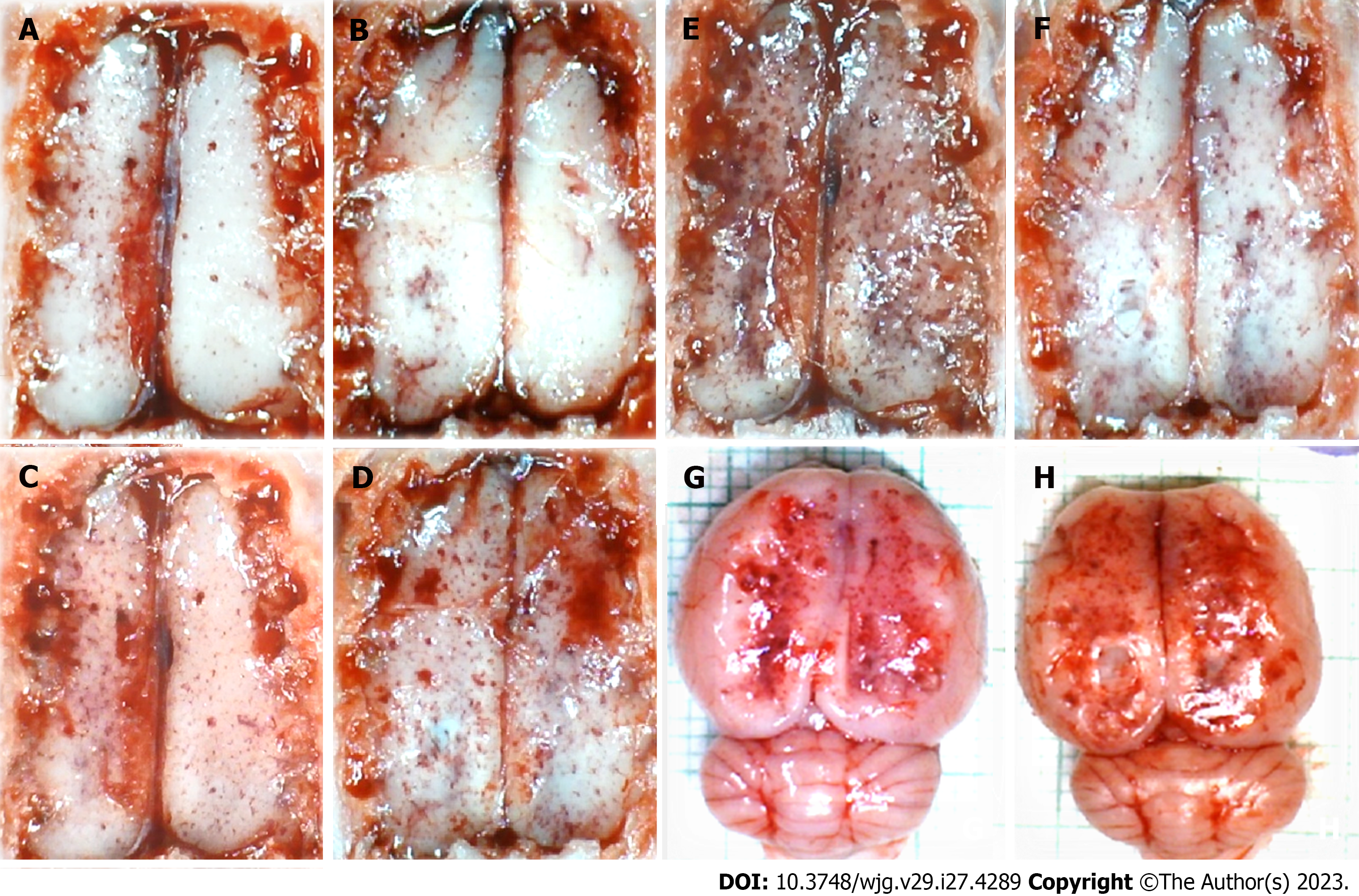
Figure 3 Brain illustrative presentation in the rats with perforated stomach.
A, C, E, G: Control rats before perforation (A); after perforation (C, E, G), before saline medication (C), specifically after saline application (5 mL/kg, 1 mL/rat into the perforated defect in the stomach; E and G), immediately thereafter (E), and later after sacrifice (G); B, D, F, H: BPC 157 rats before perforation (B); after perforation (D, F, H), before BPC 157 medication (D), specifically after BPC 157 application (10 µg or 10 ng/kg, 1 mL/rat into the perforated defect in the stomach, capitals; F, H), immediately thereafter (F), and later after sacrifice (H).
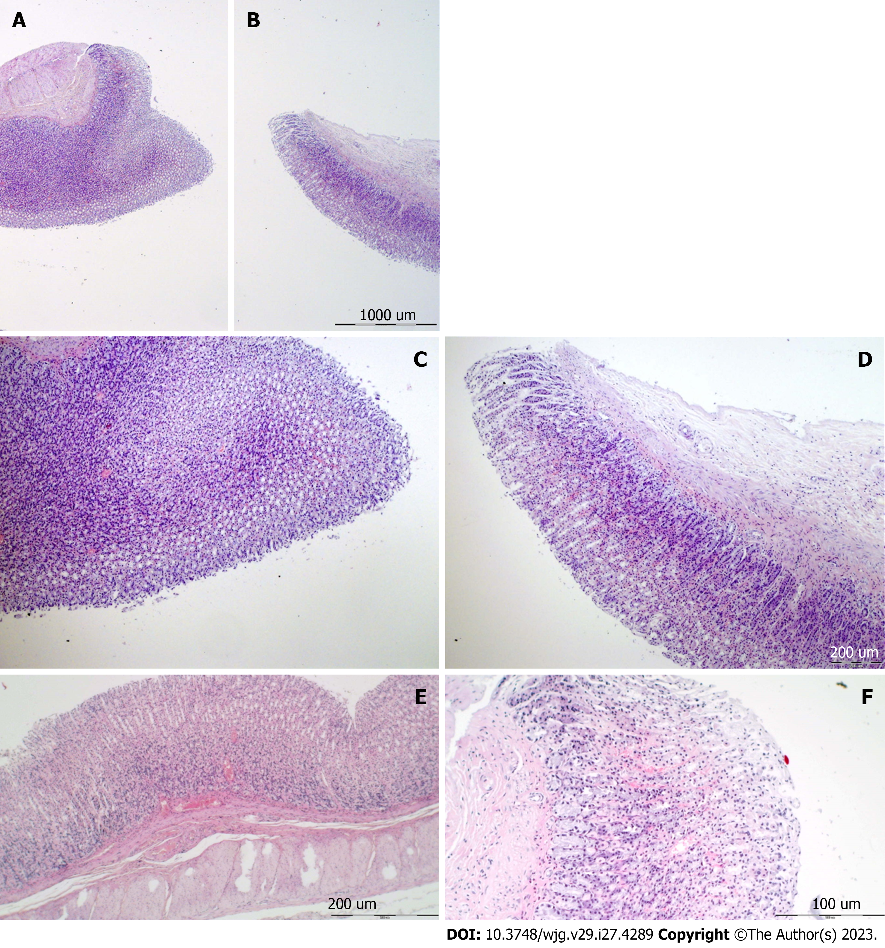
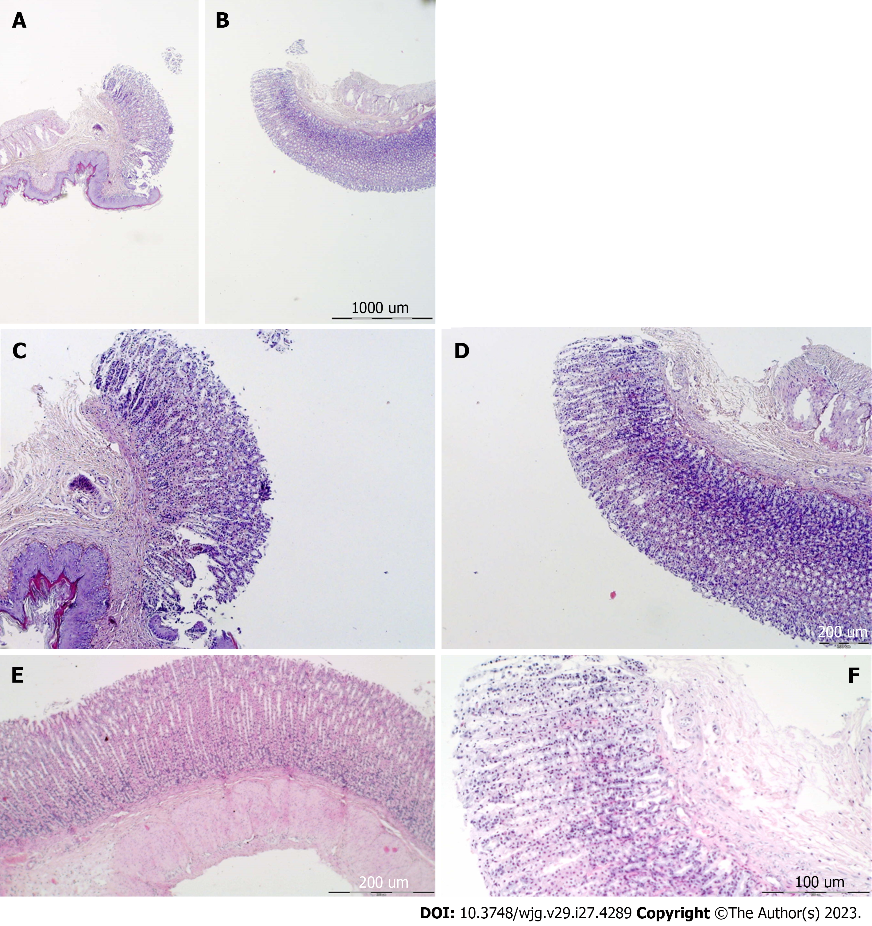
Figure 4 Stomach histology and control.
A-F: Margins of the stomach perforation showed mucosal congestion. Transmural hyperemia of the stomach wall at the margins of the stomach perforation and in the rest of the stomach wall (A, E). HE staining: Magnification 20 × (A, B), magnification 100 × (C-E), magnification 200 × (F).
Figure 5 Stomach histology, BPC 157.
A-F: Margins of the stomach perforation showed only mild mucosal congestion. No congestion was found in the rest of the stomach wall (A and E). HE staining: Magnification 20 × (A and B), magnification 40 × (C-E), and magnification 100 × (F).
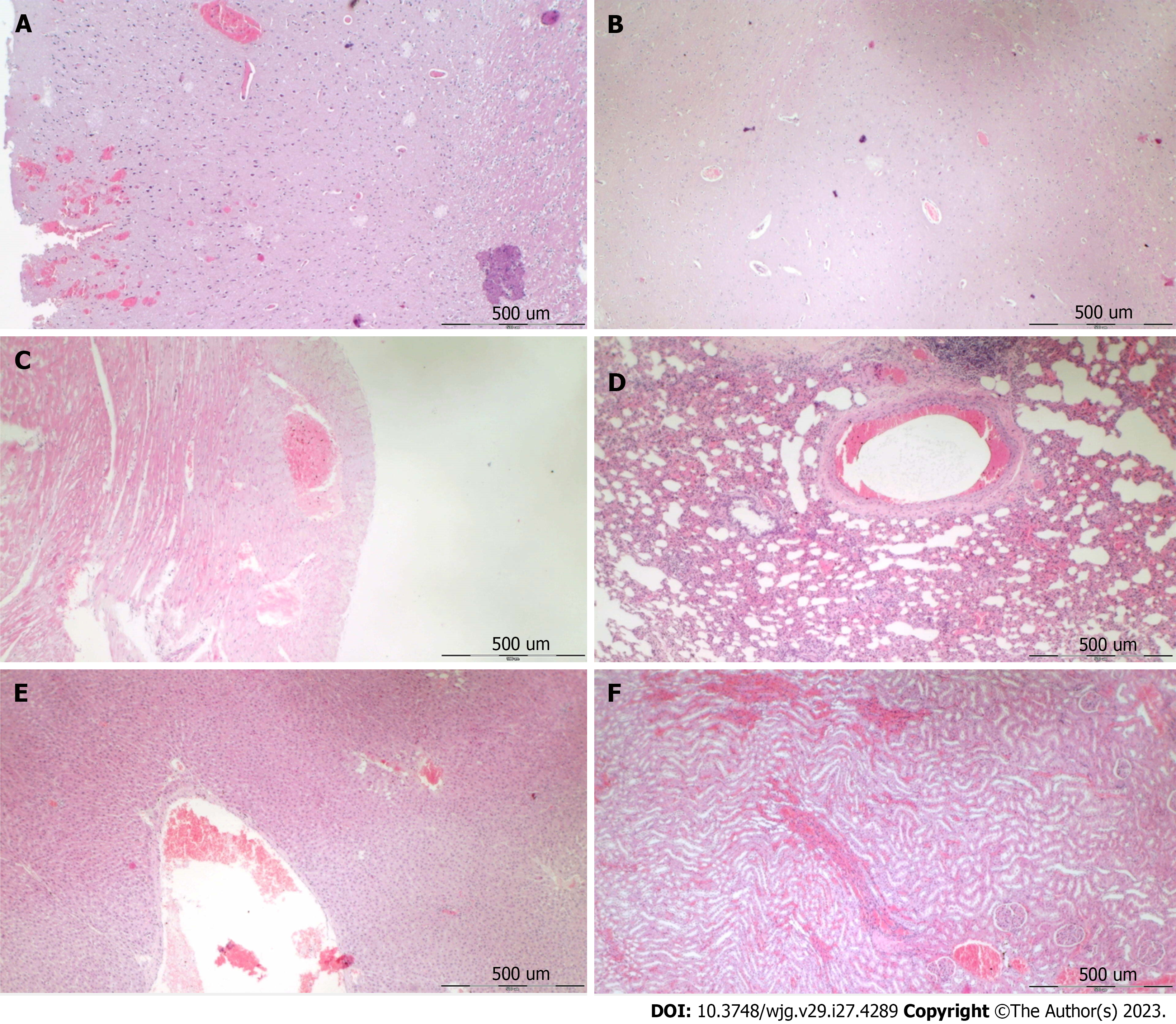
Figure 6 Following stomach perforation, illustrative microscopy presentation of the organ lesion in saline-treated rats (control).
A and B: Cerebral cortex, increased edema and congestion in the cerebral cortex. Severe cerebral hemorrhage of the neocortex; C: Heart, severe congestion within the myocardium; D: Lung, focal thickening of the alveolar membrane, lung congestion, pulmonary edema, and severe intra-alveolar hemorrhage; E: Liver, marked congestion of liver parenchyma with dilatation of blood vessels in portal tracts, central veins, and sinusoids; F: Kidney, a renal parenchyma showed severe vascular congestion and mild interstitial edema. HE staining: Magnification 40 ×.
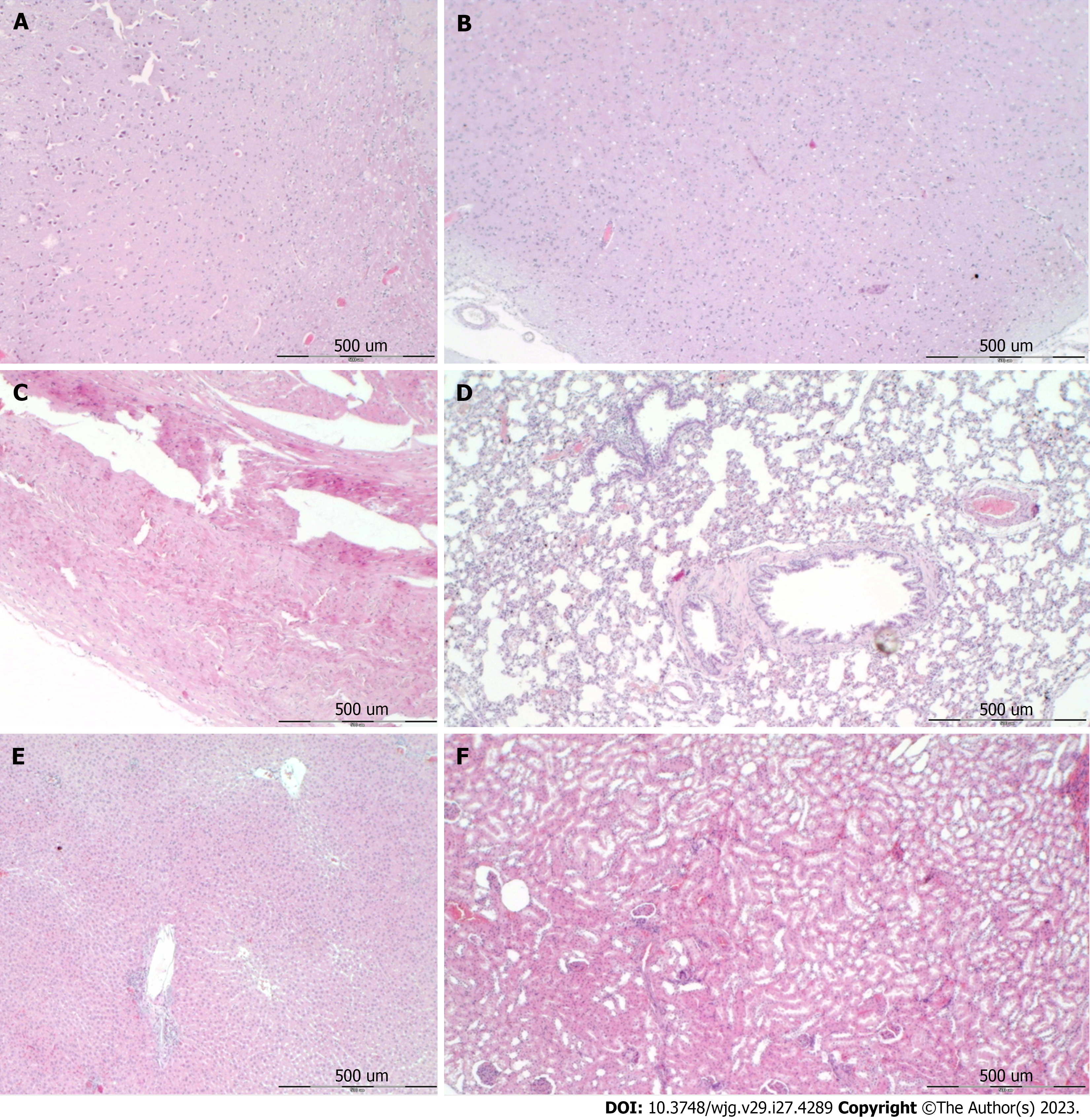
Figure 7 Following stomach perforation, illustrative microscopy presentation of the organs lesion in BPC 157 treated rats.
A and B: Cerebral cortex, no edema and congestion in the cerebral cortex. Small cerebral hemorrhage of the neocortex; C: Heart, no changes; D: Lung, only mild congestion in the lung parenchyma; E: Liver, no changes; F: Kidney, no changes. HE staining: Magnification 40 ×.
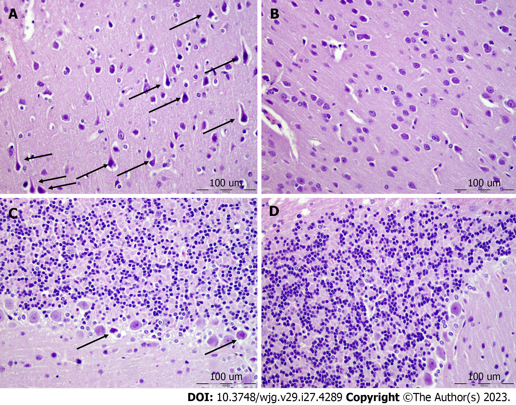
Figure 8 Neocortex, cerebellum, and neuropathological changes.
A: In the control group marked edema and congestion in the neocortical brain tissue of the control animal with an increased number of karyopycnotic nuclei of cortical neurons; B: Only mild edema was found in the neocortex in BPC 157 treated animals; C: Edema and an increased number of karyopycnotic nuclei of the cerebellar Purkinje cells in the control animals; D: No changes were found in BPC 157 treated animals. HE staining: Magnification 400 ×.
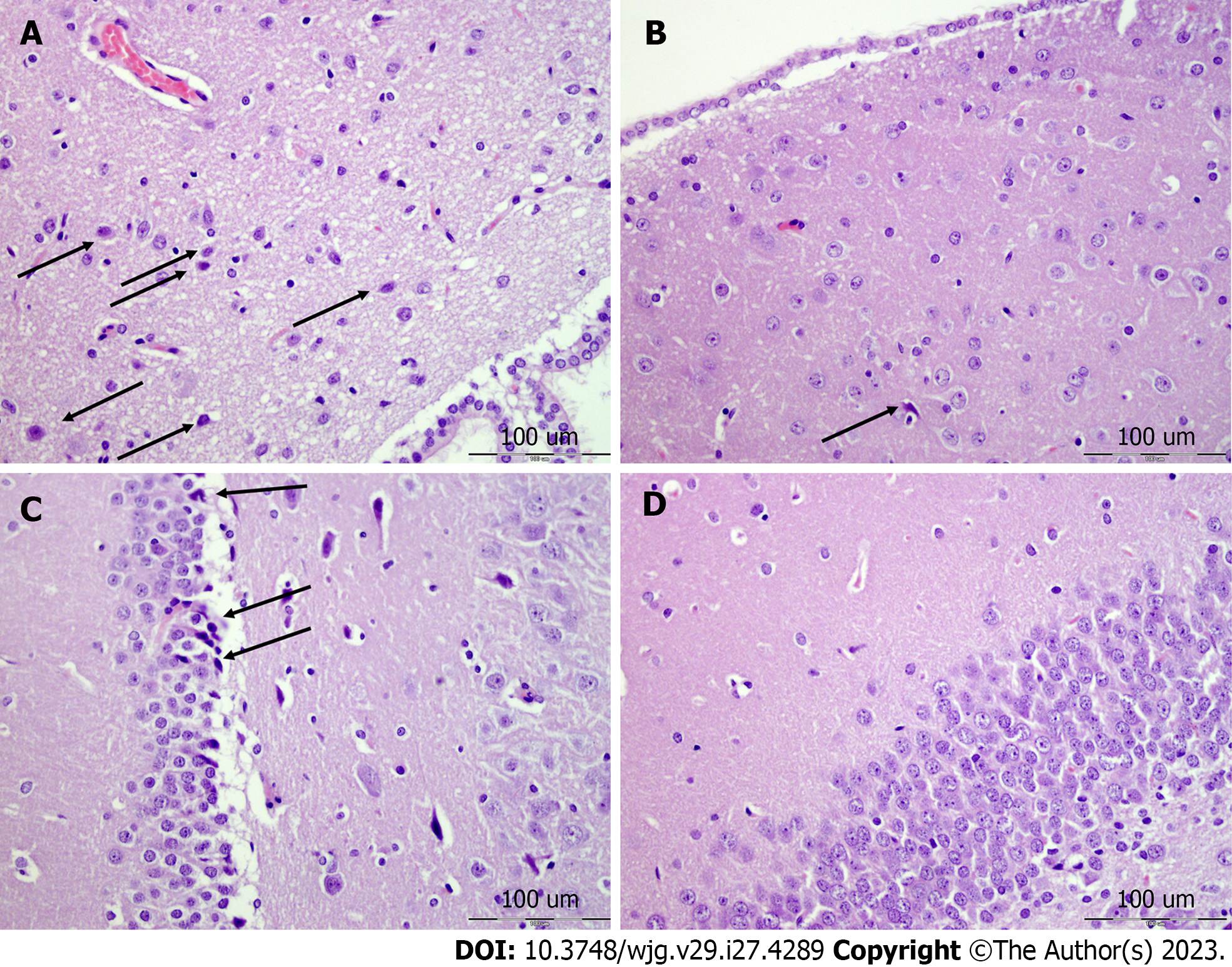
Figure 9 Hypothalamus, hippocampus, neuropathological changes.
A: In the control group marked edema and congestion in the hypothalamic area of control animals with brain tissue of control animal with an increased number of karyopycnotic cells; B: Only a few karyopycnotic cells in BPC 157 treated animals; C: Edema and increased number of karyopycnotic nuclei of the hippocampal area were noted in the control animals; D: While no changes were found in BPC 157 treated animals. HE staining, magnification 400 ×.
- Citation: Kalogjera L, Krezic I, Smoday IM, Vranes H, Zizek H, Yago H, Oroz K, Vukovic V, Kavelj I, Novosel L, Zubcic S, Barisic I, Beketic Oreskovic L, Strbe S, Sever M, Sjekavica I, Skrtic A, Boban Blagaic A, Seiwerth S, Sikiric P. Stomach perforation-induced general occlusion/occlusion-like syndrome and stable gastric pentadecapeptide BPC 157 therapy effect. World J Gastroenterol 2023; 29(27): 4289-4316
- URL: https://www.wjgnet.com/1007-9327/full/v29/i27/4289.htm
- DOI: https://dx.doi.org/10.3748/wjg.v29.i27.4289


