Copyright
©The Author(s) 2023.
World J Gastroenterol. Jul 7, 2023; 29(25): 4009-4020
Published online Jul 7, 2023. doi: 10.3748/wjg.v29.i25.4009
Published online Jul 7, 2023. doi: 10.3748/wjg.v29.i25.4009
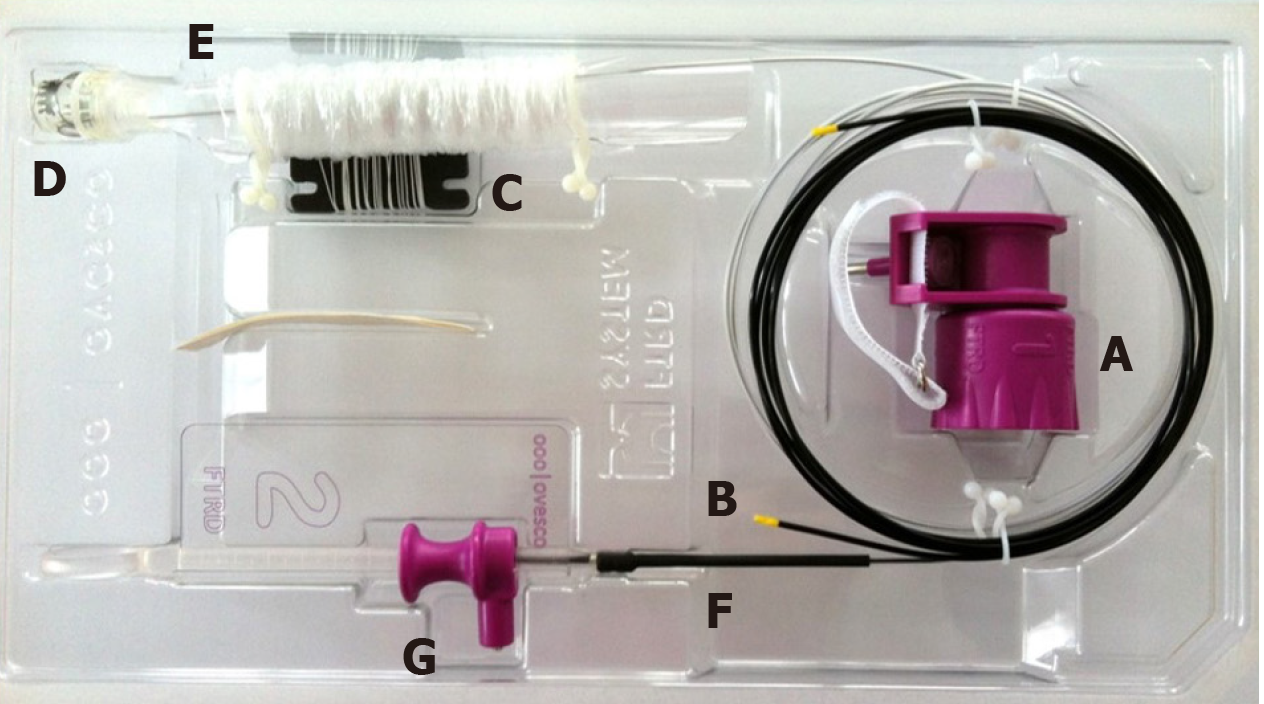
Figure 1 The colonic full-thickness resection device system.
A: Hand wheel; B: Thread Retriever; C: Thread; D: Distal attachment cap with affixed over-the-scope-clip; E: Endoscope sleeve; F: Integrated snare; G: Snare lock. Images provided by Ovesco Endoscopy.
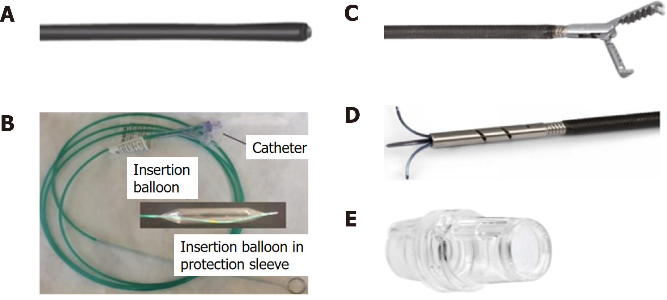
Figure 2 Full-thickness resection device separately included items.
A: Marking probe; B: Grasper; C: Guidewire and balloon (diagram) — for gastroduodenal full-thickness resection device (FTRD)®; D: Modified anchor — for gastroduodenal FTRD®; E: prOVE cap. Images courtesy of Ovesco Endoscopy.
Figure 3 Technique for accomplishing endoscopic full-thickness resection using the full-thickness resection device.
A: Borders of target lesion marked using full-thickness resection device (FTRD) Marking Probe; B: FTRD Grasper positioned over target lesion; C: FTRD Grasper opened over target lesion; D: Target lesion grasped and pulled into cap; E: Over-the-scope-clip deployed; F: Specimen resected with electrosurgical snare. Images provided by Ovesco Endoscopy.
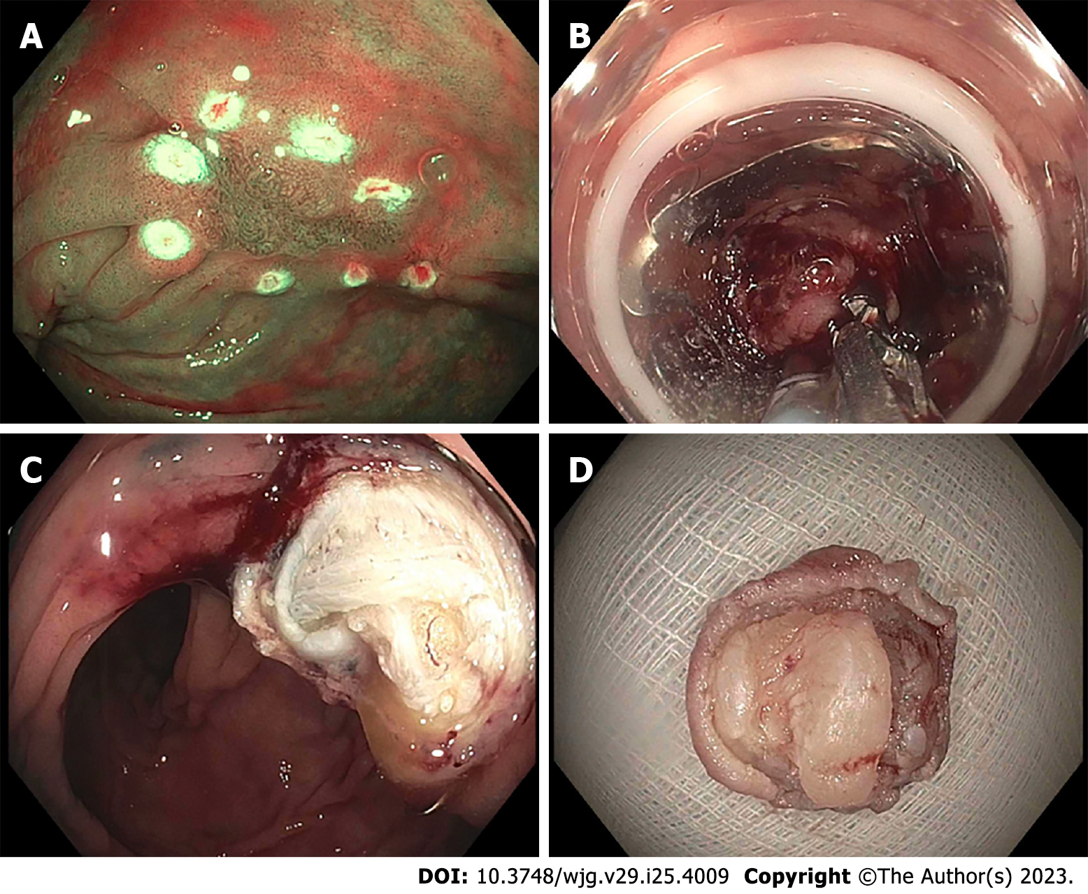
Figure 4 Endoscopic images of full-thickness resection device.
A: Full-thickness resection device (FTRD) Marking Probe used to mark the target lesion (non-lifting polyp); B: FTRD Grasper used to grasp the lesion and pull into the cap followed by successful clip deployment; C: Resection site after FTRD; D: Resected specimen. Images courtesy of Dr. Wagh MS.
- Citation: Mun EJ, Wagh MS. Recent advances and current challenges in endoscopic resection with the full-thickness resection device. World J Gastroenterol 2023; 29(25): 4009-4020
- URL: https://www.wjgnet.com/1007-9327/full/v29/i25/4009.htm
- DOI: https://dx.doi.org/10.3748/wjg.v29.i25.4009


