Copyright
©The Author(s) 2023.
World J Gastroenterol. Jun 28, 2023; 29(24): 3922-3931
Published online Jun 28, 2023. doi: 10.3748/wjg.v29.i24.3922
Published online Jun 28, 2023. doi: 10.3748/wjg.v29.i24.3922
Figure 1 Dilated and tortuous colonic varices on endoscopy without active bleeding.
A: Right transverse colon; B: Right colonic flexure.
Figure 2 Angiography images and computed tomography images.
A: Angiography of splenic artery; B: Angiography of the mesenterial vein draining the spleen; C: Computed tomography (CT)-angiography showing colonic varices; D-G: 3D-Reconstruction of CT imaging. 3D-Reconstruction with the thrombosis of the splenic vein (arrowhead) and collateral vessels to the colon indicated by arrows.
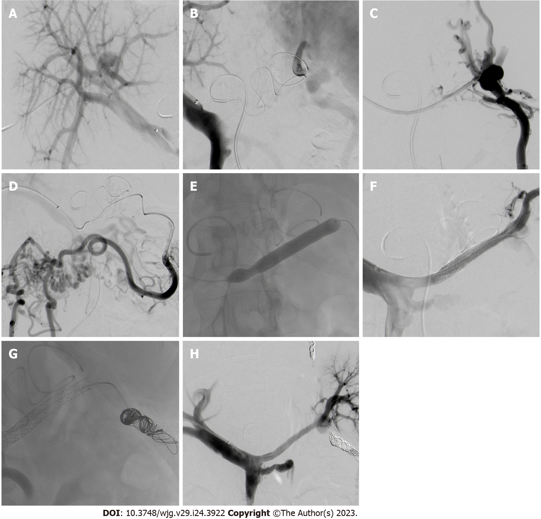
Figure 3 Angiography images.
A: Right portal vein access; B: Simultaneously performed direct portography and indirect mesentericography via the splenic artery showing complete obstruction of the splenic vein; C: Successful cannulation of the obstructed splenic vein (SV); D: Phlebography of the colonic varices (CV) arising from the splenic hilum; E: Recanalization of the SV using balloon and stentgrafts; F: Successful recanalization of the SV; G: Coiling of the aberrant vein feeding the CV; H: Patent stentgraft with drainage of the spleen through the splenic vein.
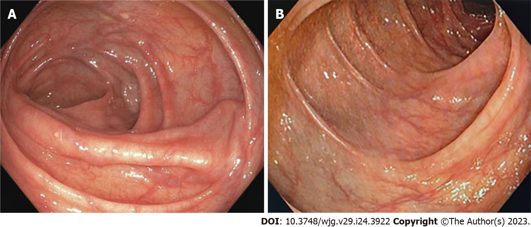
Figure 4 Follow-up colonoscopy.
A: Right colonic flexure 2 mo after intervention; B: Transverse colon 1 year after intervention.
- Citation: Füssel LM, Müller-Wille R, Dinkhauser P, Schauer W, Hofer H. Treatment of colonic varices and gastrointestinal bleeding by recanalization and stenting of splenic-vein-thrombosis: A case report and literature review. World J Gastroenterol 2023; 29(24): 3922-3931
- URL: https://www.wjgnet.com/1007-9327/full/v29/i24/3922.htm
- DOI: https://dx.doi.org/10.3748/wjg.v29.i24.3922


