Copyright
©The Author(s) 2023.
World J Gastroenterol. Jun 28, 2023; 29(24): 3883-3898
Published online Jun 28, 2023. doi: 10.3748/wjg.v29.i24.3883
Published online Jun 28, 2023. doi: 10.3748/wjg.v29.i24.3883
Figure 1 Preoperative endoscopy of prepyloric early gastric cancer.
A: Endoscopy; B: Virtual chromoendoscopy; C: Endoscopic ultrasound.
Figure 2 Endoscopic injection of indocyanine green at cardinal points 1 cm from the margins of a prepyloric early gastric cancer lesion.
A: First injection of indocyanine green (ICG); B: Second injection of ICG; C: Cardinal points of the lesion injected with ICG.
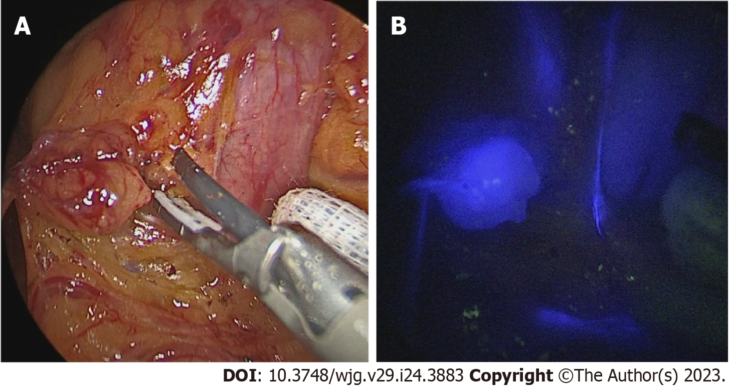
Figure 3 Sentinel lymph node biopsy.
A: Level 4 d node; B: Level 4 d fluorescent node with near-infrared vision.
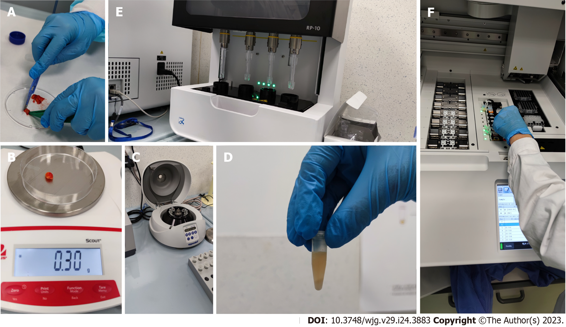
Figure 4 One-step nucleic acid amplification assay.
A-F: Lymph nodes were prepared and placed in homogenized lysis buffer (Lynorhag; Sysmex) and then centrifuged. CK19 mRNA was extracted from the lysate and analyzed by reverse transcription-loop-mediated isothermal amplification in the RD-100i system (Sysmex) using the Lynoamp (Sysmex) reagent kit[59].
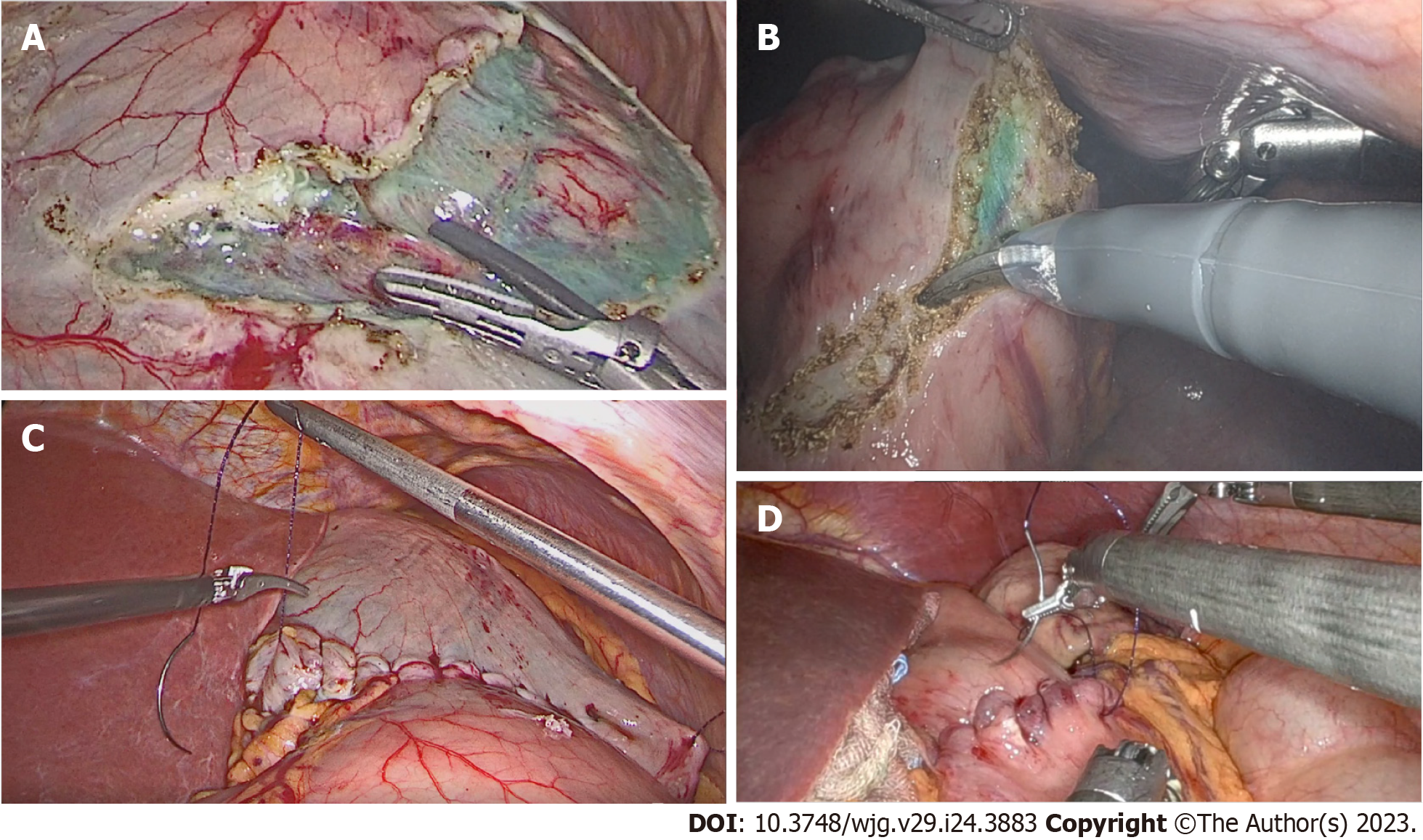
Figure 5 Laparoscopic and robotic surgical incision and reconstruction of the external gastric wall.
A and B: Incision, representative views; C and D: Reconstruction, representative views.
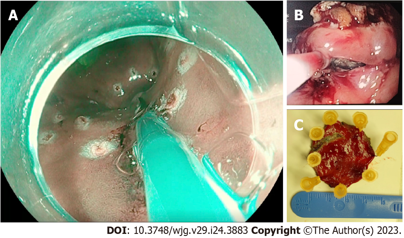
Figure 6 Endoscopic full thickness resection of early gastric cancer using the non-exposed endoscopic wall-inversion surgery procedure.
A: Mucosal markings placed around the tumor; B: Endoscopic incision of the internal layers of the gastric wall; C: Specimen.
- Citation: Crafa F, Vanella S, Morante A, Catalano OA, Pomykala KL, Baiamonte M, Godas M, Antunes A, Costa Pereira J, Giaccaglia V. Non-exposed endoscopic wall-inversion surgery with one-step nucleic acid amplification for early gastrointestinal tumors: Personal experience and literature review. World J Gastroenterol 2023; 29(24): 3883-3898
- URL: https://www.wjgnet.com/1007-9327/full/v29/i24/3883.htm
- DOI: https://dx.doi.org/10.3748/wjg.v29.i24.3883


