Copyright
©The Author(s) 2023.
World J Gastroenterol. May 14, 2023; 29(18): 2836-2849
Published online May 14, 2023. doi: 10.3748/wjg.v29.i18.2836
Published online May 14, 2023. doi: 10.3748/wjg.v29.i18.2836
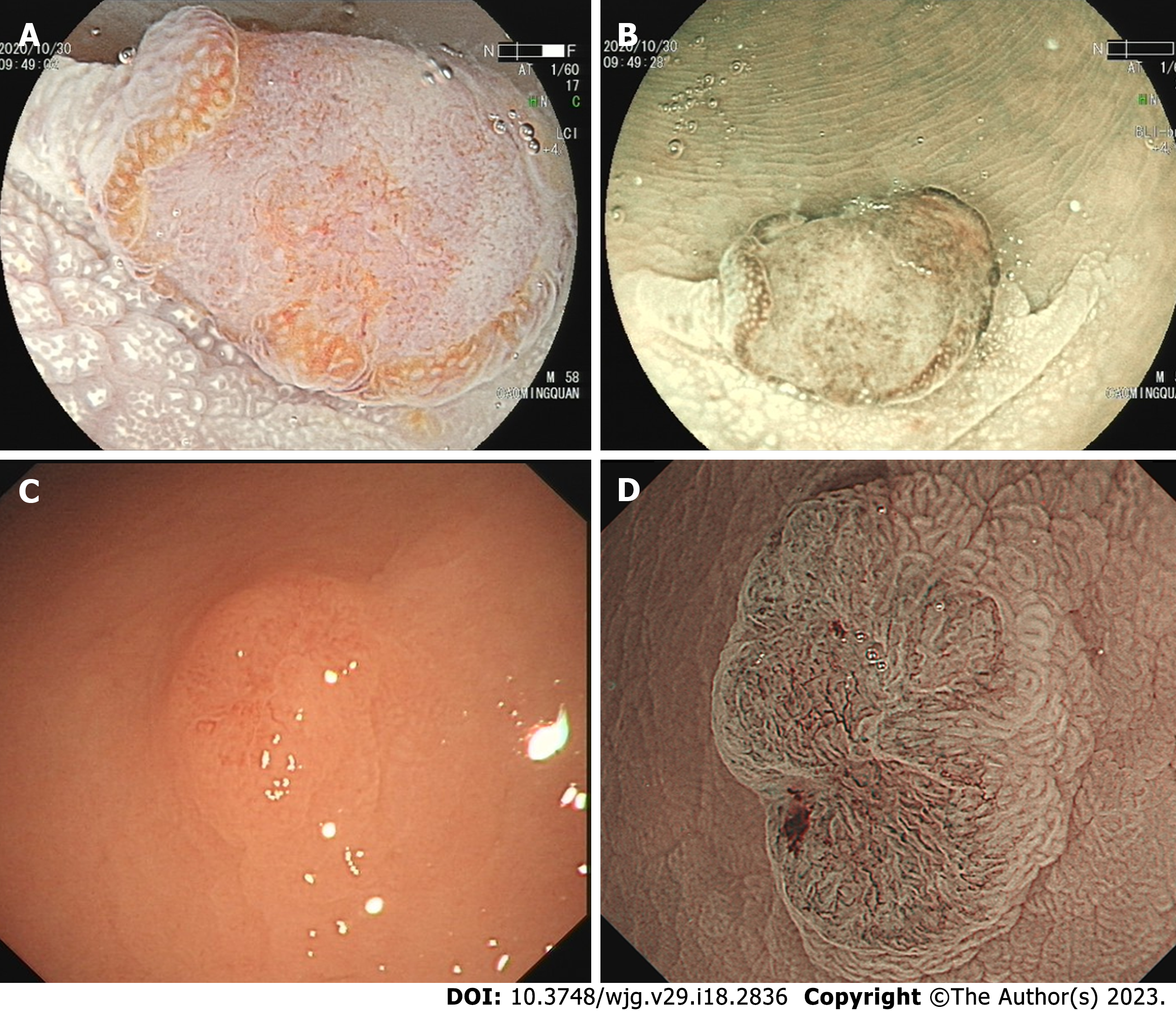
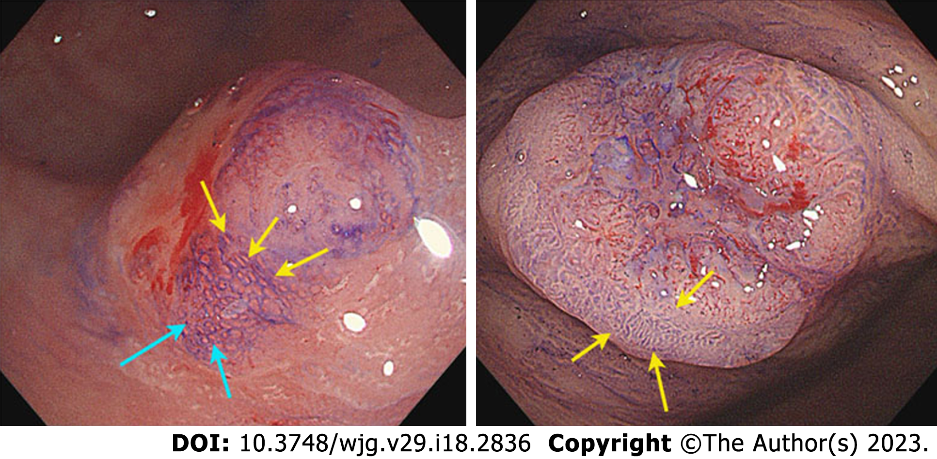
Figure 1 The de novo colorectal cancer under endoscopy.
A: A de novo colorectal cancer (CRC) under linked color imaging; B and D: The surface structure under the narrow bind imaging pattern; C: A de novo CRC under white light.
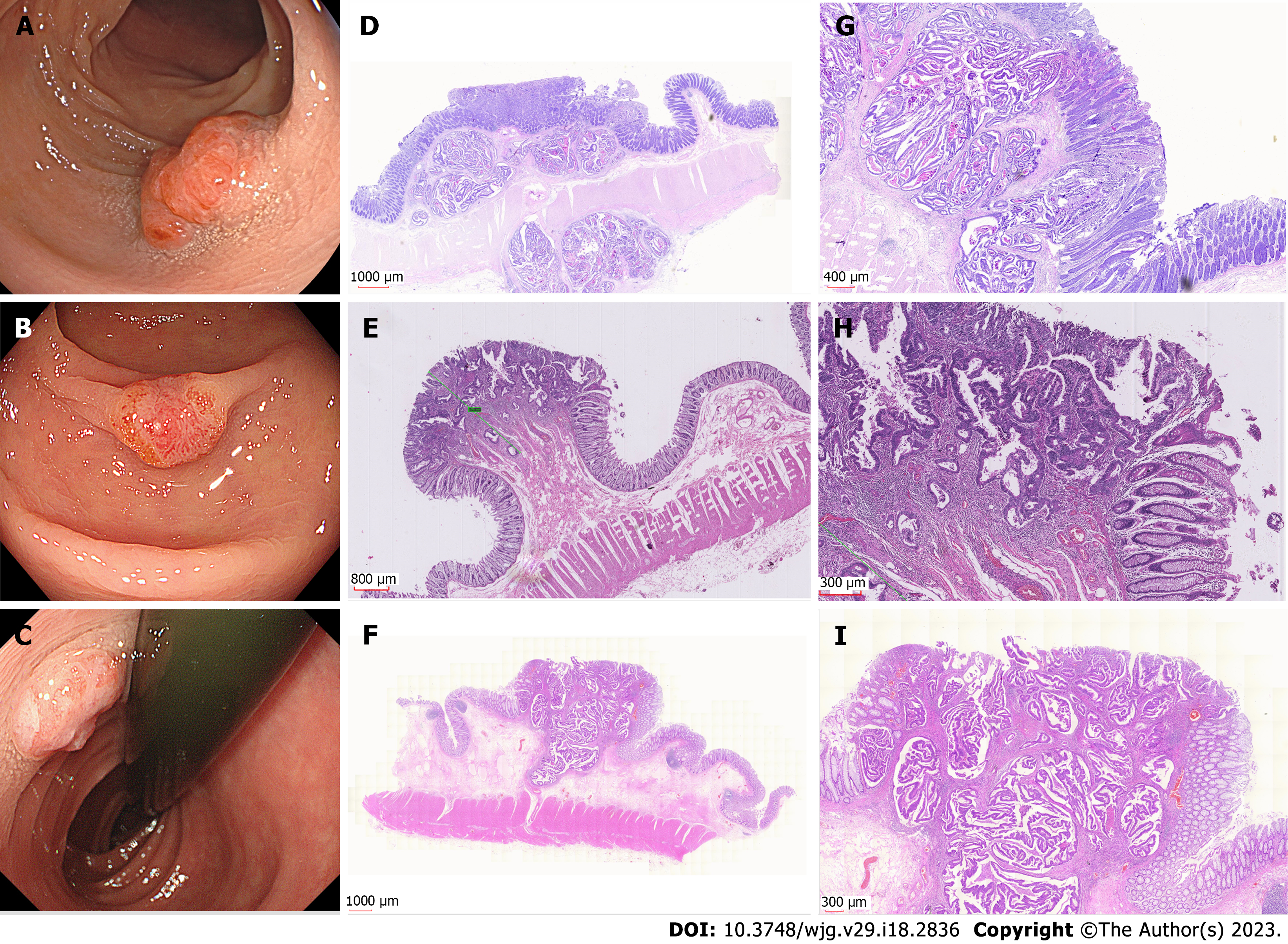
Figure 2 The examples of I-type de novo colorectal cancer.
A-C: Endoscopic images of different types de novo colorectal cancers (CRCs); D-I: Pathological images of de novo CRCs.
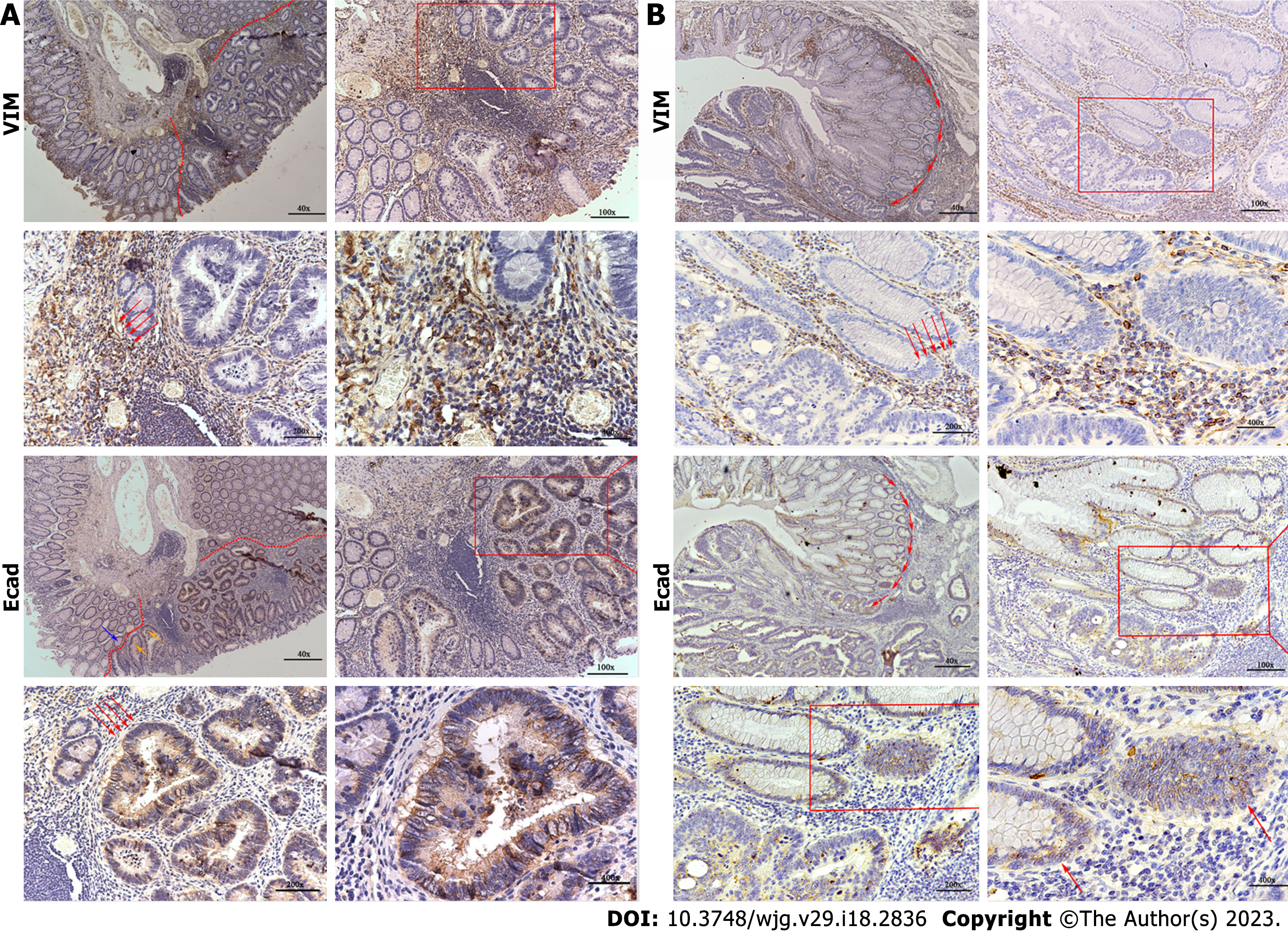
Figure 3 The immunohistory results.
A: A de novo colorectal cancer (CRC) pathological section stained with E-cad and vimentin (VIM); B: A carcinoma in adenoma CRC pathological section stained with E-cad and VIM. VIM: Vimentin.
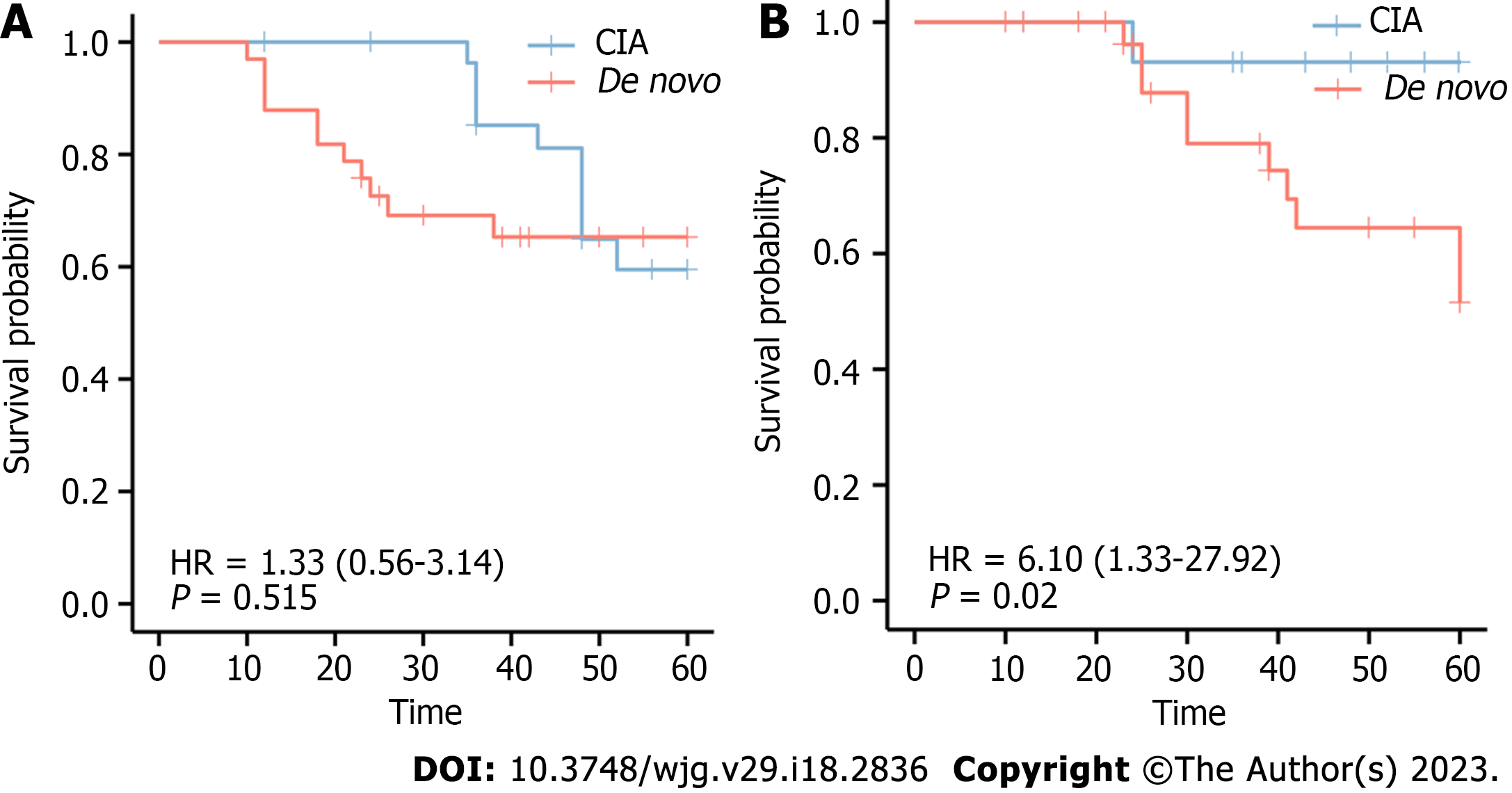
Figure 4 The association of de novo colorectal cancer and over survival.
A: Relationship between relapse rate and colorectal cancer (CRC) types; B: Relationship between rate of death and CRC types. CIA: Carcinoma in adenoma.
Figure 5 The surrounding pit of de novo colorectal cancer and carcinoma in adenoma group.
The surrounding pit around de novo colorectal cancer (CRC) is elongated I-type and IIIL-type around carcinoma in adenoma CRC.
- Citation: Li SY, Yang MQ, Liu YM, Sun MJ, Zhang HJ. Endoscopic and pathological characteristics of de novo colorectal cancer: Retrospective cohort study. World J Gastroenterol 2023; 29(18): 2836-2849
- URL: https://www.wjgnet.com/1007-9327/full/v29/i18/2836.htm
- DOI: https://dx.doi.org/10.3748/wjg.v29.i18.2836


