Copyright
©The Author(s) 2023.
World J Gastroenterol. May 7, 2023; 29(17): 2657-2665
Published online May 7, 2023. doi: 10.3748/wjg.v29.i17.2657
Published online May 7, 2023. doi: 10.3748/wjg.v29.i17.2657
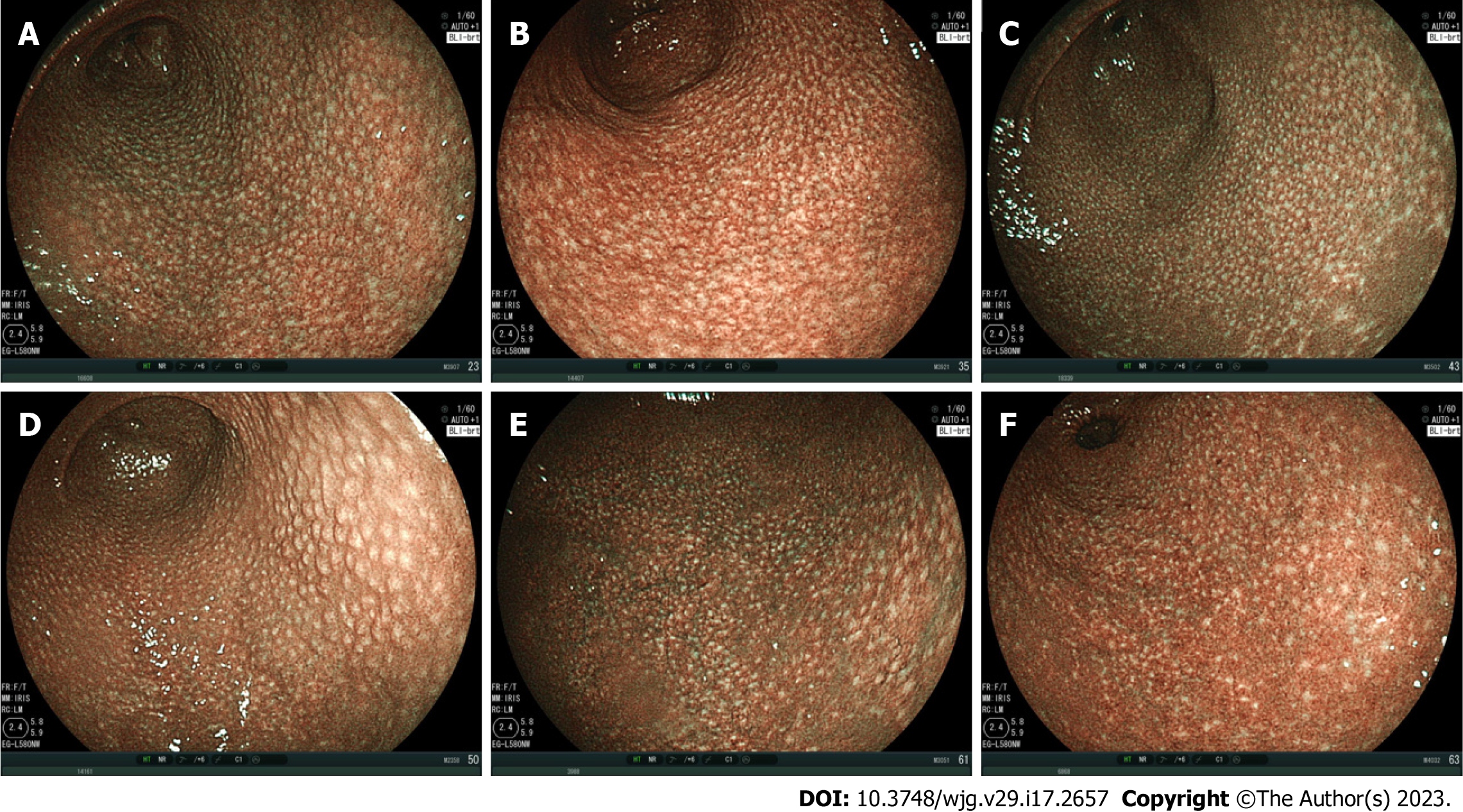
Figure 1 Spotty pattern.
Representative images of the spotty pattern shown as white spots of 1-2 mm in diameter in the gastric antrum of 6 patients infected with Helicobacter pylori (H. pylori) observed using blue laser imaging-bright mode. A: 45-year-old female, H. pylori-positive; B: 51-year-old female, H. pylori-positive; C: 27-year-old female, H. pylori-positive; D: 59-year-old female, H. pylori-positive, nodular gastritis; E: 49-year-old male, H. pylori-positive; F: 66-year-old female, H. pylori-positive.
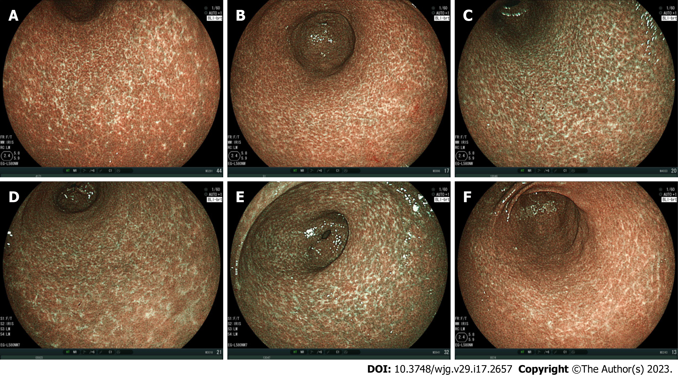
Figure 2 Cracked pattern.
Representative images of the cracked pattern as white net-like cracks consisting of lines in the gastric antrum of 6 patients after Helicobacter pylori (H. pylori) eradication observed using blue laser imaging-bright mode. A: 69-year-old female, H. pylori-negative, 2 years, 11 mo after eradication; B: 75-year-old female, H. pylori-negative, 6 years, 0 mo after eradication; C: 63-year-old female, H. pylori-negative, 1 year, 2 mo after eradication; D: 72-year-old female, H. pylori-negative, 1 year, 0 mo after eradication; E: 63-year-old female, H. pylori-negative, 1 year, 2 mo after eradication; F: 52-year-old male, H. pylori-negative, 2 years, 1 mo after eradication.
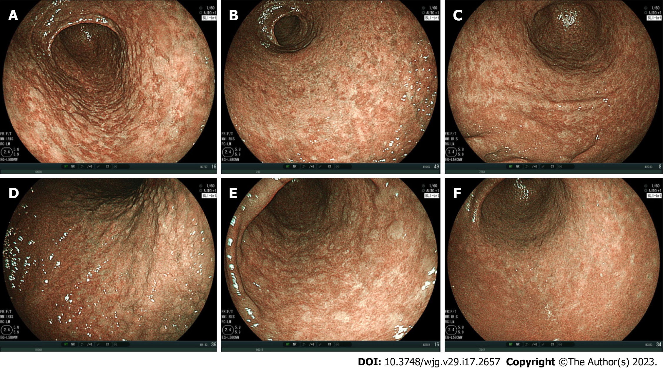
Figure 3 Mottled pattern.
Representative images of the mottled pattern observed using blue laser imaging-bright mode in the gastric antrum of 6 patients. The whitish-colored mottled areas varied from small to large. Only panel A shows the pattern of a patient infected with Helicobacter pylori (H. pylori). A: 67-year-old male, H. pylori-positive, before eradication; B: 74-year-old male, H. pylori-negative, no eradication; C: 73-year-old female, H. pylori-negative, 2 years, 11 mo after eradication; D: 87-year-old male, H. pylori-negative, no eradication; E: 83-year-old male, H. pylori-negative, 5 years, 2 mo after eradication; F: 60-year-old female, H. pylori-negative, 2 years, 2 mo after eradication.
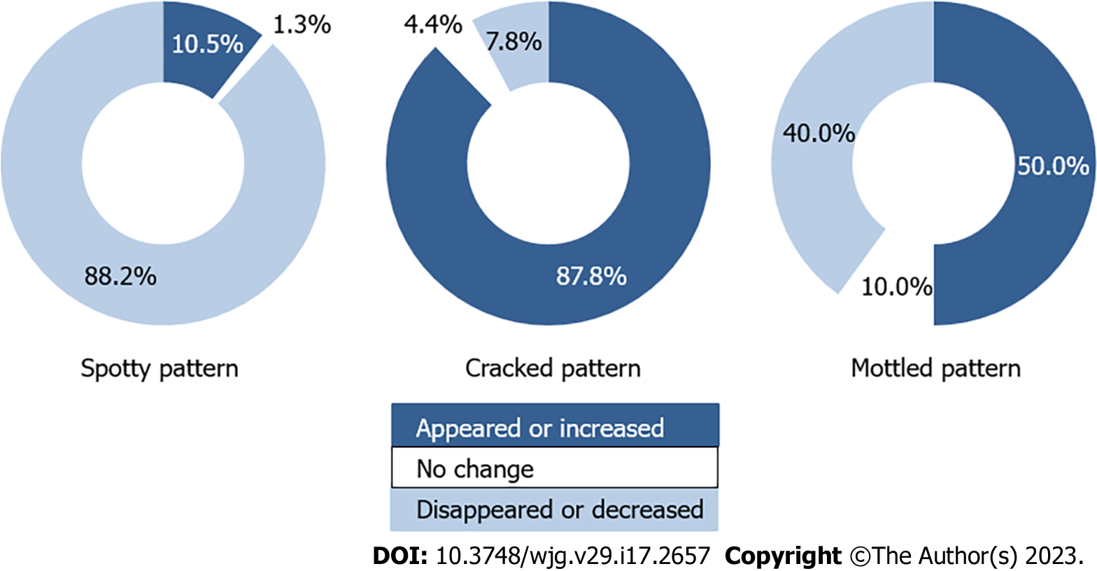
Figure 4 Change in mucosal pattern after Helicobacterpylori eradication.
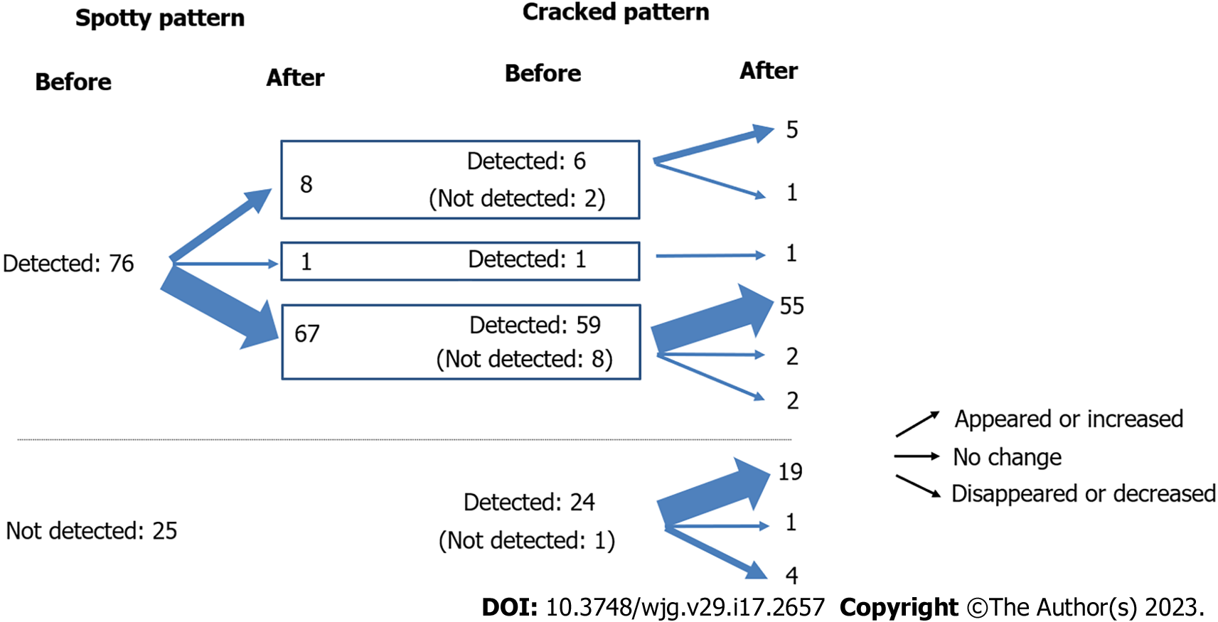
The spotty pattern tended to disappear or decrease, the cracked pattern appeared or increased, and the mottled pattern exhibited no specific tendency.
Figure 5 Relationship between the spotty and cracked patterns before and after Helicobacterpylori eradication.
- Citation: Nishikawa Y, Ikeda Y, Murakami H, Hori SI, Yoshimatsu M, Nishikawa N. Mucosal patterns change after Helicobacter pylori eradication: Evaluation using blue laser imaging in patients with atrophic gastritis. World J Gastroenterol 2023; 29(17): 2657-2665
- URL: https://www.wjgnet.com/1007-9327/full/v29/i17/2657.htm
- DOI: https://dx.doi.org/10.3748/wjg.v29.i17.2657


