Copyright
©The Author(s) 2023.
World J Gastroenterol. Apr 21, 2023; 29(15): 2349-2358
Published online Apr 21, 2023. doi: 10.3748/wjg.v29.i15.2349
Published online Apr 21, 2023. doi: 10.3748/wjg.v29.i15.2349
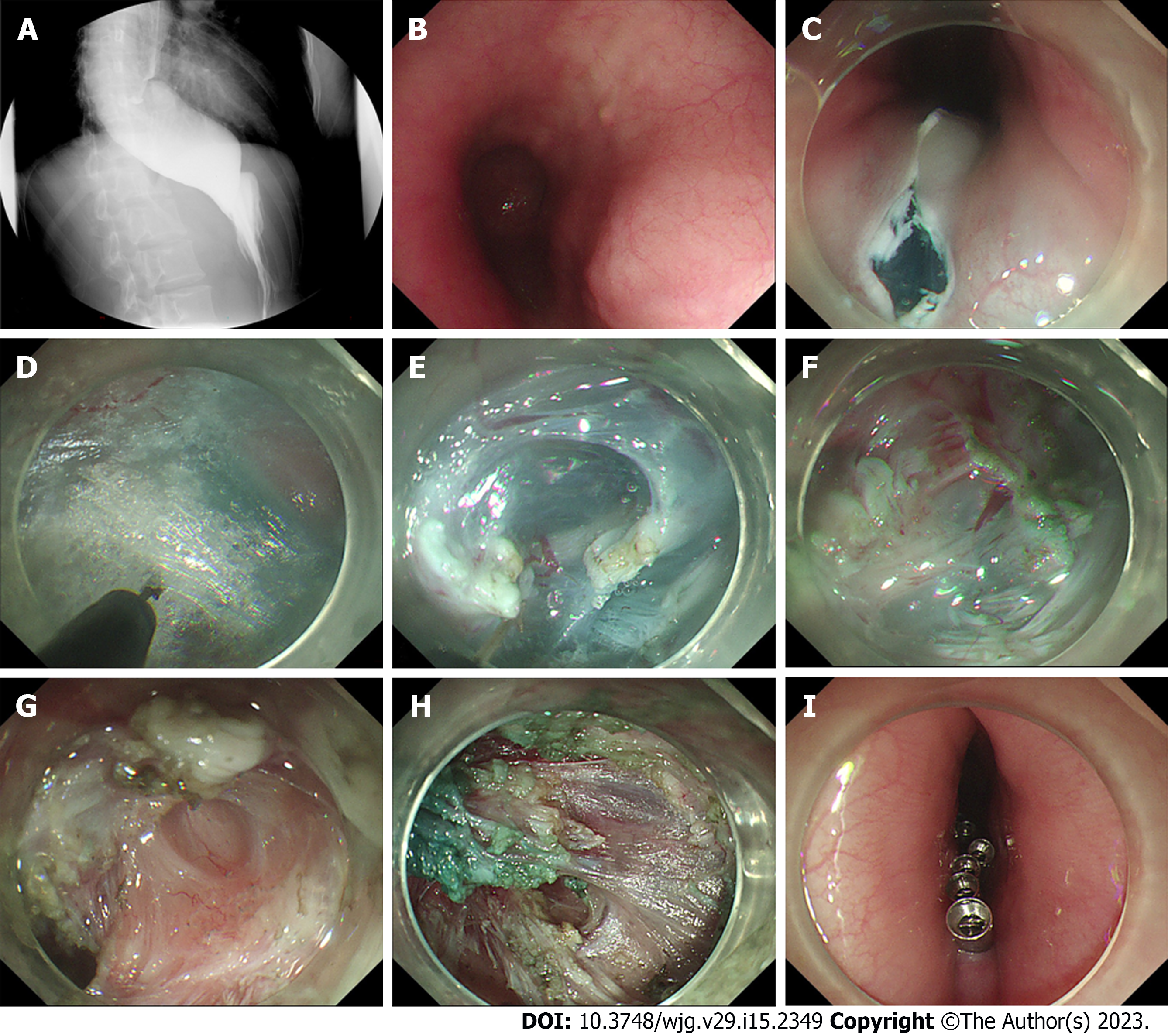
Figure 1 Repeat peroral endoscopic myotomy with simultaneous submucosal and muscle dissection in a 28-year-old woman with recurrence of symptoms after previous peroral endoscopic myotomy.
A: Radiographic image showing the difficulty of barium to pass through the pipe into the stomach; B: The mucosal scar of previous peroral endoscopic myotomy; C: Reverse T incision of submucosal entry near the scarred area; D: Construction of the submucosal tunnel; E: Obvious submucosal fibrosis disconnected by electrocoagulation forceps; F: Simultaneous submucosal and muscle dissection started from the muscular scar; G: Full-thickness myotomy of the esophagus; H: Exact hemostasis using hemostatic forceps; I: Closure of the mucosal entry point with metal clips.
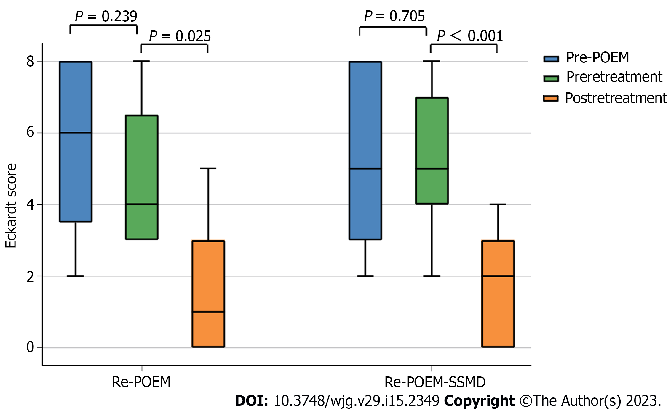
Figure 2 Eckardt score changes in patients with recurrent achalasia.
POEM: Peroral endoscopic myotomy; Re-POEM: Repeat peroral endoscopic myotomy; Re-POEM-SSMD: Repeat peroral endoscopic myotomy with simultaneous submucosal and muscle dissection.
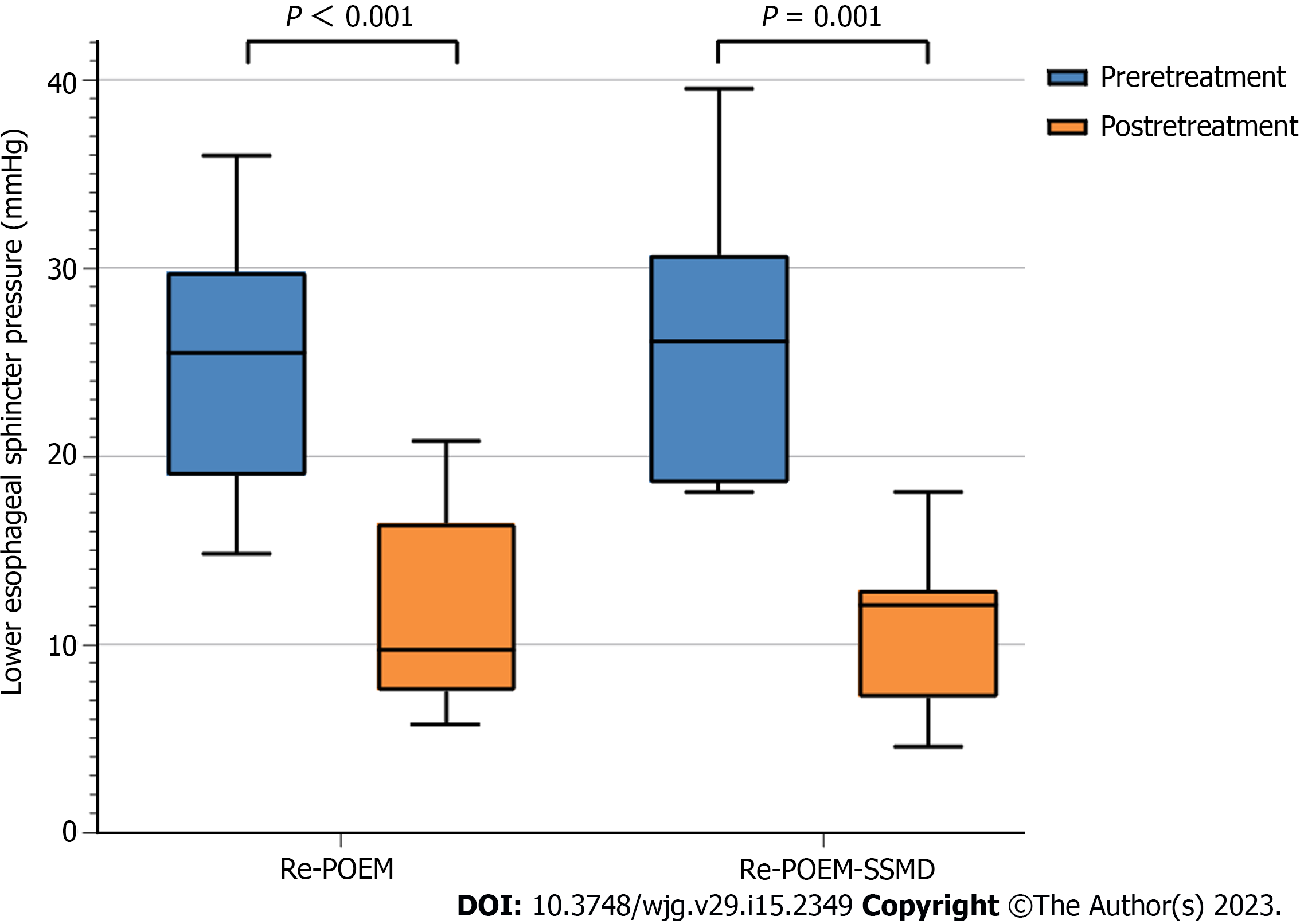
Figure 3 Lower esophageal sphincter pressure changes in patients with recurrent achalasia.
Re-POEM: Repeat peroral endoscopic myotomy; Re-POEM-SSMD: Repeat peroral endoscopic myotomy with simultaneous submucosal and muscle dissection.
- Citation: Lin YJ, Liu SZ, Li LS, Han K, Shao BZ, Linghu EQ, Chai NL. Repeat peroral endoscopic myotomy with simultaneous submucosal and muscle dissection as a salvage option for recurrent achalasia. World J Gastroenterol 2023; 29(15): 2349-2358
- URL: https://www.wjgnet.com/1007-9327/full/v29/i15/2349.htm
- DOI: https://dx.doi.org/10.3748/wjg.v29.i15.2349


