Copyright
©The Author(s) 2022.
World J Gastroenterol. Feb 28, 2022; 28(8): 794-810
Published online Feb 28, 2022. doi: 10.3748/wjg.v28.i8.794
Published online Feb 28, 2022. doi: 10.3748/wjg.v28.i8.794
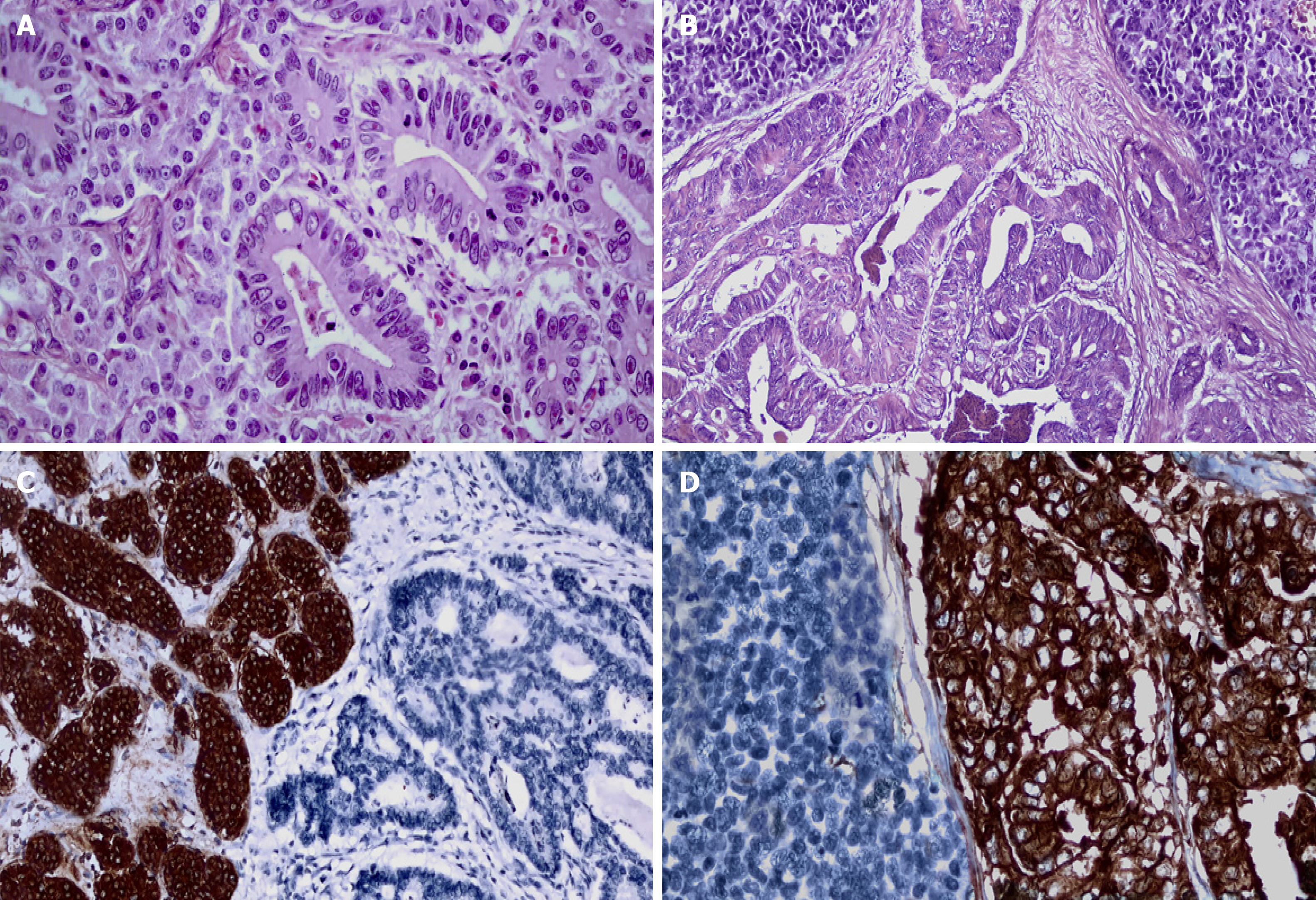
Figure 1 Examples of histopathological and immunohistochemical findings in gastrointestinal mixed neuroendocrine-nonneuroendo
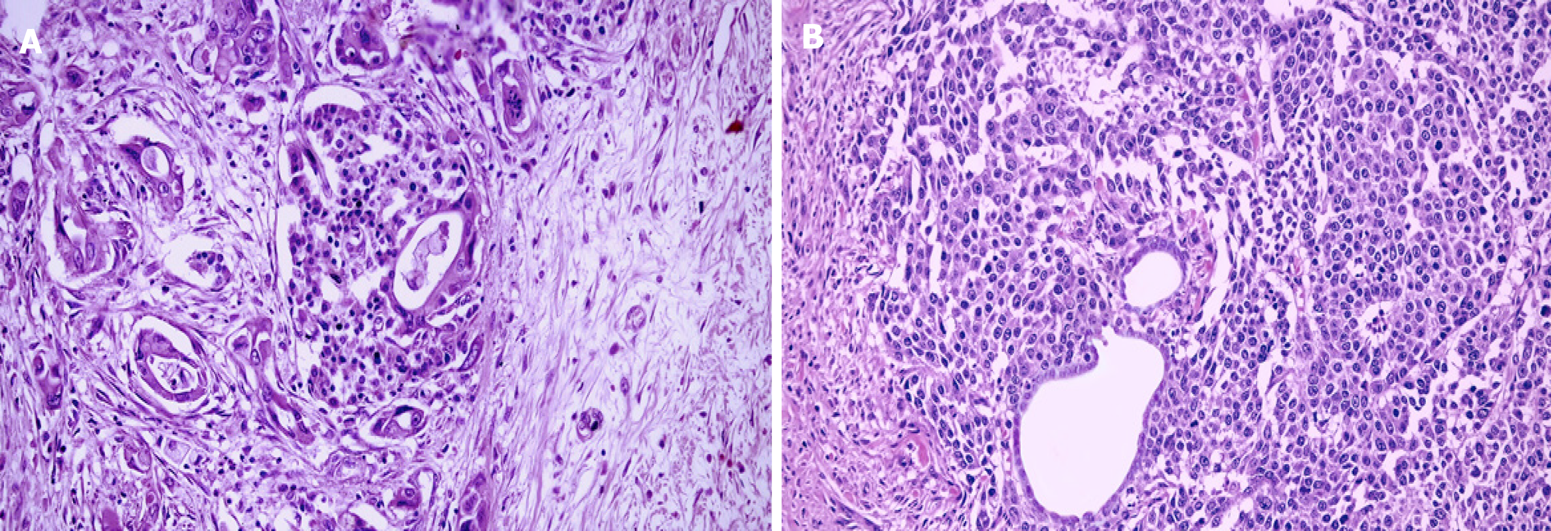
Figure 2 Histopathological pitfalls in the diagnosis of mixed neuroendocrine-nonneuroendocrine neoplasms of the pancreas.
A: A ductal adenocarcinoma of the pancreas surrounding and invading an islet in the background of chronic pancreatitis (x 200). The islet has regular contours despite an invasion; B: A neuroendocrine tumor of the pancreas with entrapped two ductulus without atypia. Such areas should be evaluated carefully to avoid a misdiagnosis of mixed neuroendocrine-nonneuroendocrine neoplasms (x 200).
Figure 3 Acinar carcinoma of the pancreas.
A: The tumor is composed of cells that demonstrate the presence of monomorphic nuclei, sometimes forming minute lumens. Tumor cells are in a monolayer with basally located nuclei and have a granular eosinophilic cytoplasm (x 400); B: Bcl-10 expression with higher staining in the apical portion of tumor cells (x 400).
- Citation: Elpek GO. Mixed neuroendocrine–nonneuroendocrine neoplasms of the gastrointestinal system: An update. World J Gastroenterol 2022; 28(8): 794-810
- URL: https://www.wjgnet.com/1007-9327/full/v28/i8/794.htm
- DOI: https://dx.doi.org/10.3748/wjg.v28.i8.794


