Copyright
©The Author(s) 2022.
World J Gastroenterol. Feb 14, 2022; 28(6): 624-634
Published online Feb 14, 2022. doi: 10.3748/wjg.v28.i6.624
Published online Feb 14, 2022. doi: 10.3748/wjg.v28.i6.624
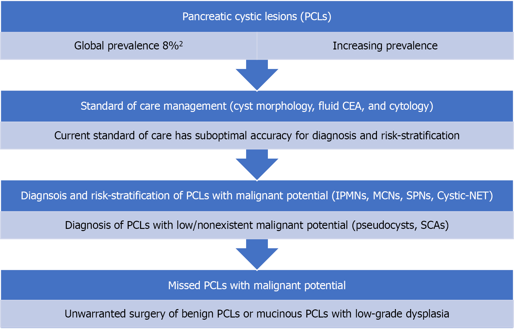
Figure 1 Current standard of care diagnostic methods are suboptimal in the diagnosis of specific types of pancreatic cystic lesions and risk-stratification of mucinous cysts.
PCL: Pancreatic cystic lesion, CEA: Carcinoembryonic antigen, IPMN: Intraductal papillary mucinous neoplasms, MCN: Mucinous cystic neoplasm, SPN: Solid pseudopapillary neoplasm, Cystic-NET: Cystic neuroendocrine tumors, SCA: Serous cystadenoma.
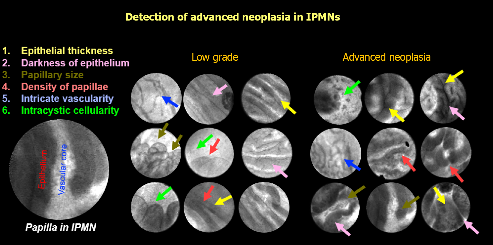
Figure 2 Features identified on endoscopic ultrasound guided needle confocal laser endomicroscopy.
IPMN: Intraductal papillary mucinous neoplasms.
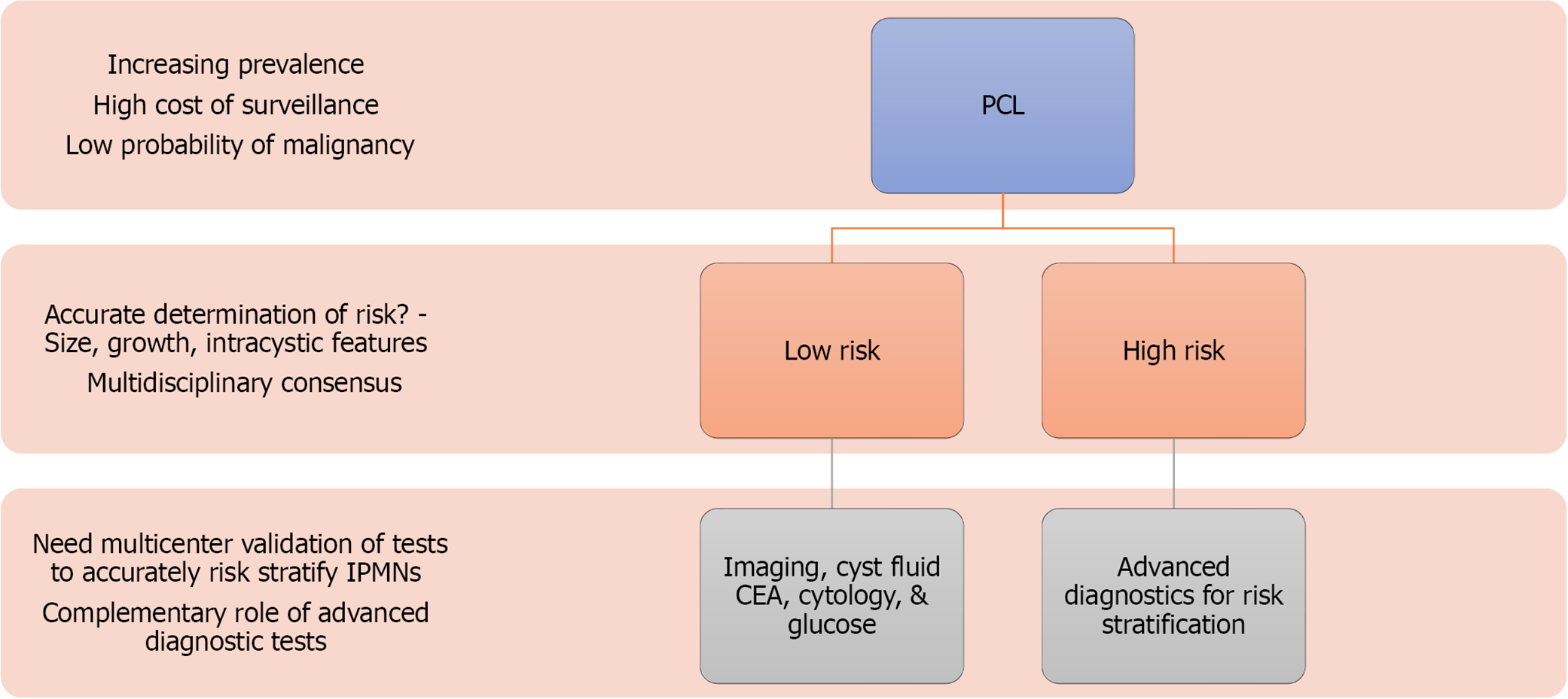
Figure 3 Future directions of detection and risk stratification of pancreatic cystic lesion to guide clinical management.
PCL: Pancreatic cystic lesion, CEA: Carcinoembryonic antigen, IPMN: Intraductal papillary mucinous neoplasms.
- Citation: Ardeshna DR, Cao T, Rodgers B, Onongaya C, Jones D, Chen W, Koay EJ, Krishna SG. Recent advances in the diagnostic evaluation of pancreatic cystic lesions. World J Gastroenterol 2022; 28(6): 624-634
- URL: https://www.wjgnet.com/1007-9327/full/v28/i6/624.htm
- DOI: https://dx.doi.org/10.3748/wjg.v28.i6.624


