Copyright
©The Author(s) 2022.
World J Gastroenterol. Sep 14, 2022; 28(34): 5086-5092
Published online Sep 14, 2022. doi: 10.3748/wjg.v28.i34.5086
Published online Sep 14, 2022. doi: 10.3748/wjg.v28.i34.5086
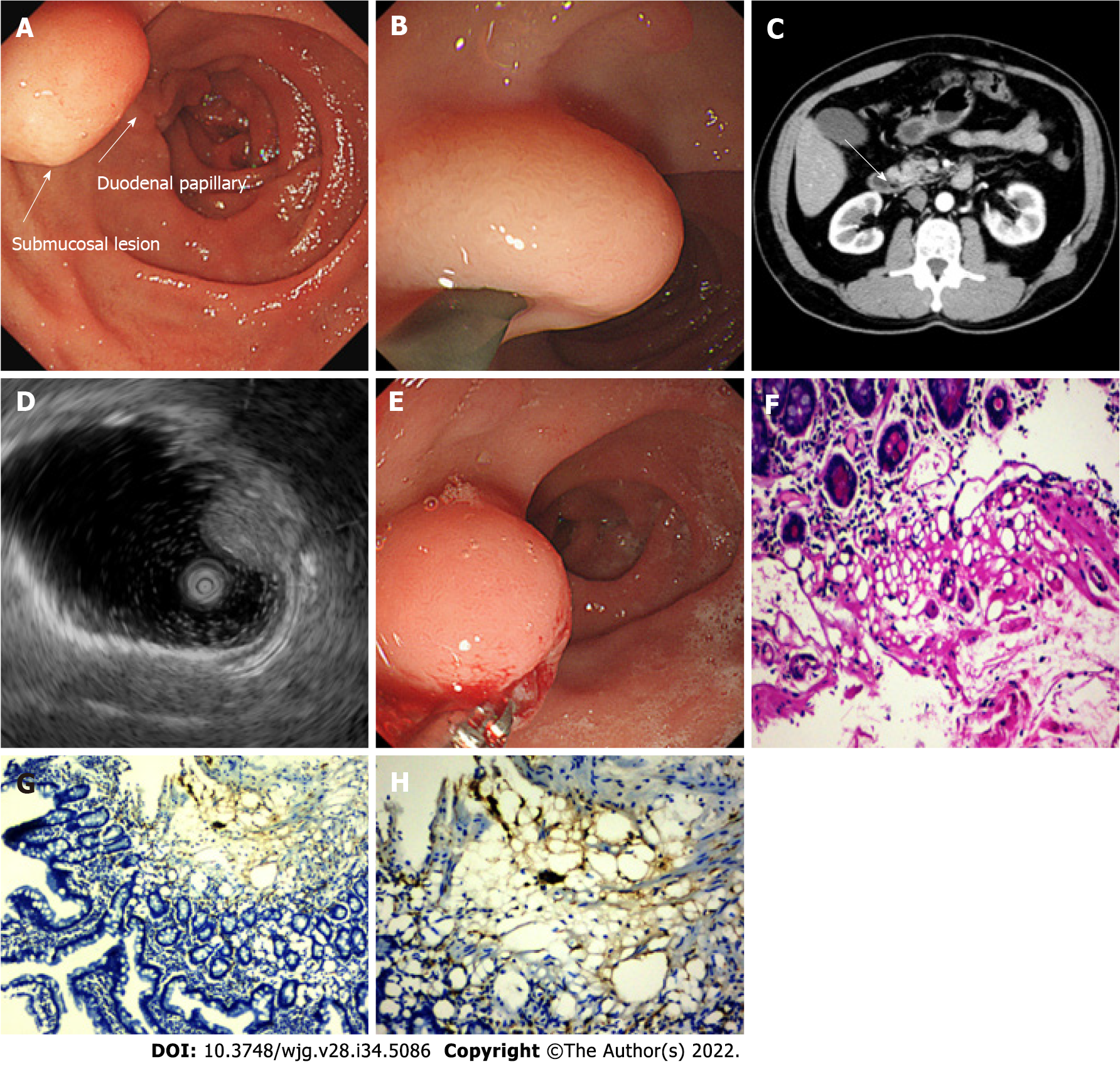
Figure 1 Imaging findings at the time of diagnosis.
A and B: Esophagogastroduodenoscopy revealed a 10-mm soft yellowish submucosal lesion with the “pillow sign,” located in the second portion of duodenum, immediately upon the duodenal papillary; C: Abdominal contrast-enhanced computed tomography shows a suspicious low-density lesion in the periampullary region without enhancement (white arrow); D: Endoscopic ultrasonography with a 12-MHz catheter probe showed a hyperechoic, homogenous, and round solid lesion with echo attenuation, arising from the submucosal layer; E: Deep biopsy via bite-on-bite technique with forceps was performed; F-H: Microscopic examination showed a small amount of roundish adipocyte in the submucosa layer, expressing S-100. Tiny lipid droplets were observed in cell cytoplasm. The glands of epithelium were neatly arranged on top (F: Hematoxylin and eosin staining, × 200; G: Immunohistochemical S-100 stain, × 100; H: IHC S-100 stain, × 200).
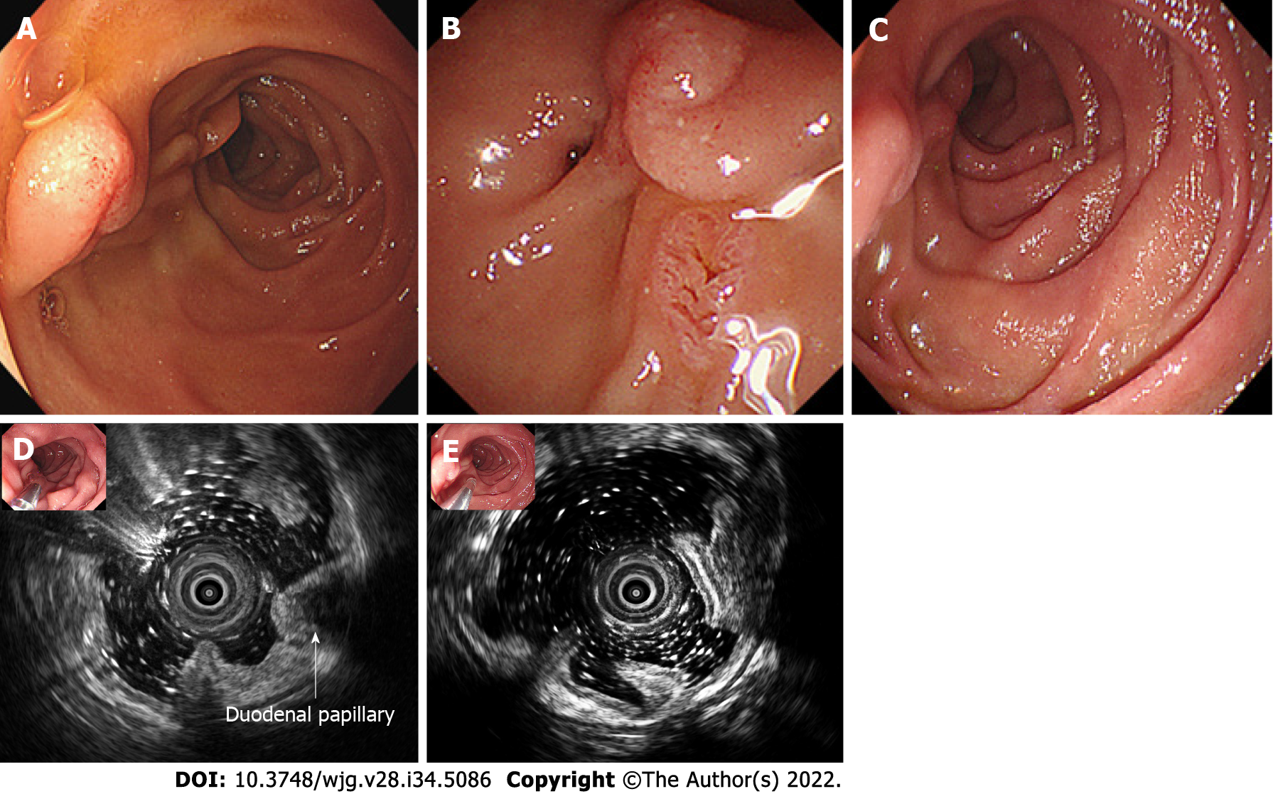
Figure 2 Follow-up endoscopic view of the lesion.
A and B: 12 d after the biopsy, the lipoma was spontaneously expelled, with red scar and inflammatory mucosa residue in situ of the lesion; C-E: Follow-up endoscopic ultrasonography after 2 mo revealed that the in situ mucosa was smooth, and the former lesion no longer existed in the surrounding duodenal wall or periduodenal papilla region.
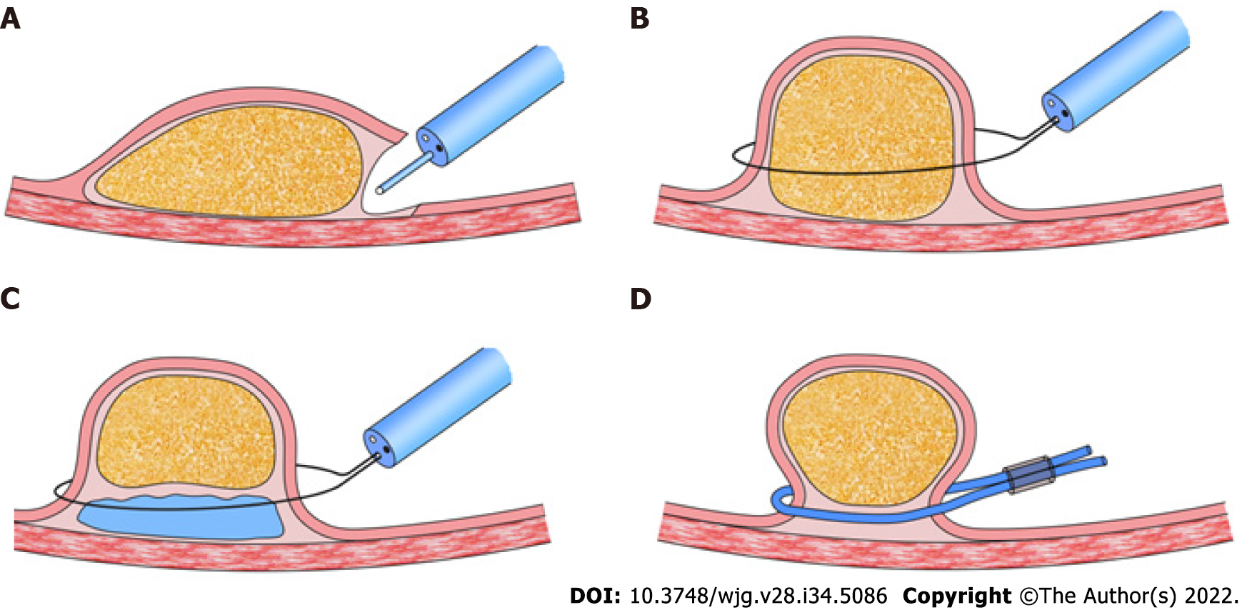
Figure 3 Techniques for endoscopic excision of gastrointestinal lipomas[14].
A: Dissection-based resection technique; B: Unroofing technique; C: Endoscopic mucosal resection; D: Loop-assisted resection technique.
- Citation: Chen ZH, Lv LH, Pan WS, Zhu YM. Spontaneous expulsion of a duodenal lipoma after endoscopic biopsy: A case report. World J Gastroenterol 2022; 28(34): 5086-5092
- URL: https://www.wjgnet.com/1007-9327/full/v28/i34/5086.htm
- DOI: https://dx.doi.org/10.3748/wjg.v28.i34.5086


