Copyright
©The Author(s) 2022.
World J Gastroenterol. Aug 14, 2022; 28(30): 4075-4088
Published online Aug 14, 2022. doi: 10.3748/wjg.v28.i30.4075
Published online Aug 14, 2022. doi: 10.3748/wjg.v28.i30.4075
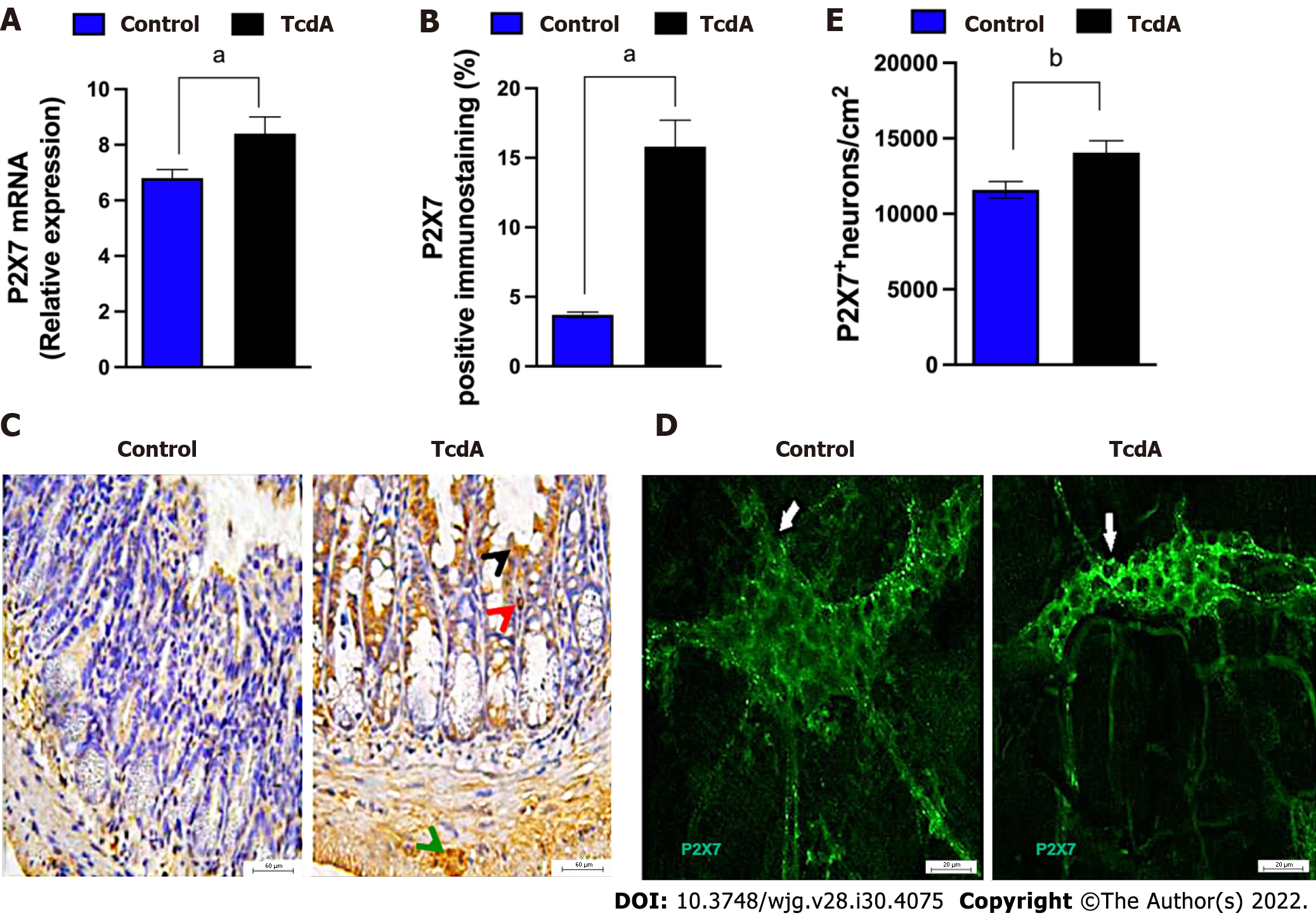
Figure 1 Clostridioides difficile toxin A increases the expression of the P2X7 receptor in the ileum myenteric plexus of mice.
A: The expression of the P2RX7 gene [mean ± standard error of the mean (SEM)] assayed by quantitative real-time polymerase chain reaction in the ileal samples of mice injected with TcdA [TcdA, 50 μg in phosphate-buffered saline (PBS)] or PBS alone in the ileal loops (Control) (n = 4); B: Quantification of the percentage (mean ± SEM) of the P2X7 receptor-immunopositive area in the ileum from control and TcdA-challenged mice in 5-6 microscope fields per sample (n = 4 animals per group); C: Representative immunohistochemical images of the expression of the P2X7 receptor in the ileum of control and TcdA-challenged mice. Increased expression of the P2X7 receptor (arrowhead) was detected in the intestinal epithelial layer (black arrowhead), lamina propria (red arrowhead), and myenteric plexus (green arrowhead). Scale bars, 50 μm; D: Representative photomicrographs ofimmunostaining of the P2X7 receptor (arrow indicates the region stained green) in the ileum myenteric plexus from control and TcdA-challenged mice; E: Quantification of the number of P2RX7+ neurons/cm2 (mean ± SEM) in the ileum of control and TcdA-challenged mice (n = 4 animals per group); A, B and E: Unpaired two-tailed Student's t test (aP < 0.05; bP = 0.01).
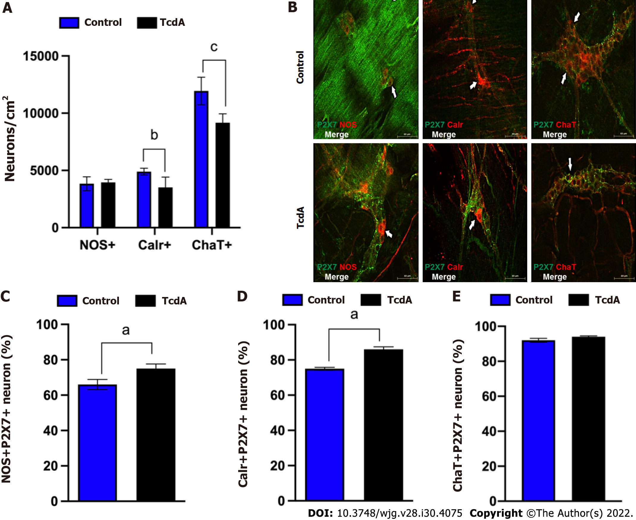
Figure 2 Clostridioides difficile toxin A induces alterations in enteric neuronal coding in the myenteric plexus in the ileum of mice.
A: Quantification of the number of neuronal nitric oxide synthase+ (nNOS+), calretinin+ (Calr+), and choline acetyltransferase+ (ChaT+) neurons/cm2 [mean ± standard error of the mean (SEM)] in the ileum myenteric plexus in control and TcdA-challenged mice; B: Representative photomicrographs of the P2X7 receptor (green), nNOS (left panels, red), Calr (center panels, red), and ChaT (right panel, red) immunostaining and DAPI (blue) nuclear staining in control and TcdA-challenged mice. White arrows indicate positive immunostaining. Scale bars, 50 μm; C-E: Percentage of NOS+P2X7+ neurons (C), Calr+P2RX7+ neurons (D), and ChaT+P2RX7+ neurons (E) (mean ± SEM) in the ileum myenteric plexus of control and TcdA-challenged mice (n = 4); A, C, D and E: Unpaired two-tailed Student's t test (aP < 0.05; bP < 0.03; cP < 0.002 ). NOS: Nitric oxide synthase; Calr: Calretinin; ChaT: Choline acetyltransferase.
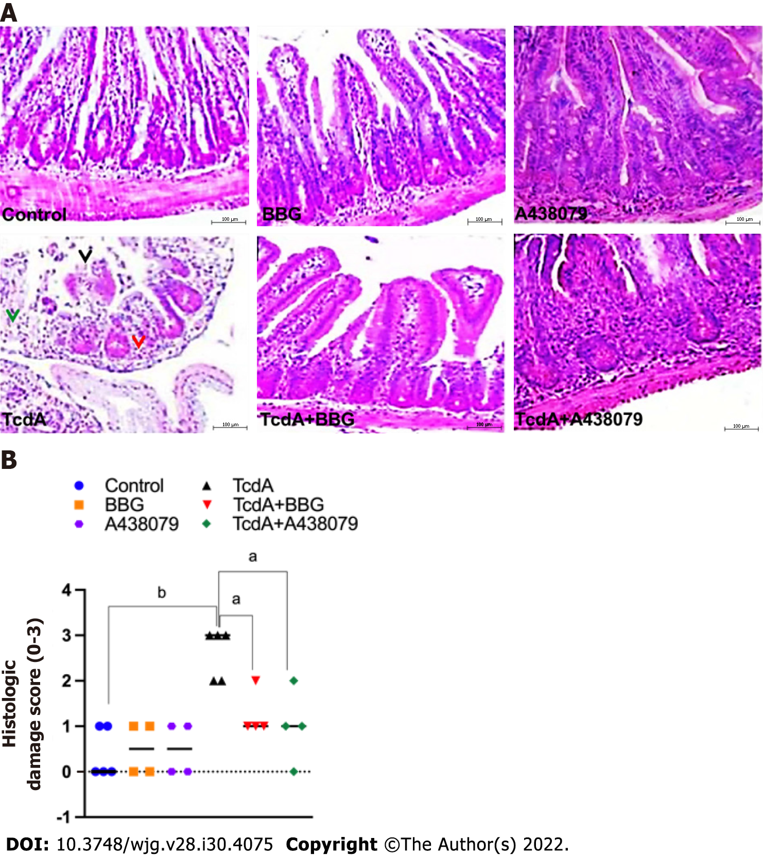
Figure 3 Inhibition of the P2X7 receptor decreases Clostridioides difficile toxin A-induced ileal damage in mice.
Mouse ileal loops were injected with TcdA [TcdA, 50 μg in phosphate-buffered saline (PBS)] or PBS alone (control) (n = 5), and the animals were pretreated with a P2X7 receptor antagonist [Brilliant Blue G (BBG) or A438079] one hour prior to TcdA challenge. A: Representative histological images (magnification of 200 ×) of TcdA-unchallenged (control, BBG, and A438079) and challenged (TcdA, TcdA + BBG, and TcdA + A438079) mice. TcdA induced epithelial disruption (black arrowhead), edema (green arrowhead), and neutrophil infiltration (red arrowhead); B: Histopathological score (median; 0 corresponds to-no damage and 3 corresponds to intense damage) performed by a blinded histopathological expert and based on epithelial damage, submucosal edema, and infiltration of inflammatory cells. Kruskal–Wallis nonparametric test followed by Dunn’s test. aP < 0.04; bP < 0.007. BBG: Brilliant Blue G.
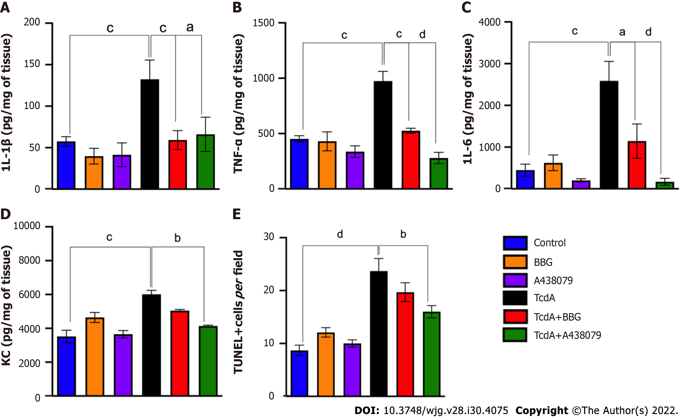
Figure 4 P2X7 receptor inhibition abrogates Clostridioides difficile toxin A-induced inflammation and cell death in mice.
A-D: The levels of interleukin (IL)-1β (A), tumor necrosis factor-α (B), IL-6 (C), and keratinocyte chemoattractant (D) in the ileal samples of TcdA-unchallenged [control, Brilliant Blue G (BBG), and A438079] and challenged (TcdA, TcdA + BBG, and TcdA + A438079) mice measured by ELISA (n = 5); E: The number of TUNEL+ cells per field in the ileal samples of TcdA-unchallenged (control, BBG, and A438079) and challenged (TcdA, TcdA + BBG, and TcdA + A438079) mice. The data are expressed as the mean ± standard error of the mean. ANOVA followed by Tukey’s test was used (aP = 0.03; bP = 0.01; cP < 0.008; dP < 0.0001). IL: Interleukin; TNF: Tumor necrosis factor; BBG: Brilliant Blue G; KC: Keratinocyte chemoattractant; TUNEL: Terminal deoxynucleotidyltransferase-mediated dUTP-biotin nick end labeling.
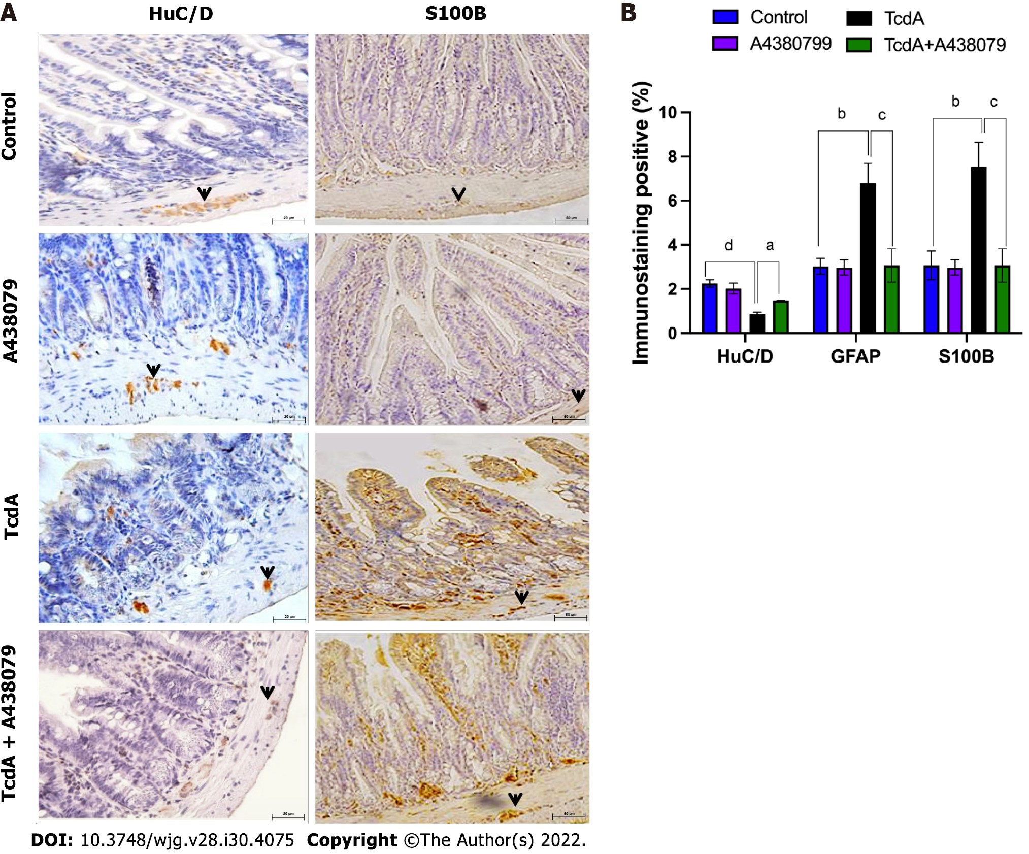
Figure 5 P2X7 receptor inhibition attenuates Clostridioides difficile toxin A-induced enteric neuron loss and S100B synthesis in mice.
A: Representative immunohistochemical images of the expression of HuC/D (neuronal marker) and S100B (enteric glia marker) in the ileum of TcdA-unchallenged (control and A438079) and challenged (TcdA and TcdA + A438079) mice. Black arrows indicate positive immunostaining. Scale bars, 50 μm; B: Quantification of the percentage (mean ± standard error of the mean) of HuC/D- and S100B-immunopositive areas in the ileum of TcdA-unchallenged (control and A438079) and challenged (TcdA and TcdA + A438079) mice (n = 4 animals per group). ANOVA followed by Tukey’s test was used (aP = 0.04; bP = 0.01; cP < 0.009; dP < 0.0001). GFAP: Glial fibrillary acidic protein.
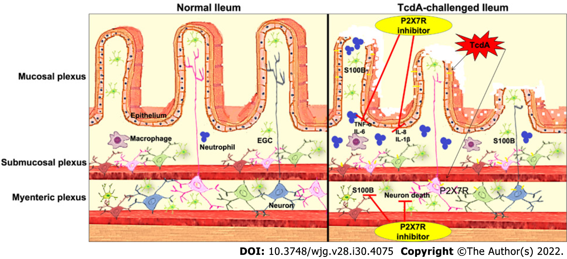
Figure 6 Scheme of the hypothetical role of the P2X7 receptor during Clostridioides difficile toxin A-induced ileitis.
Clostridioides difficile TcdA promotes epithelial damage and the upregulation of the P2X7 receptor in enteric neurons, intestinal epithelial cells, and immune cells. Once activated, the P2X7 receptor promotes the death of enteric neurons and the release of proinflammatory mediators by epithelial/immune cells [interleukin (IL)-1β, IL-6, keratinocyte chemoattractant, and tumor necrosis factor-α]. These proinflammatory mediators induce S100B synthesis by enteric glial cells. Blockade of the P2X7 receptor using a pharmacological approach attenuates these deleterious effects of TcdA. IL: Interleukin; TNF: Tumor necrosis factor; EGC: Enteric glial cell.
- Citation: Santos AAQA, Costa DVS, Foschetti DA, Duarte ASG, Martins CS, Soares PMG, Castelucci P, Brito GAC. P2X7 receptor blockade decreases inflammation, apoptosis, and enteric neuron loss during Clostridioides difficile toxin A-induced ileitis in mice. World J Gastroenterol 2022; 28(30): 4075-4088
- URL: https://www.wjgnet.com/1007-9327/full/v28/i30/4075.htm
- DOI: https://dx.doi.org/10.3748/wjg.v28.i30.4075


