Copyright
©The Author(s) 2022.
World J Gastroenterol. Jun 7, 2022; 28(21): 2396-2402
Published online Jun 7, 2022. doi: 10.3748/wjg.v28.i21.2396
Published online Jun 7, 2022. doi: 10.3748/wjg.v28.i21.2396
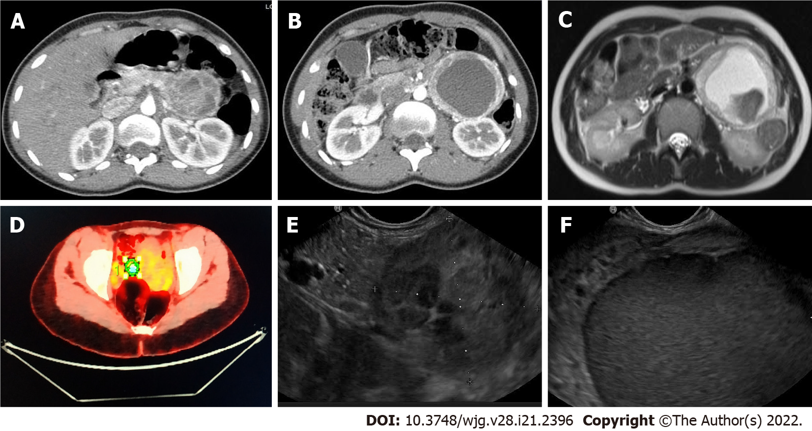
Figure 1 Radiologic findings.
A and B: Computed tomography-scan showed a 95-mm cystic lesion with no cleavage plane from the pancreas; C: Nuclear magnetic resonance evidenced that the lesion was hyper-intense in T1; D: Positron emission tomography demonstrated a 4.7% standard uptake value; E and F: Endoscopic ultrasonography identified a hypoechoic mass close to the pancreatic tail.
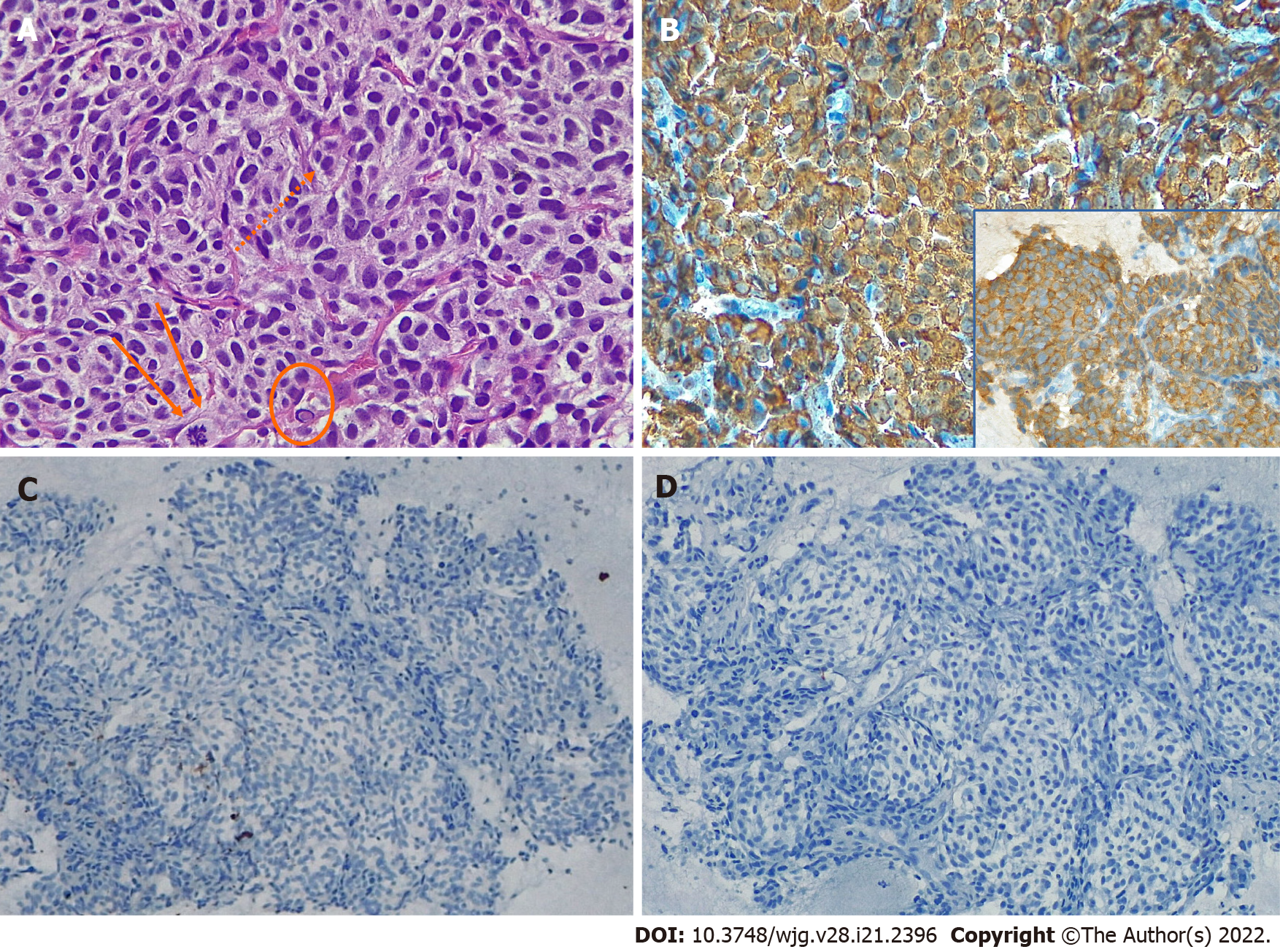
Figure 2 Fine needle biopsy findings.
A: Proliferation of small to medium-sized cells arranged in a nest pattern was evident, the cells occasionally showed small nucleoli (dots), mitotic figures (arrows), and intra-cytoplasmic hyaline globules (encircled), hematoxylin and eosin original magnification (O.M) × 40; B: Chromogranin A (CgA) positivity of neoplastic cells; inset: Synaptophysin positivity of neoplastic cells, CgA stain, O.M. × 20; B inset, synaptophysin stain, O.M. × 20; C: AE1/AE3 cytokeratins expression in scattered cells, cytokeratin AE1/AE3 stain, O.M. × 20; D: S100 negativity, S100 stain, O.M. × 20).
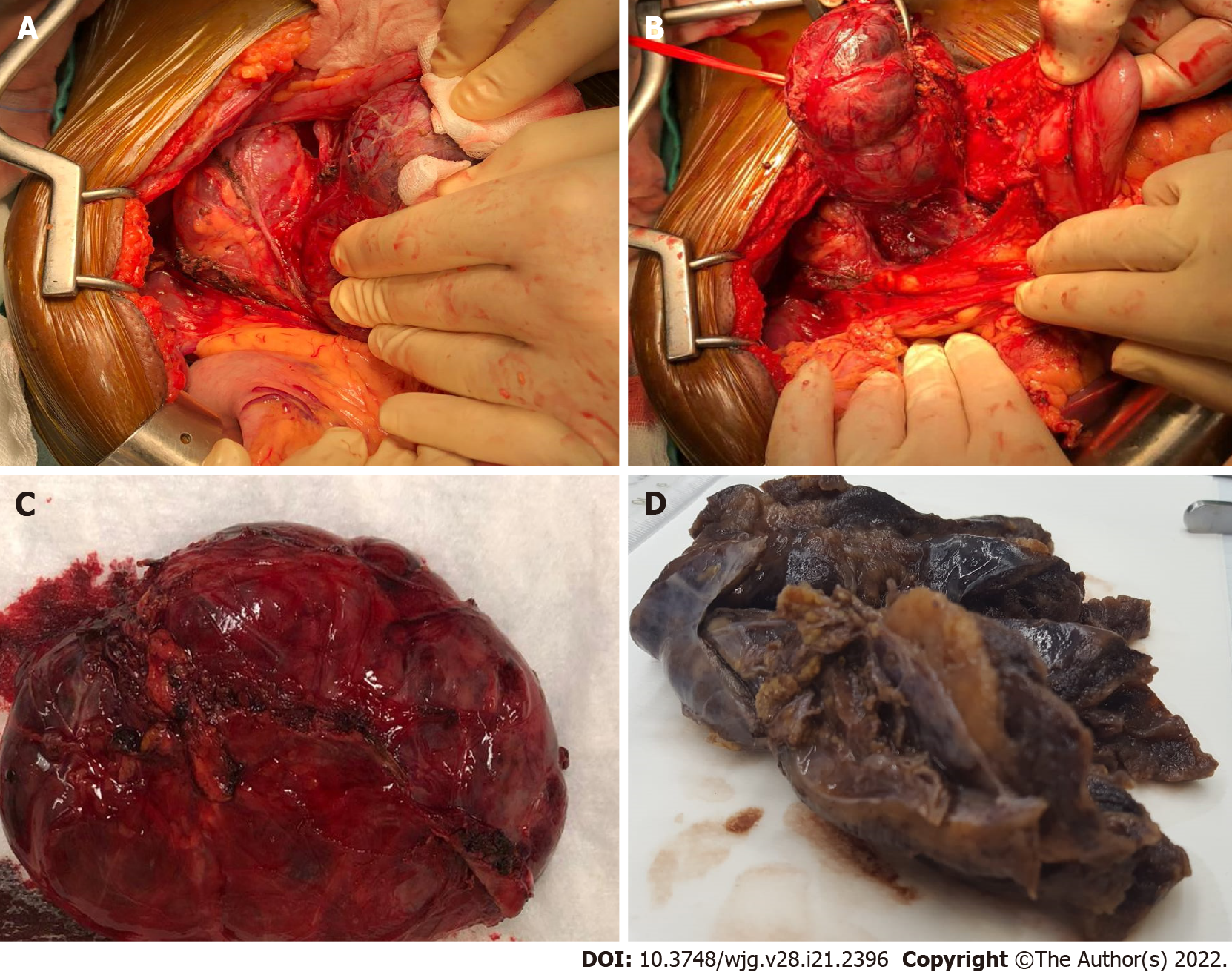
Figure 3 Macroscopic findings.
A and B: At surgical exploration a large well-defined cystic mass located underneath the mesocolon plane was found; C: Radical enucleation of the lesion; D: Grossly, the cystic lesion showed a thick fibrous wall with a solid component and a yellowish, lobulated appearance on cut surface.
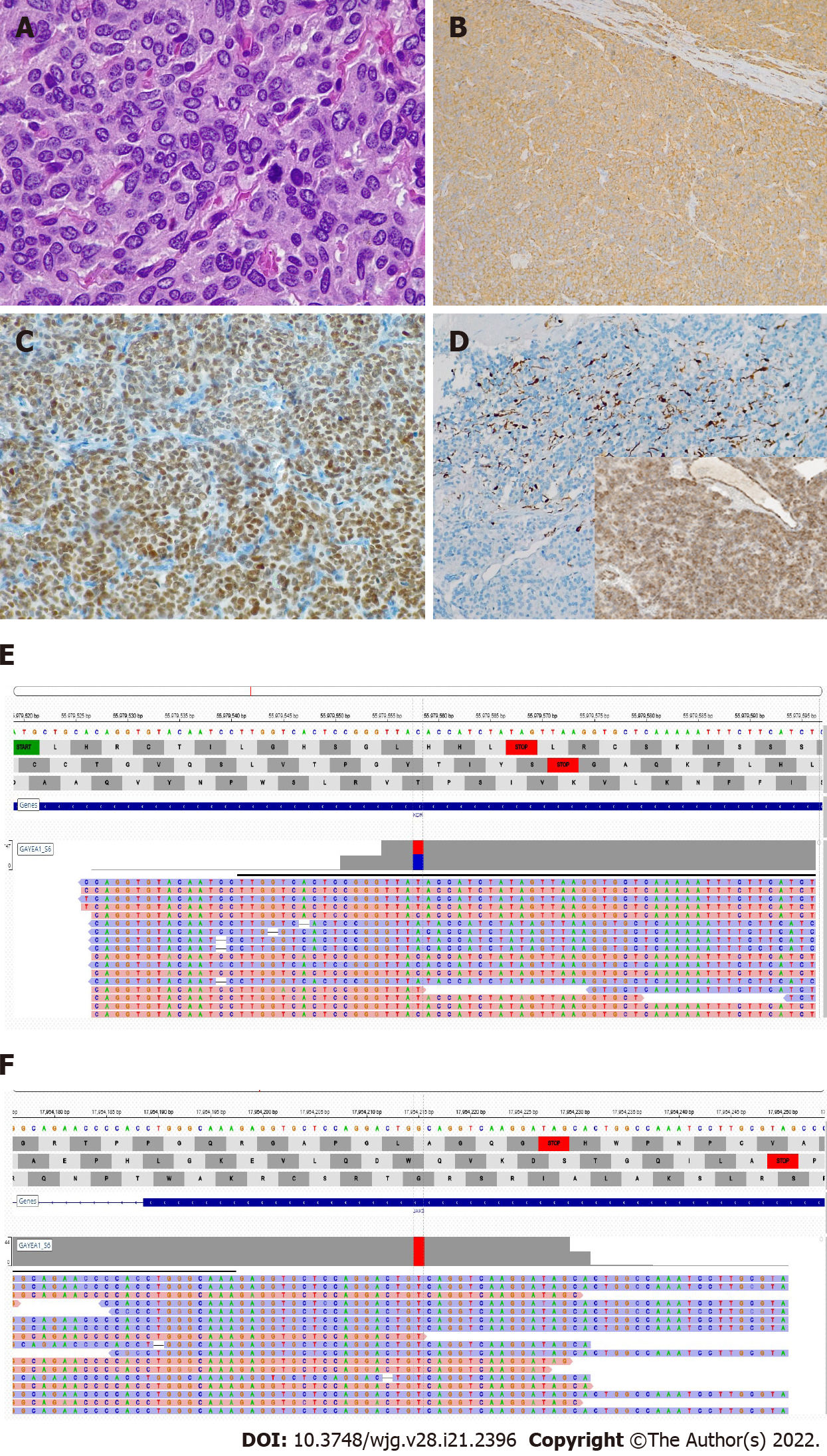
Figure 4 Histologic and molecular findings.
A: The lesion was composed of nests and cords of polygonal cells with abundant granular cytoplasm, and occasional mitotic figures in the more cellular areas, hematoxylin and eosin original magnification (O.M) × 60; B: Chromogranin A (CgA) positivity in neoplastic cells; inset: Synaptophysin positivity in neoplastic cells, CgA stain, O.M × 10; C: GATA-3 positivity in neoplastic cells, GATA-3 stain, O.M. × 10; D: S100-positive cells were scattered; inset: Succinate dehydrogenase subunit B (SDHB) expression was preserved, S100 stain, O.M. × 10; inset, SDHB stain, O.M. × 10; E: KDR mutation; F: JAK3 mutation.
- Citation: Petrelli F, Fratini G, Sbrozzi-Vanni A, Giusti A, Manta R, Vignali C, Nesi G, Amorosi A, Cavazzana A, Arganini M, Ambrosio MR. Peripancreatic paraganglioma: Lesson from a round table. World J Gastroenterol 2022; 28(21): 2396-2402
- URL: https://www.wjgnet.com/1007-9327/full/v28/i21/2396.htm
- DOI: https://dx.doi.org/10.3748/wjg.v28.i21.2396


