Copyright
©The Author(s) 2022.
World J Gastroenterol. Jan 14, 2022; 28(2): 199-215
Published online Jan 14, 2022. doi: 10.3748/wjg.v28.i2.199
Published online Jan 14, 2022. doi: 10.3748/wjg.v28.i2.199
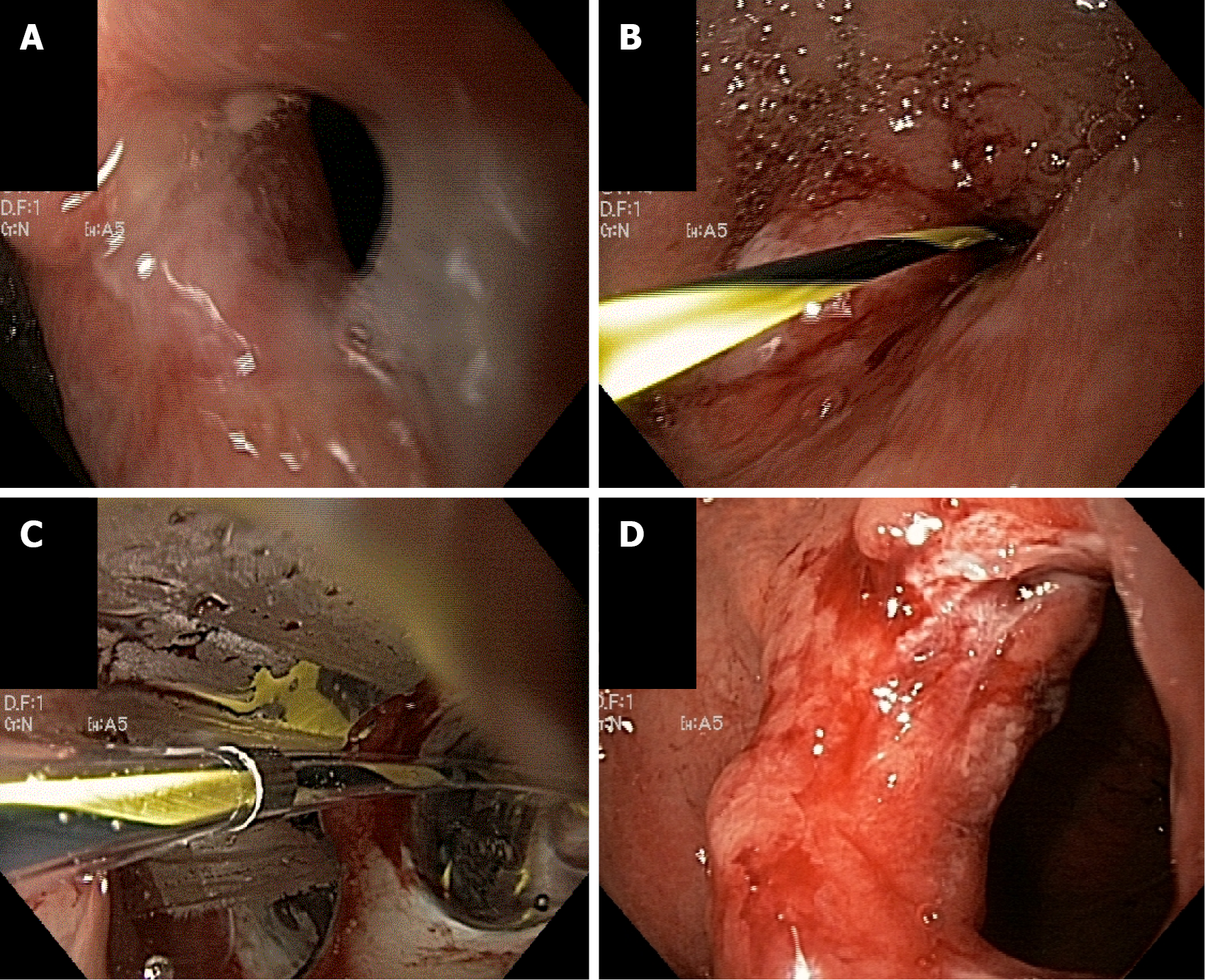
Figure 1 Anastomotic stricture dilation.
A: Tight stricture of gastrojejunostomy; B: Wire placement through stenosis; C: Balloon dilation; D: Stricture appearance after dilation.
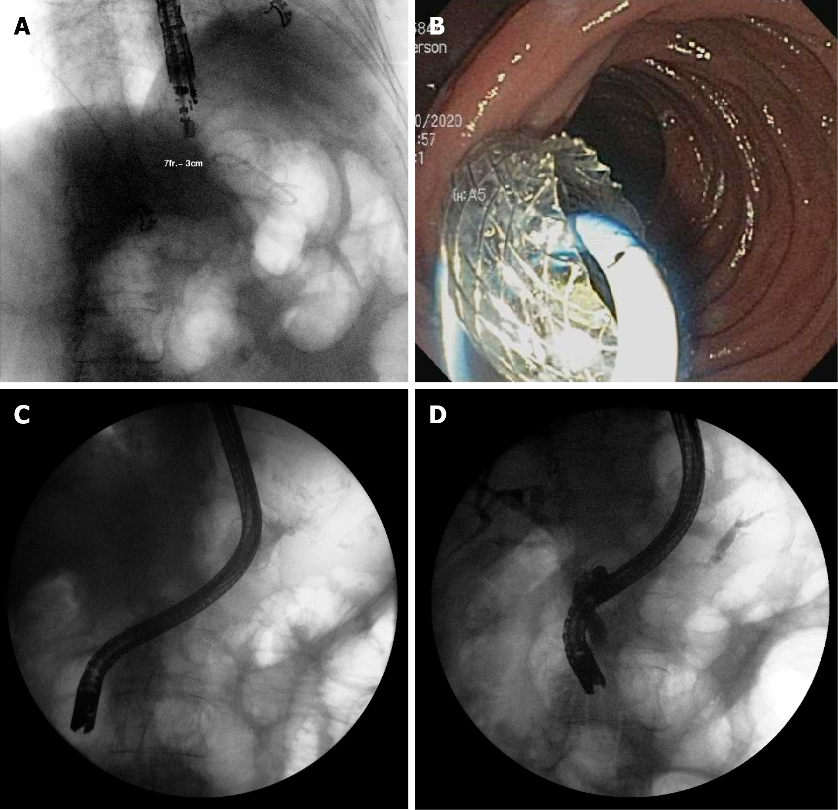
Figure 2 Endoscopic ultrasonography-directed transgastric endoscopic retrograde cholangiopancreatography.
A: Endoscopic ultrasonography placement of lumen apposing metal stents (LAMS) gastrogastric fistula; B: Endoscopic view of LAMS; C: Duodenoscope passing through LAMS for endoscopic retrograde cholangiopancreatography; D: Successful cholangiogram and pancreatogram.
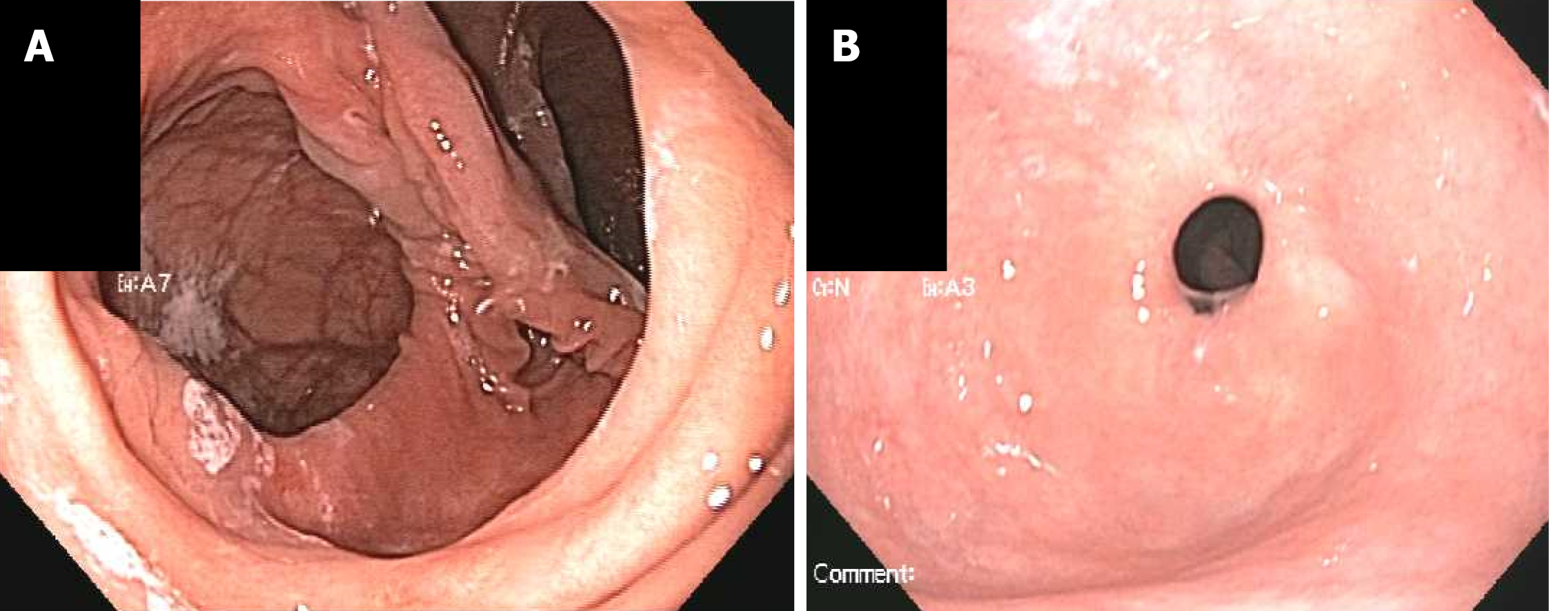
Figure 3 Dilated gastrojejunostomy treated with endoscopic suturing.
A: Dilated gastrojejunostomy; B: Gastrojejunostomy several months after suturing for stoma reduction.
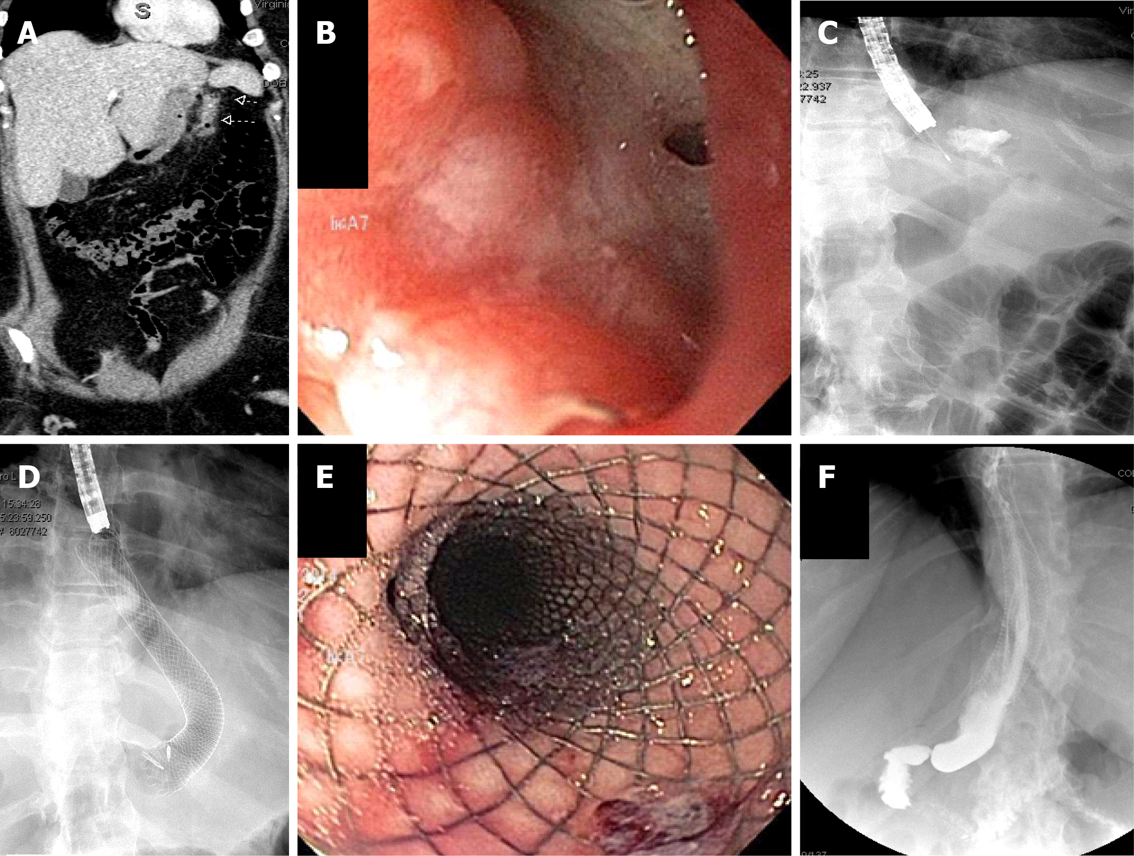
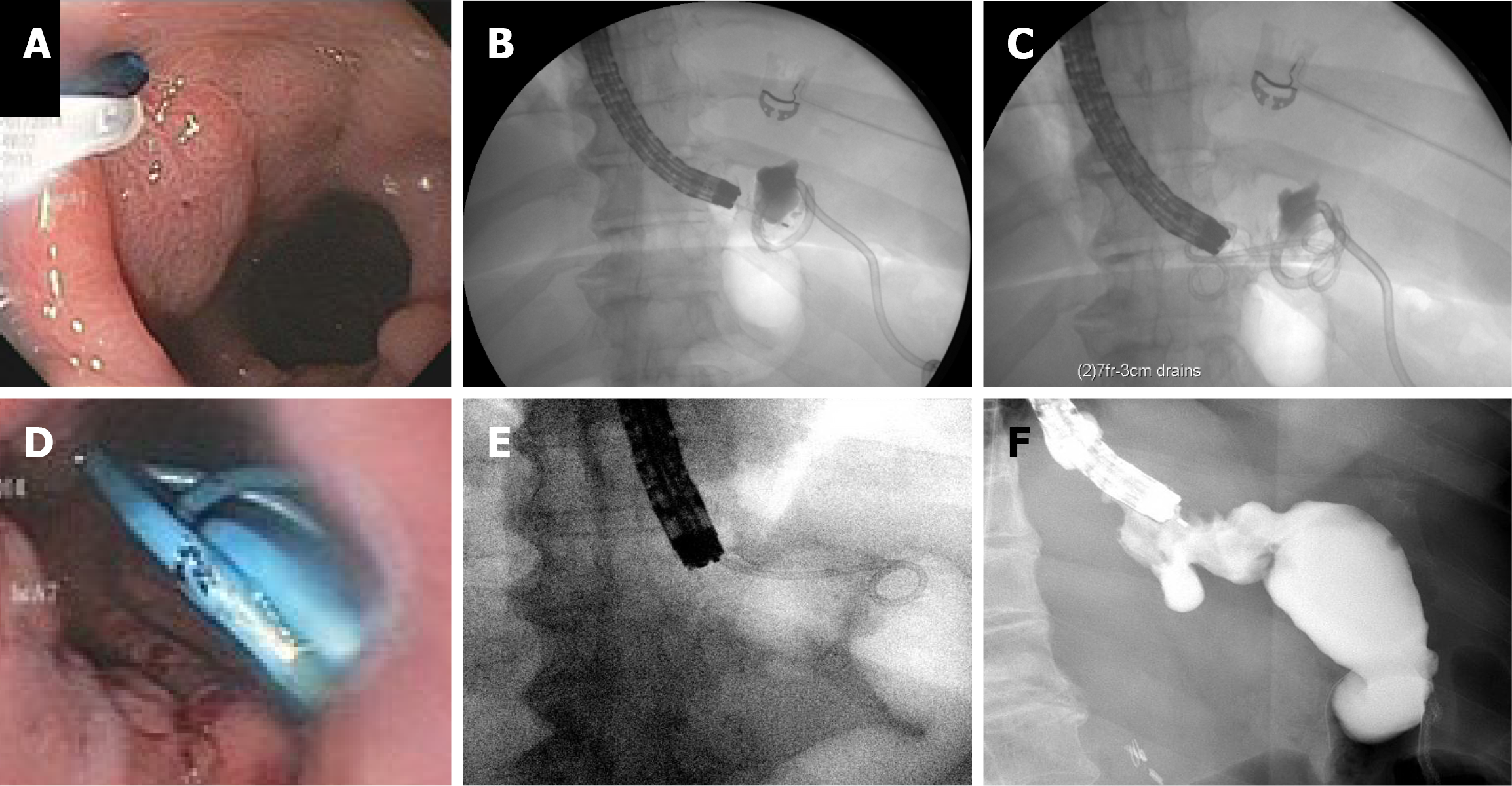
Figure 4 Sleeve leak treated with covered esophageal stent.
Computed tomography imaging demonstrating sleeve leak; B: Endoscopic appearance of leak site; C: Contrast injection to confirm leak site; D: Placement of covered self-expandable metal stents; E: Endoscopic appearance of stent; F: Subsequent upper gastrointestinal series showing no residual leak after stent placement.
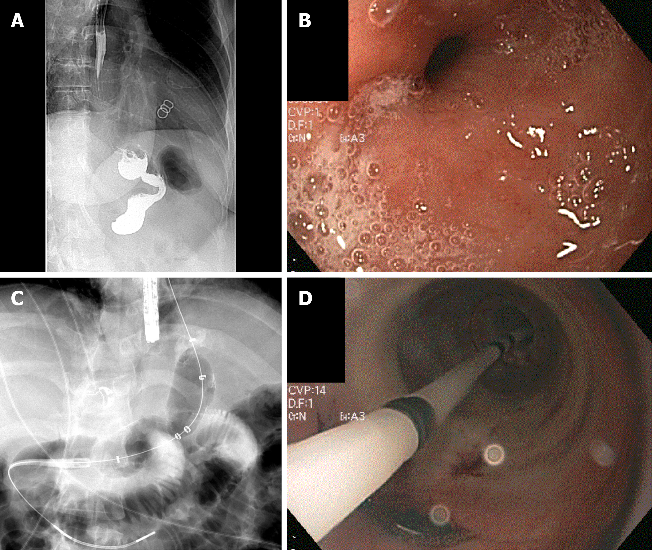
Figure 5 Sleeve gastrectomy leak treated with internal drainage.
A: Endoscopic appearance of leak site; B: Contrast injection to confirm leak site; C: Placement of transgastric double pigtail stents; D: Endoscopic appearance of stents; E: Repeat endoscopy for stent removal; F: Contrast injection after stent removal confirming no residual leak.
Figure 6 Sleeve stenosis treated with balloon dilation.
A: Upper gastrointestinal series demonstrating stenosis at the level of the incisura; B: Endoscopic appearance of the stenosis; C: Pneumatic balloon dilation of stricture; D: Endoscopic appearance of balloon dilation.
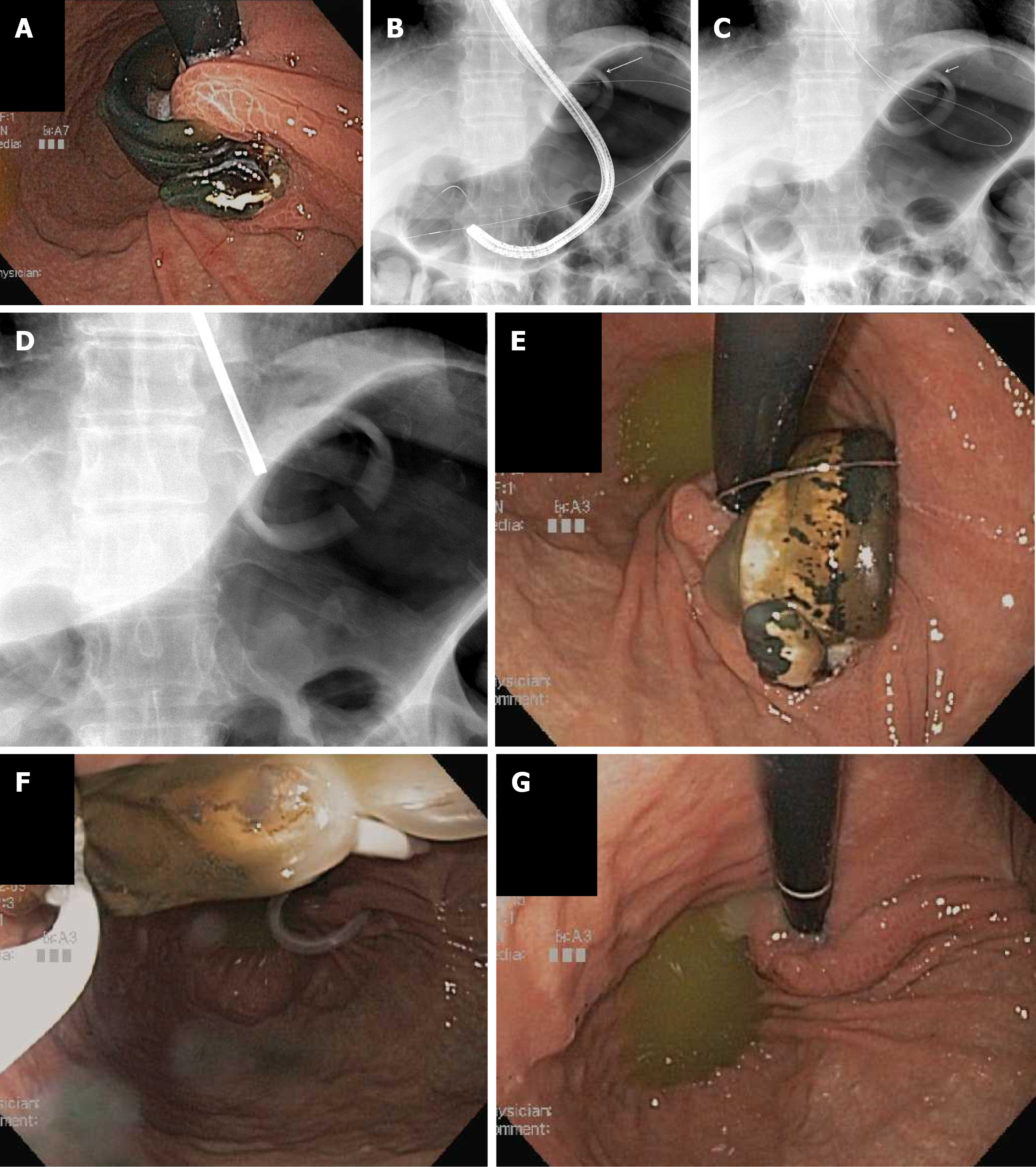
Figure 7 Eroded lap band removed endoscopically.
A: Eroded lap band; B: Endoscope passage through the band with distal deployment of wire; C: Wire looped around band; D: Fluoroscopic view of cut lapband; E: Endoscopic view of cut lap band; F: Cut band grasped by snare; G: Endoscopic view after band removal.
- Citation: Larsen M, Kozarek R. Therapeutic endoscopy for the treatment of post-bariatric surgery complications. World J Gastroenterol 2022; 28(2): 199-215
- URL: https://www.wjgnet.com/1007-9327/full/v28/i2/199.htm
- DOI: https://dx.doi.org/10.3748/wjg.v28.i2.199


