Copyright
©The Author(s) 2022.
World J Gastroenterol. Jan 7, 2022; 28(1): 1-22
Published online Jan 7, 2022. doi: 10.3748/wjg.v28.i1.1
Published online Jan 7, 2022. doi: 10.3748/wjg.v28.i1.1
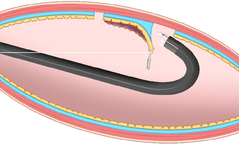
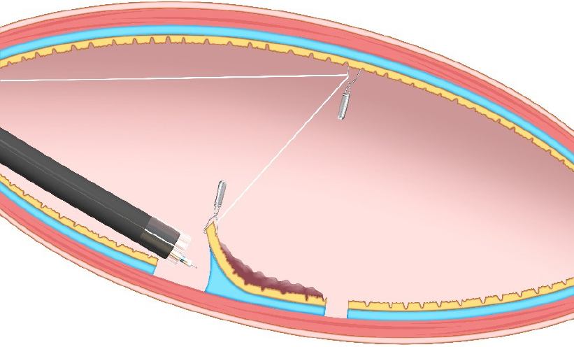
Figure 1 Clip-with-line method.
This method provides traction for the lesion by pulling the line. The traction direction is limited to the direction in which the line is pulled.
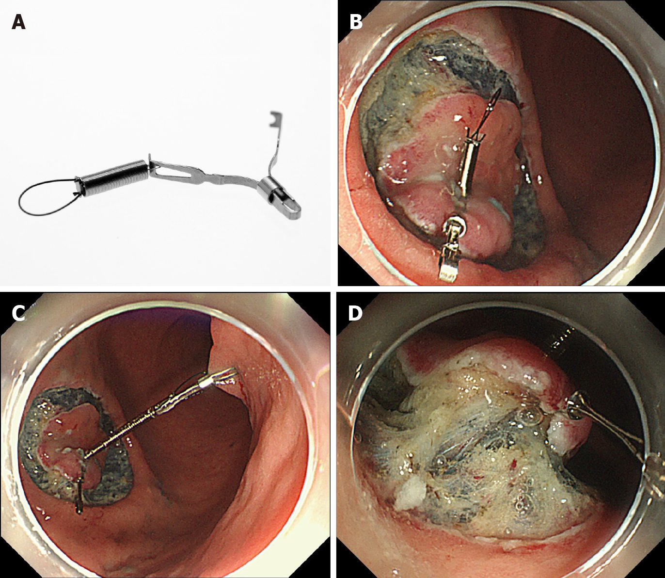
Figure 2 Internal traction method using the spring-and-loop with clip (Zeon Medical, Tokyo, Japan).
A: The spring-and-loop with clip (S–O clip) has a 5-mm long spring and a 4-mm long loop at one side of the clip claws; B: The S–O clip is attached to the lesion; C: The regular clip anchors the loop of the S–O clip on the opposite side of the lesion; D: The extension of the spring provides traction on the lesion. Citation: Mitsuru Nagata. Internal traction method using a spring-and-loop with clip (S–O clip) allows countertraction in gastric endoscopic submucosal dissection. Surg Endosc 2020; 34(8): 3722–3733. Copyright © 2020 Mitsuru Nagata[10].
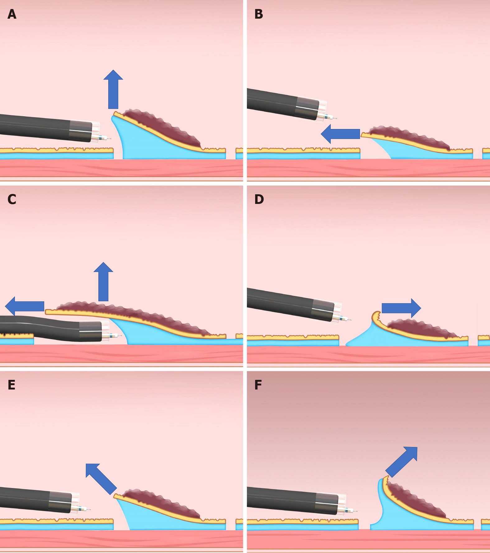
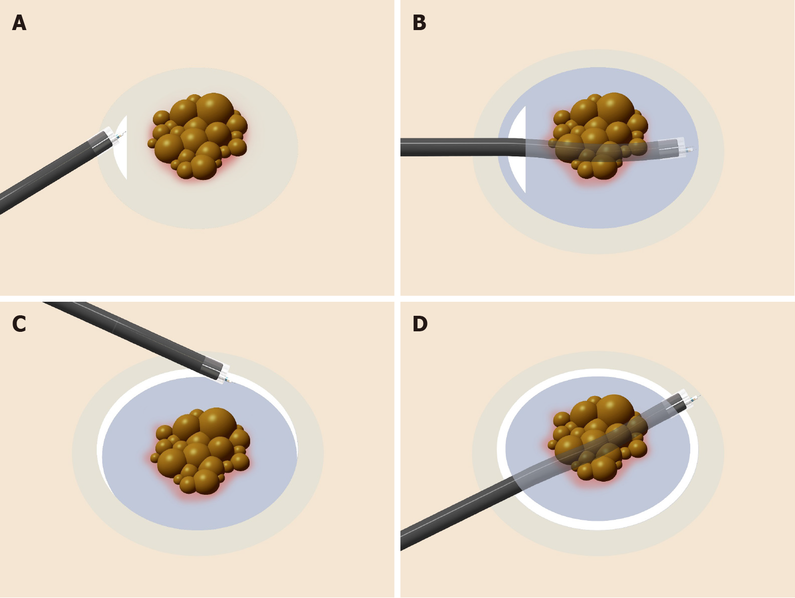
Figure 3 Classification of the traction direction.
A: Vertical traction; B: Proximal traction; C: Proximal traction combined with hood traction; D: Distal traction; E: Diagonally proximal traction; F: Diagonally distal traction.
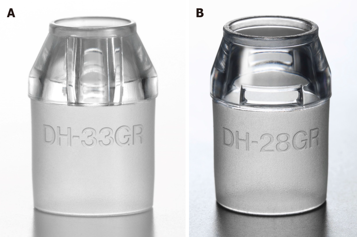
Figure 4 Small-caliber tip transparent hood (Fujifilm, Tokyo, Japan).
A: DH-33GR [small-caliber tip transparent (ST) hood] with 7-mm tip opening diameter and 7-mm tip protruding length; B: DH-28GR (short ST hood) with 8-mm tip opening diameter and 7-mm tip protruding length.
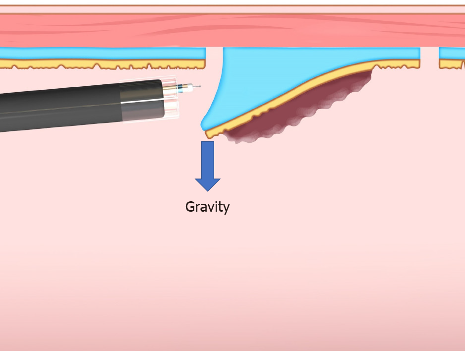
Figure 5 Gravity provides favorable traction when the lesion is located at the upper side of the gravitational force.
Figure 6 Procedure for the pocket creation method of endoscopic submucosal dissection.
A: A minimal initial mucosal incision (approximately 20-mm in width) is made for entry into the submucosa; B: Submucosal dissection is performed, followed by creation of a submucosal pocket under the lesion; C: Additional mucosal incision of the gravitational lower side of the submucosal pocket; D: Opening around the submucosal pocket, then, en bloc resection is achieved.
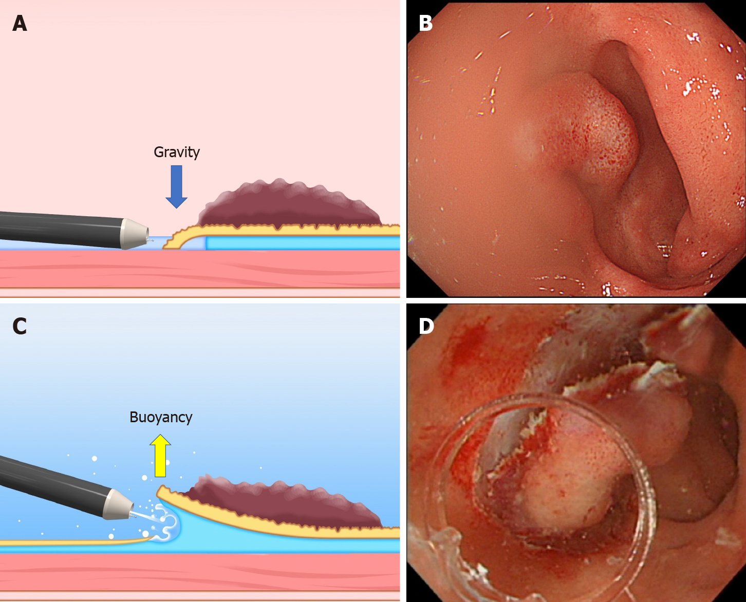
Figure 7 The difference between the conventional endoscopic submucosal dissection and the underwater endoscopic submucosal dissection.
A and B: The conventional endoscopic submucosal dissection for the lesion is located at the gravitational lower side. Gravity obstructs the opening of the mucosal flap. Incomplete submersion deteriorates the visual field; C and D: The underwater condition aids the opening of the mucosal flap by buoyancy. Water pressure from the endoscope (using its water supply function) also assists in opening the mucosal flap. Complete submersion improves the visual field. Citation: Mitsuru Nagata. Underwater endoscopic submucosal dissection in saline solution using a bent-type knife for duodenal tumor. VideoGIE 2018; 3(12): 375–377. Copyright © 2018 American Society for Gastrointestinal Endoscopy. Published by Elsevier Inc[21].
Figure 8 Pulley method.
This method is the modified clip-with-line method, which can provide traction in any direction depending on the pulley site.
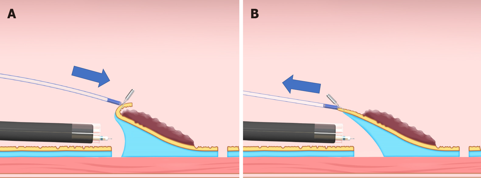
Figure 9 Sheath traction method.
This method has two traction directions by pushing or pulling the sheath. A: Pushing the sheath; B: Pulling the sheath.
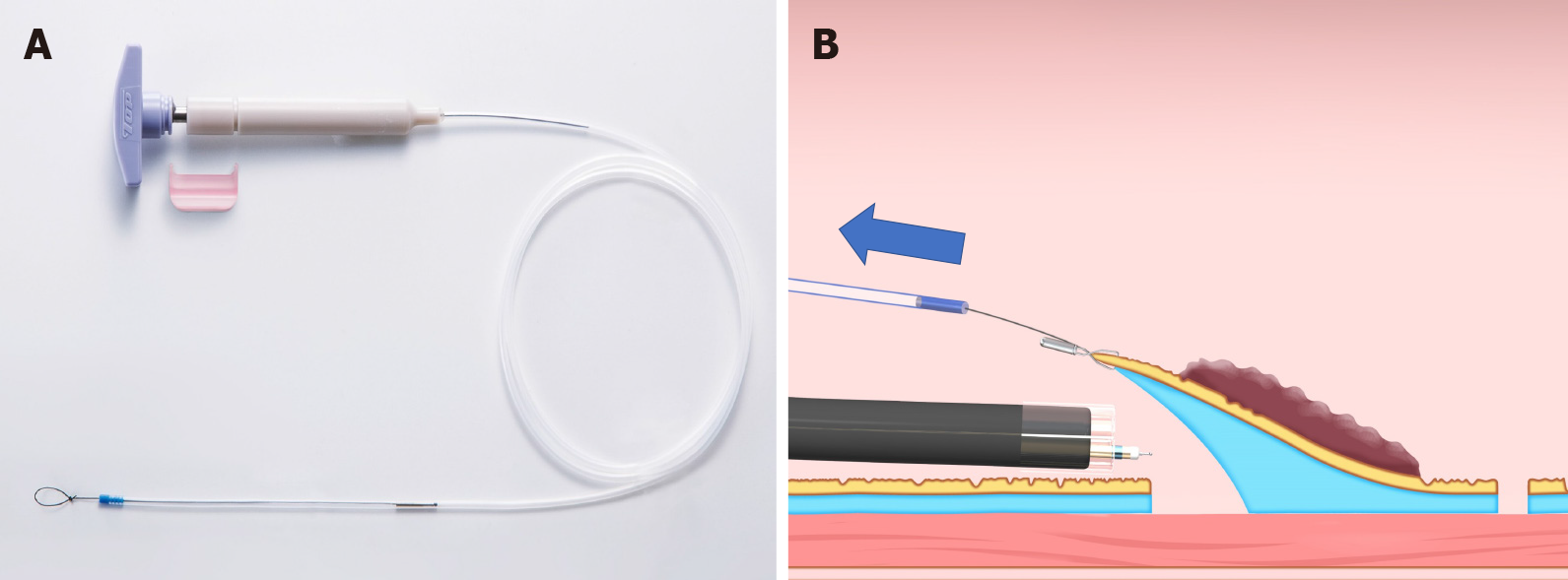
Figure 10 Endo Trac (TOP, Tokyo, Japan).
A: This device can be used for the sheath traction method; B: This device has a structure that can release the sheath from the lesion to avoid interference between the sheath and the endoscope tip.
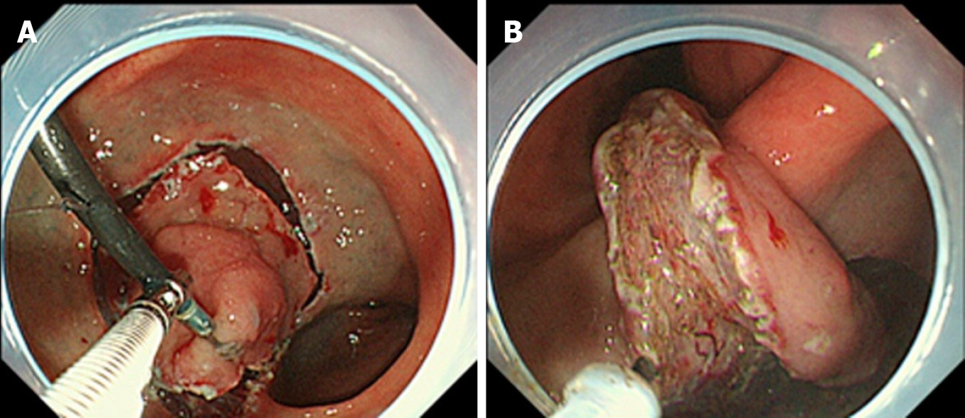
Figure 11 Endoscopic submucosal dissection using external forceps.
A: External grasping forceps was anchored at the distal margin of the lesion in the lesser curvature of the antrum under the control of the endoscope and a second grasping forceps; B: With gentle oral traction applied with the external grasping forceps, the submucosal layer was dissected in retroversion from the aboral side. Citation: Imaeda H, Hosoe N, Kashiwagi K, Ohmori T, Yahagi N, Kanai T, Ogata H. Advanced endoscopic submucosal dissection with traction. World J Gastrointest Endosc 2014; 6(7): 286-295. Copyright © 2014 Baishideng Publishing Group Inc. Published by Baishideng Publishing Group Inc[35].
Figure 12 Internal traction method using the spring-and-loop with clip (Zeon Medical, Tokyo, Japan).
A: The spring-and-loop with clip (S–O clip) is attached to the lesion; B: The regular clip anchors the loop part of the S–O clip on the gastrointestinal wall; C: The extension of the spring provides traction on the lesion. The traction direction can be controlled by the anchor site. Citation: Nagata M, Fujikawa T, Munakata H. Comparing a conventional and a spring-and-loop with clip traction method of endoscopic submucosal dissection for superficial gastric neoplasms: a randomized controlled trial (with videos). Gastrointest Endosc 2021; 93(5): 1097-1109. Copyright © 2021 American Society for Gastrointestinal Endoscopy. Published by Elsevier Inc[11].
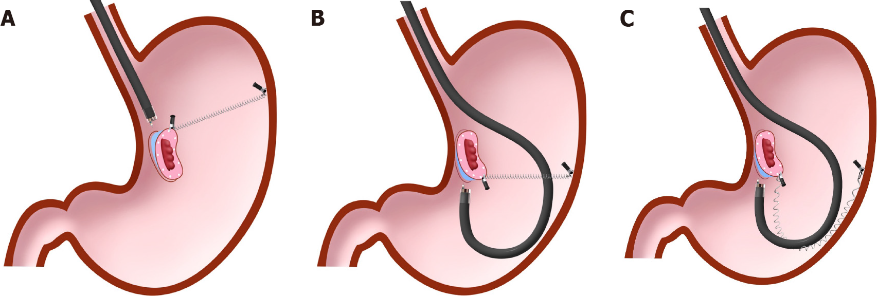
Figure 13 Spring-and-loop with clip-assisted gastric endoscopic submucosal dissection.
A: Forward endoscopic position; B: Retroflexed endoscopic position; C: The endoscope has the possibility to stretch the spring, resulting in a loss of spring elasticity in the retroflexed endoscopic position. In this situation, the modified attachment method that we described in detail in the previous papers[9-11] is required to avoid this problem. Citation: Nagata M, Fujikawa T, Munakata H. Comparing a conventional and a spring-and-loop with clip traction method of endoscopic submucosal dissection for superficial gastric neoplasms: a randomized controlled trial (with videos). Gastrointest Endosc 2021; 93(5): 1097-1109. Copyright © 2021 American Society for Gastrointestinal Endoscopy. Published by Elsevier Inc[11].
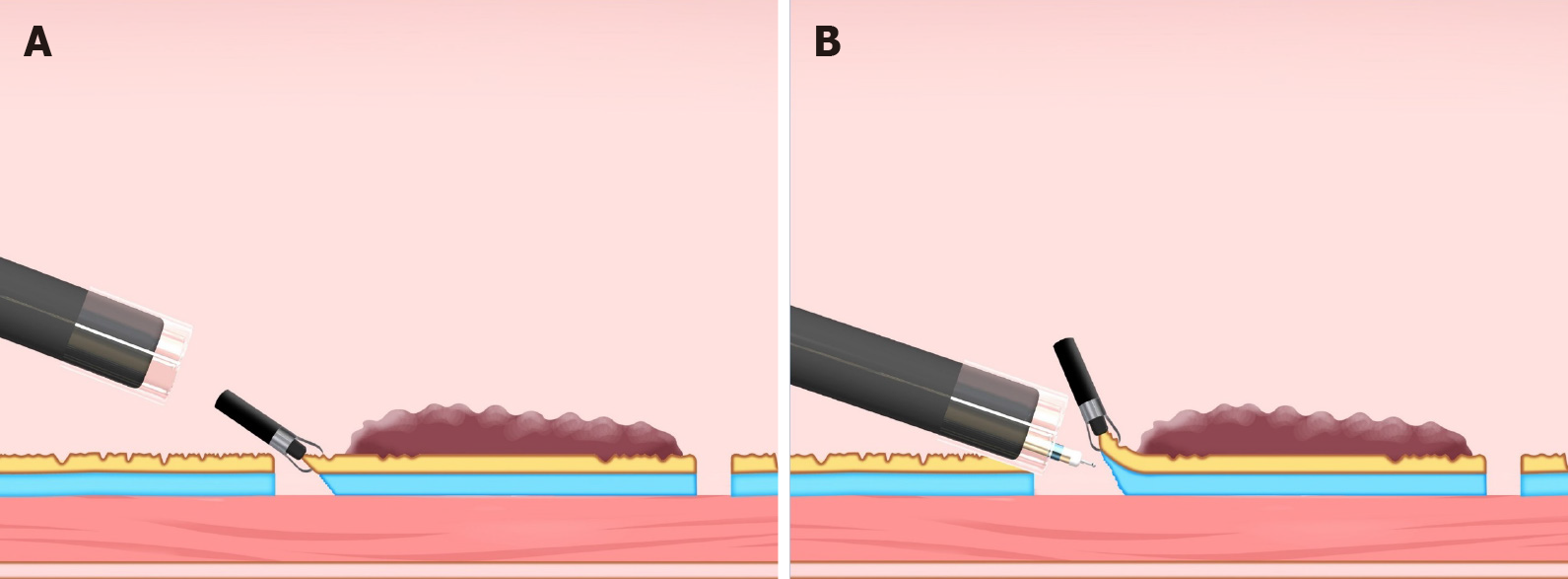
Figure 14 Clip flap method.
A: The clip is attached to the edge of the lesion; B: The clip can be used as a substitute for the mucosal flap.
Figure 15 Difference in traction direction depending on the lesion location in the clip-with-line method.
A: Distal traction; B: Proximal traction; C: Vertical traction.
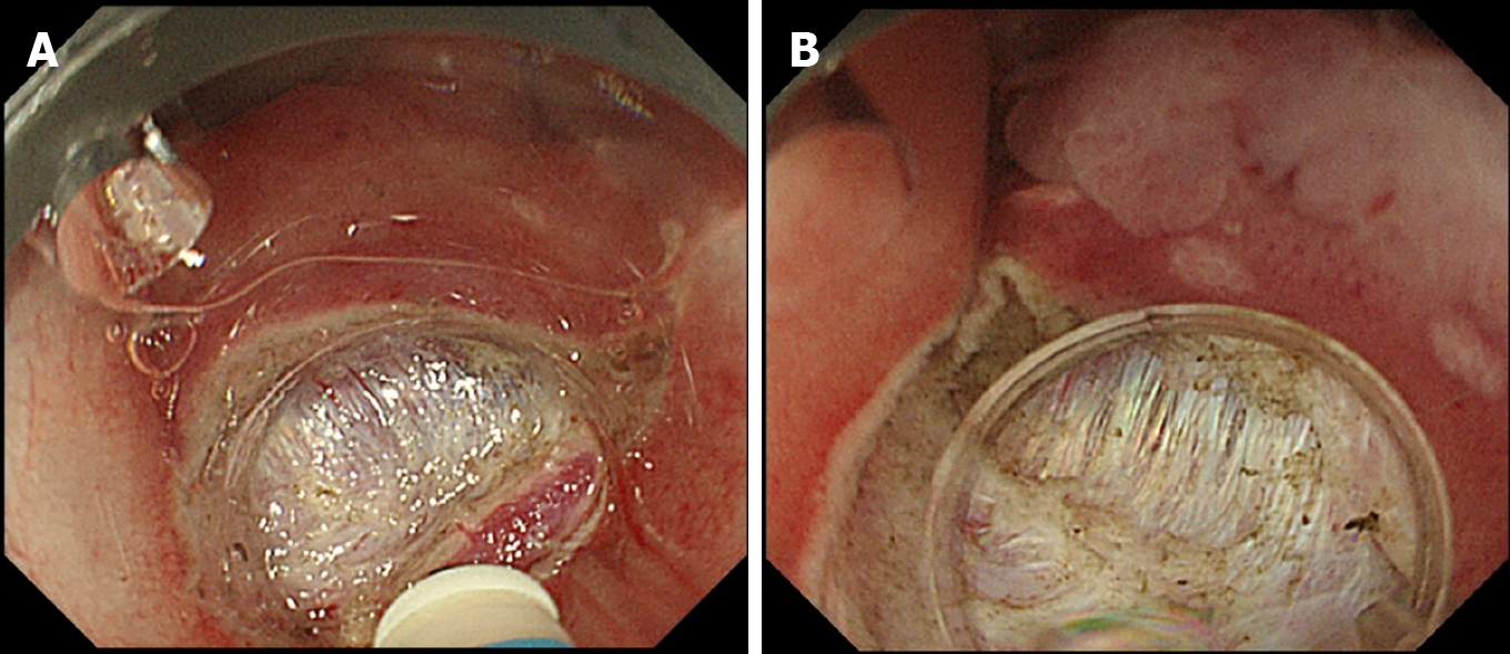
Figure 16 Comparison of aerial and underwater endoscopic view.
A: Severe fibrosis and halation makes the boundary between the submucosal layer and muscle layer unclear; B: Submergence enables a detailed observation through a natural zoom effect and causes halation disappearance, thereby clarifying the boundary between the submucosal layer and muscle layer. Citation: Nagata M. Usefulness of underwater endoscopic submucosal dissection in saline solution with a monopolar knife for colorectal tumors (with videos). Gastrointest Endosc 2018; 87(5): 1345-1353. Copyright © 2018 American Society for Gastrointestinal Endoscopy. Published by Elsevier Inc[20].
- Citation: Nagata M. Advances in traction methods for endoscopic submucosal dissection: What is the best traction method and traction direction? World J Gastroenterol 2022; 28(1): 1-22
- URL: https://www.wjgnet.com/1007-9327/full/v28/i1/1.htm
- DOI: https://dx.doi.org/10.3748/wjg.v28.i1.1


