Copyright
©The Author(s) 2021.
World J Gastroenterol. Dec 28, 2021; 27(48): 8302-8322
Published online Dec 28, 2021. doi: 10.3748/wjg.v27.i48.8302
Published online Dec 28, 2021. doi: 10.3748/wjg.v27.i48.8302
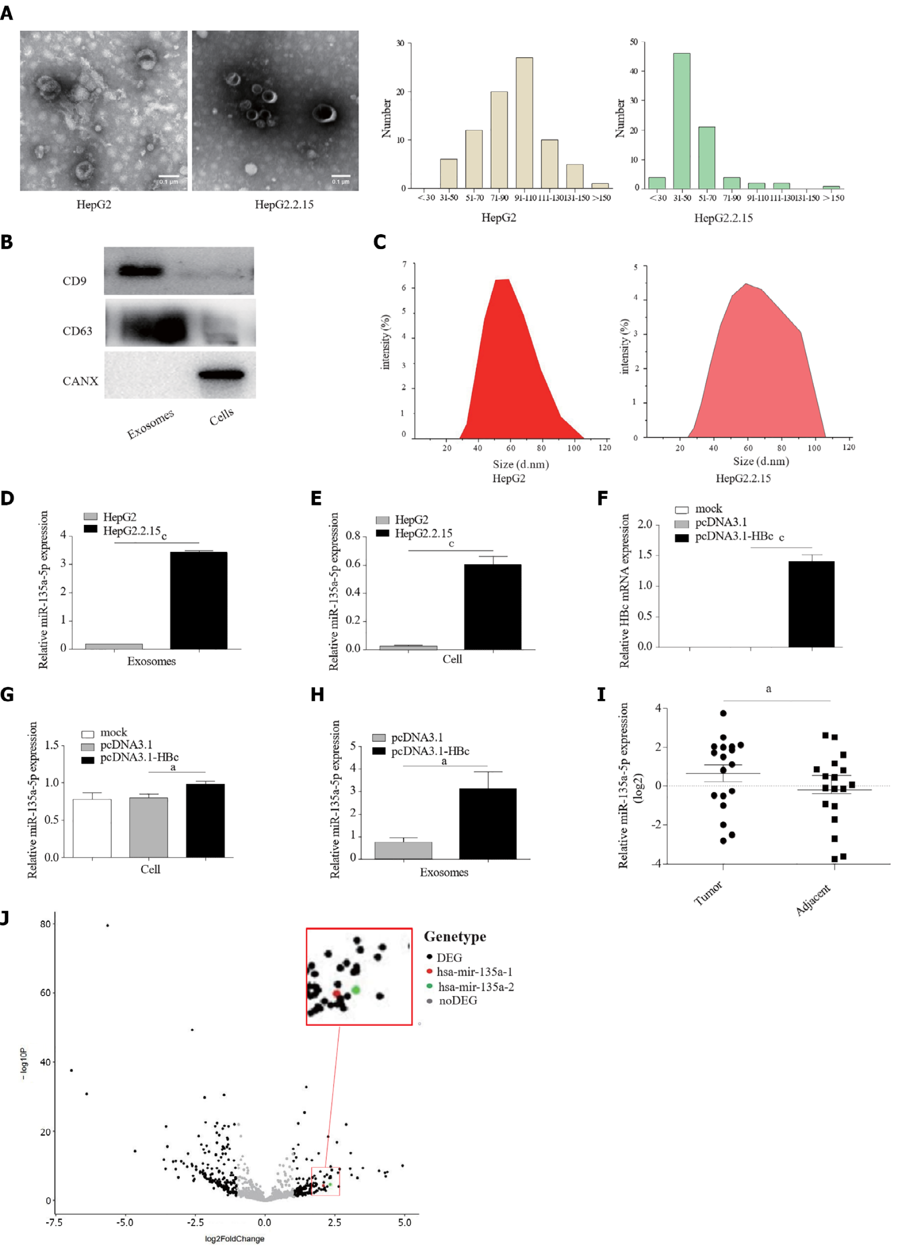
Figure 1 Hepatitis B virus upregulated the expression levels of miR-135a-5p in exosomes.
A: Transmission electron microscopy image of exosomes; B: Western blotting indicated proteins in exosomes; C: Analysis of particle size distribution of exosomes; D and E: Quantitative polymerase chain reaction (qPCR) assay examined the expression of miR-135a-5p in exosomes derived from cancer cells and in HCC cell lines; F: Overexpression of Hepatitis B core antigen (HBc) in HepG2 cells was detected by qPCR; G and H: The qRCP assay identified the level of miR-135a-5p in HepG2 cells overexpressed HBc and exosomes isolated from HepG2 cells after transfected with pcDNA3.1-HBc plasmids; I: Detection of miR-135a-5p in adjacent and tumor tissues from 18 patients; J: Expression of miR-135a-5p obtained from TCGA in HCC. aP < 0.05; cP < 0.001. HBc: Hepatitis B core antigen.
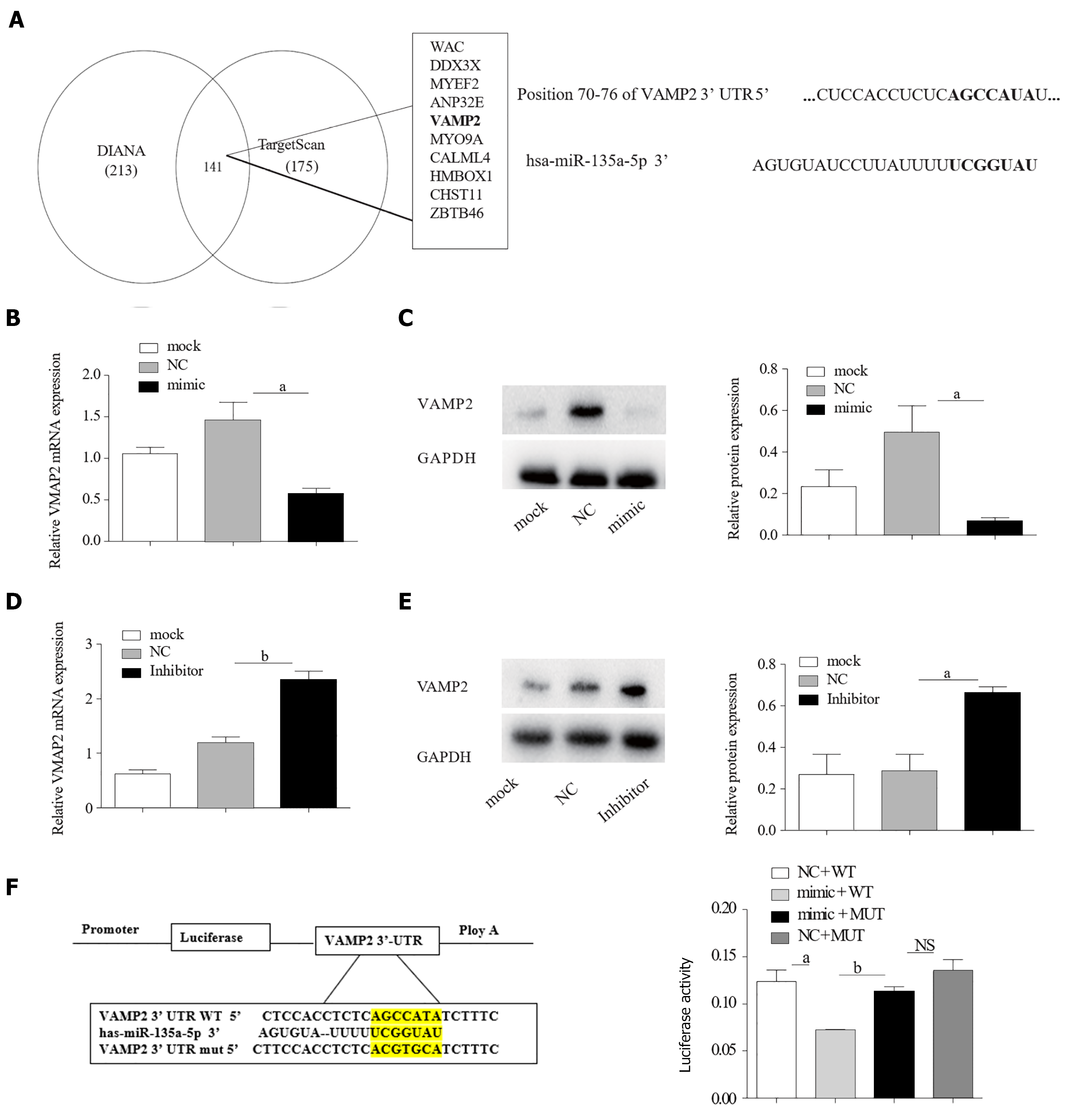
Figure 2 miR-135a-5p targeted vesicle-associated membrane protein 2 in hepatocellular carcinoma cells.
A: Prediction results of target gene of miR-135a-5p; B: The expression of miR-135a-5p was measured by quantitative polymerase chain reaction (qPCR) in HepG2 cells transfected with miR-135a-5p mimics; C and D: The mRNA and protein levels of vesicle-associated membrane protein 2 (VAMP2) were detected in the overexpressed miR-135a-5p cells; E: Quantification of miR-135a-5p in HepG2 cells transfected with miR-135a-5p inhibitors; F and G: qPCR and western blot analyses of VAMP2 level in HepG2 cells transfected with miR-135a-5p inhibitors; H: Luciferase assay in HepG2 cells. aP < 0.05; bP < 0.01; NS: Not Statistically Significant. GAPDH: glyceraldehyde-3-phosphate dehydrogenase; VAMP2: Vesicle-associated membrane protein 2; WT: Wild-type; MUT: Mutant.
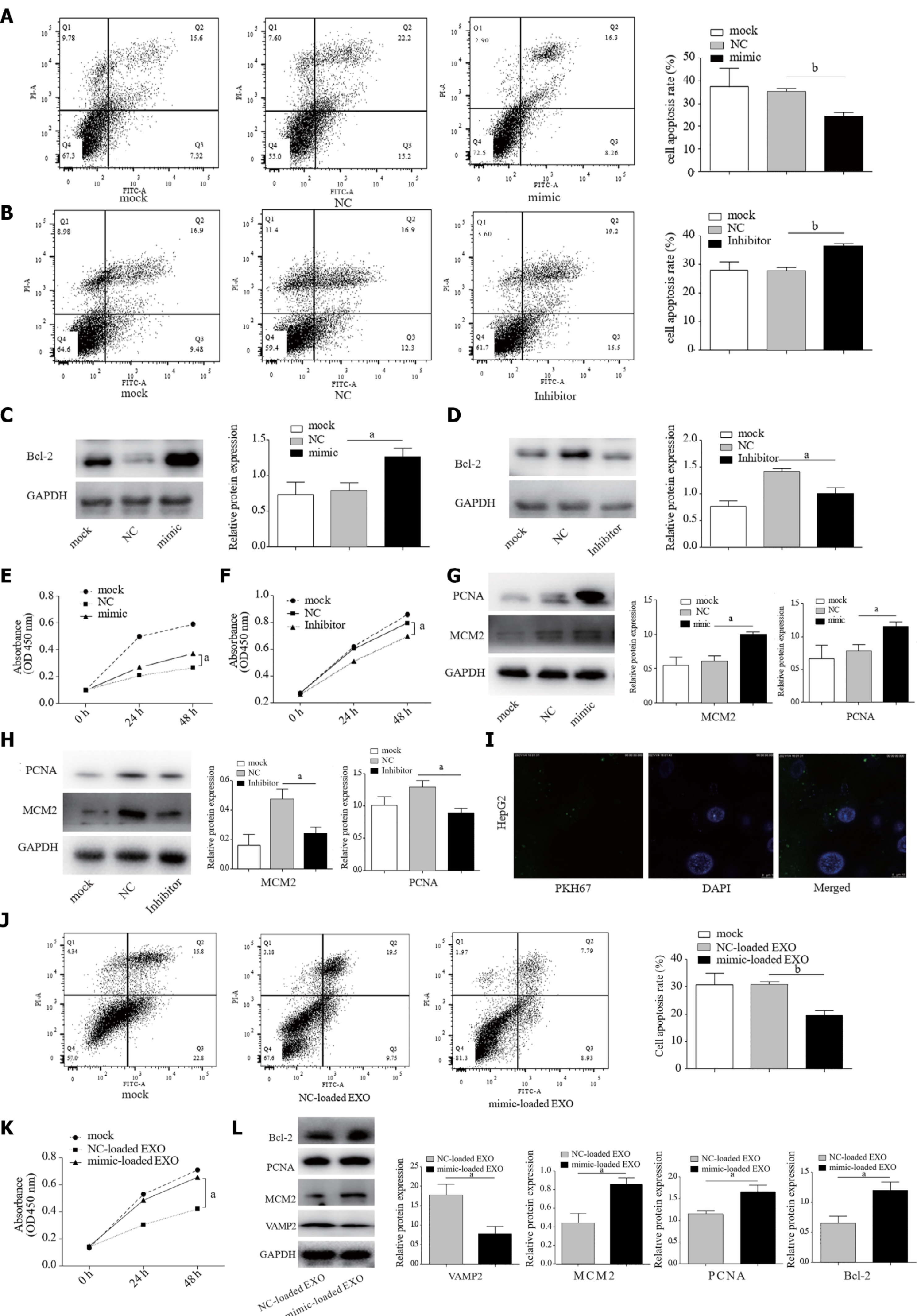
Figure 3 miR-135a-5p suppressed apoptosis and promoted proliferation.
A and B: Annexin V-FITC/PI assay for the effect of overexpression or knockdown of miR-135a-5p on apoptosis of HepG2 cells; C and D: The protein expression of B-cell lymphoma-2 (Bcl-2) in the group with overexpression of miR-135a-5p and the miR-135a-5p inhibited group; E and F: Cell counting kit 8 assays were used to determine the proliferation of HepG2 cells transfected with miR-135a-5p mimics and miR-135a-5p inhibitors; G and H: Western blot analyses of the level of mini-chromosome maintenance protein-2 (MCM2) and proliferating cell nuclear antigen (PCNA) in the group with overexpression of miR-135a-5p and the miR-135a-5p inhibited group; I: Confocal image showing that HepG2 cells were treated with exosomes rich in miR-135a-5p; J: Flow cytometry analysis of the effect of exosomal miR-135a-5p on cell apoptosis; K: Cell counting assay was performed to determine the proliferation of HepG2 cells treated with exosomes with overexpressed miR-135a-5p; L: Western blot analyses of Bcl-2, MCM2, PCNA and vesicle-associated membrane protein 2 in HepG2 cells incubated with mimic-loaded EXO or NC-loaded EXO. aP < 0.05; bP < 0.01. Bcl-2: B-cell lymphoma-2; MCM2: Mini-chromosome maintenance protein-2; PCNA: Proliferating cell nuclear antigen.
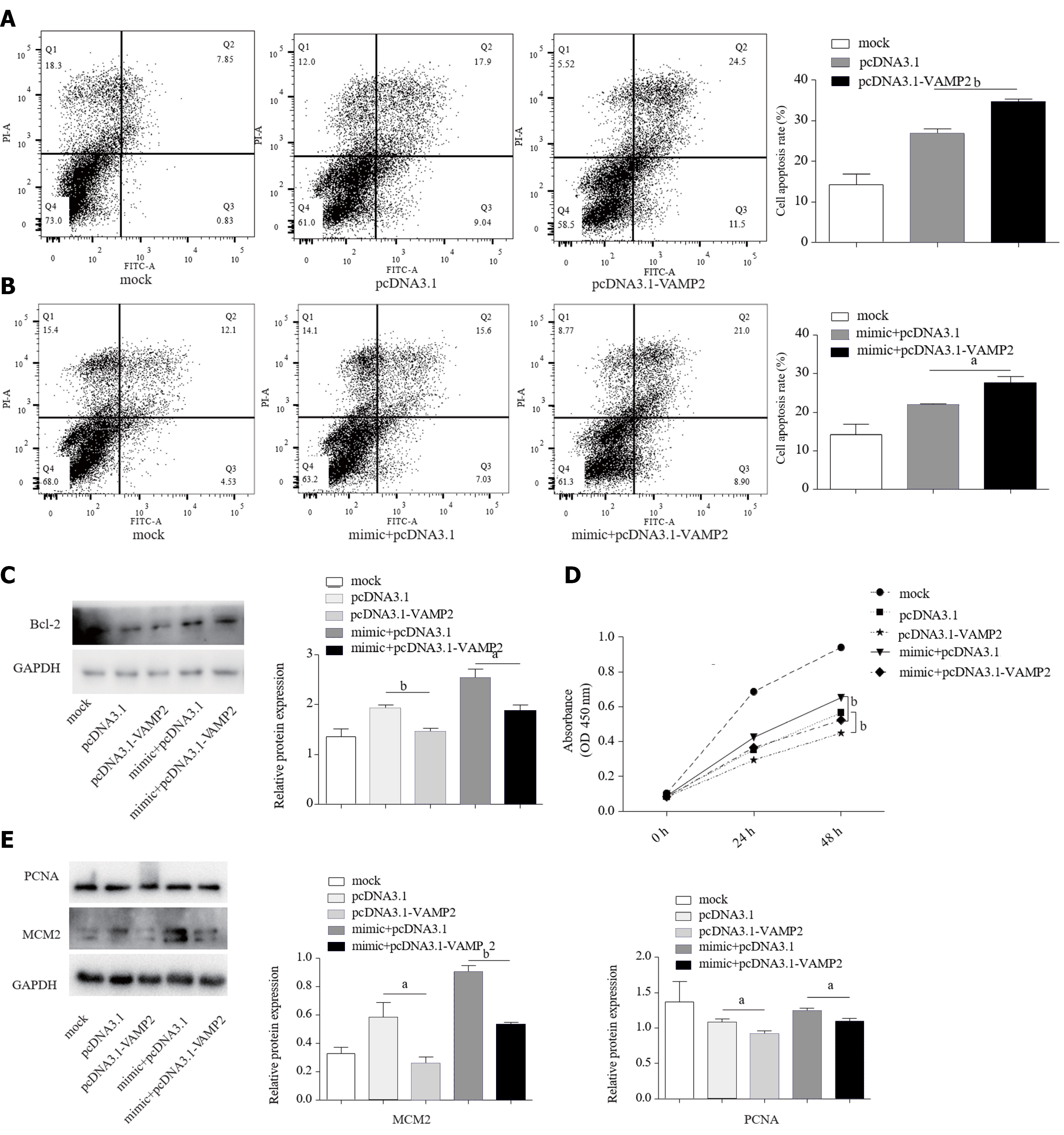
Figure 4 Vesicle-associated membrane protein 2 contributed to anti-apoptosis and proliferation induced by miR-135a-5p.
A and B: Cell apoptosis was examined by flow cytometry in HepG2 cells transfected with the specific plasmid combinations; C: B-cell lymphoma-2 expression was detected by Western blot in HepG2 cells after transfection with the indicated plasmids; D: Cell counting kit 8 assay showed the proliferation of HepG2 cells after transfection with the plasmid combination shown above; E: The protein level of mini-chromosome maintenance protein-2 and proliferating cell nuclear antigen was measured by Western blot in HepG2 cells transfected with the plasmid group shown in the figure above. aP < 0.05; bP < 0.01. Bcl-2: B-cell lymphoma-2; MCM2: Mini-chromosome maintenance protein-2; PCNA: Proliferating cell nuclear antigen; GAPDH: glyceraldehyde-3-phosphate dehydrogenase; VAMP2: Vesicle-associated membrane protein 2.
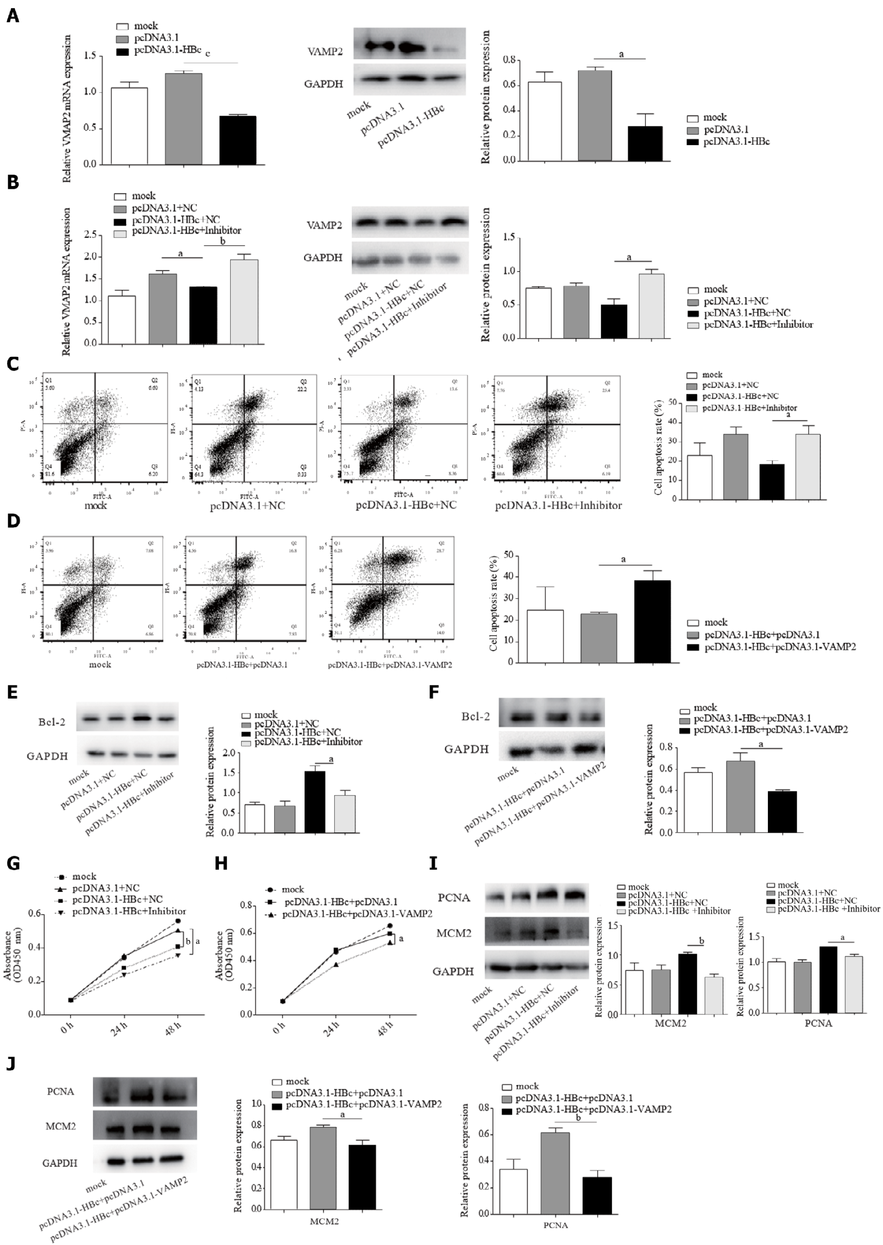
Figure 5 Hepatitis B core antigen induced anti-apoptosis and proliferation via miR-135a-5p and its target gene vesicle-associated membrane protein 2.
A and B: Quantitative polymerase chain reaction and western blot analyses of the level of vesicle-associated membrane protein 2 in HepG2 cells after transfection with the specific plasmid combinations; C and D: Annexin V-FITC/PI assay was performed to assess cell apoptosis in HepG2 cells after transfected with the indicated plasmids; E and F: Western blot was performed to analyze the level of B-cell lymphoma-2 in HepG2 cells after transfection with the plasmid combination shown above; G and H: Cell counting kit 8 assay was performed to assess cell proliferation in HepG2 cells transfected with the specific plasmid combinations; I and J: Western blot analyses of mini-chromosome maintenance protein-2 and proliferating cell nuclear antigen in HepG2 cells transfected with the plasmid group shown in the figure above. aP < 0.05; bP < 0.01. Bcl-2: B-cell lymphoma-2; MCM2: Mini-chromosome maintenance protein-2; PCNA: Proliferating cell nuclear antigen; GAPDH: glyceraldehyde-3-phosphate dehydrogenase; VAMP2: Vesicle-associated membrane protein 2.
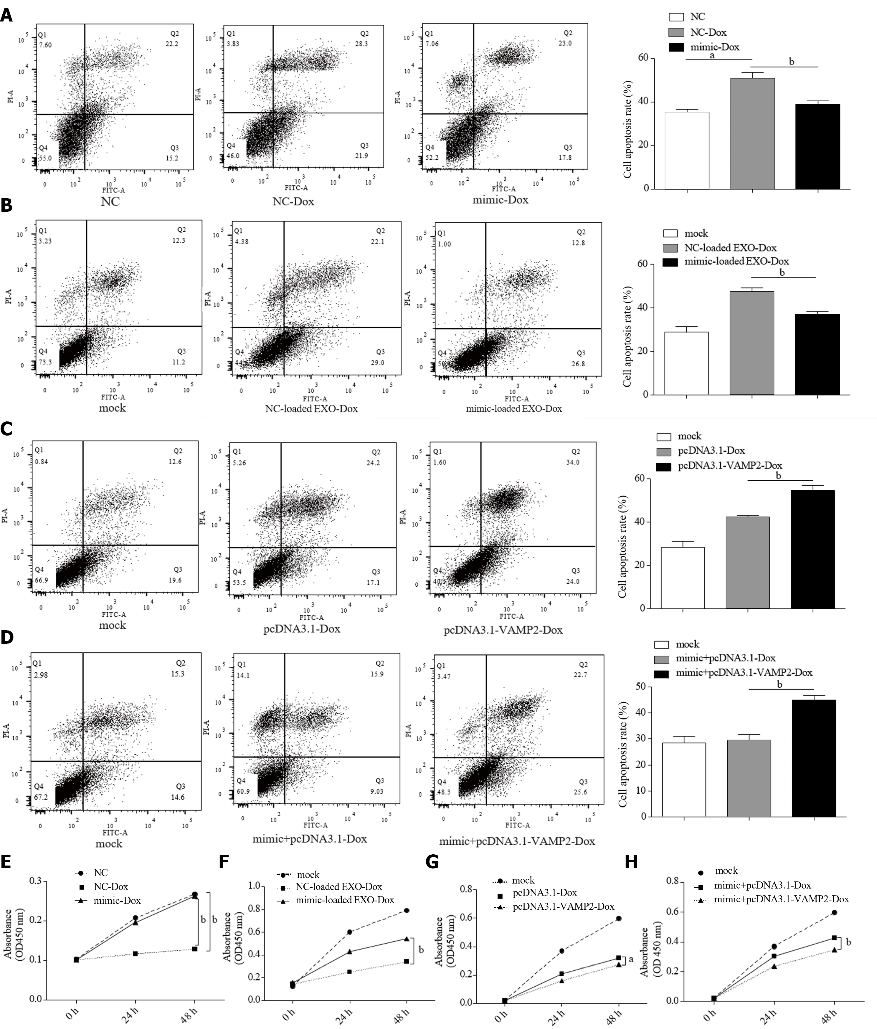
Figure 6 miR-135a-5p enhanced Dox-resistance and reduced cell apoptosis of hepatocellular carcinoma cells by down-regulating vesicle-associated membrane protein 2.
A: The apoptosis rate of HepG2 cells after treatment with Doxorubicin hydrochloride (Dox). Flow cytometry was used to detected the effect of HepG2 cells with overexpressed miR-135a-5p after treatment with Dox; B: Annexin V-FITC/PI assay was used to discover the rate of apoptosis in HepG2 cells cultured with mimic-loaded exosomes; C and D: Flow cytometry was used to detect the rate of Dox-induced apoptosis in HepG2 cells after transfected with the plasmid group shown in the figure above; E and F: Cell counting kit 8 assay was used to determine the proliferation rate of HepG2 cells transfected with miR-135a-5p mimics after treated with Dox; G and H: Cell counting assay was performed to determine the proliferation of HepG2 cells transfected with pcDNA3.1-vesicle-associated membrane protein 2. aP < 0.05; bP < 0.01. Dox: Doxorubicin hydrochloride; VAMP2: Vesicle-associated membrane protein 2.
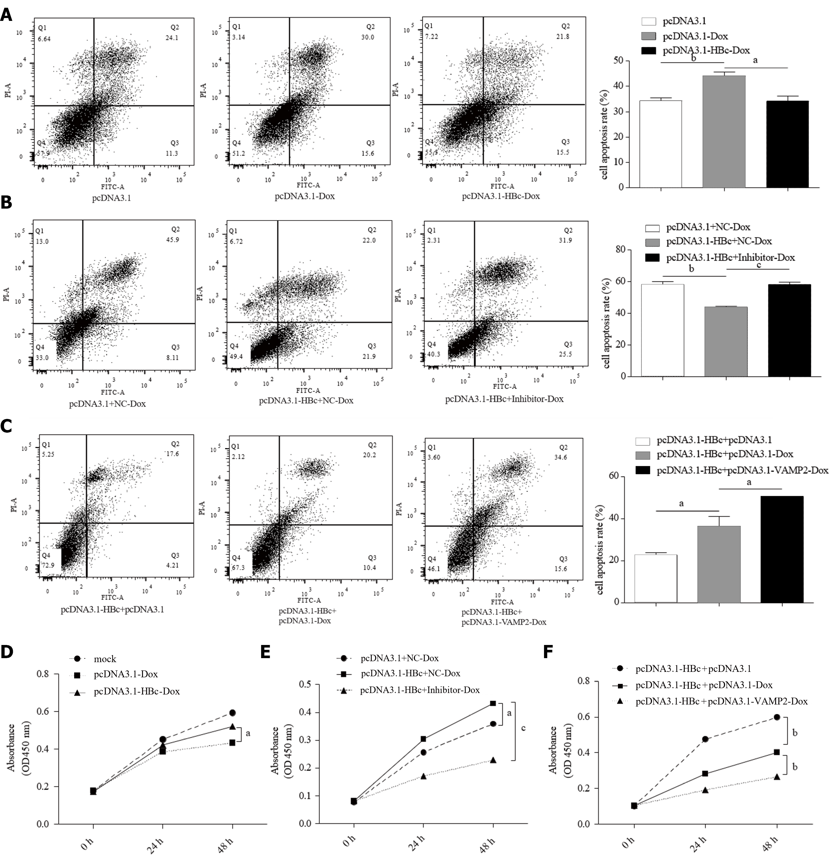
Figure 7 Hepatitis B core antigen mediated resistance of hepatocellular carcinoma cells to Doxorubicin hydrochloride via miR-135a-5p/vesicle-associated membrane protein 2.
A: HepG2 cells were transfected with pcDNA3.1-hepatitis B core antigen (HBc) plasmids, flow cytometry was used to determine the rate of Doxorubicin hydrochloride (Dox)-induced apoptosis; B and C: Cell apoptosis rate was measured in HepG2 cells treated with Dox by flow cytometry after transfection with the indicated plasmid; D: Cell counting assay was performed to determine the proliferation of HepG2 cells transfected with pcDNA3.1-HBc plasmids after treatment with Dox; E: Cell proliferation in HepG2 cells co-transfected with pcDNA3.1-HBc plasmids and miR-135a-5p inhibitors assessed by the cell counting kit 8 assay; F: Cell counting assay used to determine the proliferation of HepG2 cells co-transfected with pcDNA3.1-HBc and pcDNA3.1-vesicle-associated membrane protein 2 plasmids after treatment with Dox. aP < 0.05; bP < 0.01; cP < 0.001. HBc: Hepatitis B core antigen; VAMP2: Vesicle-associated membrane protein 2; Dox: Doxorubicin hydrochloride.
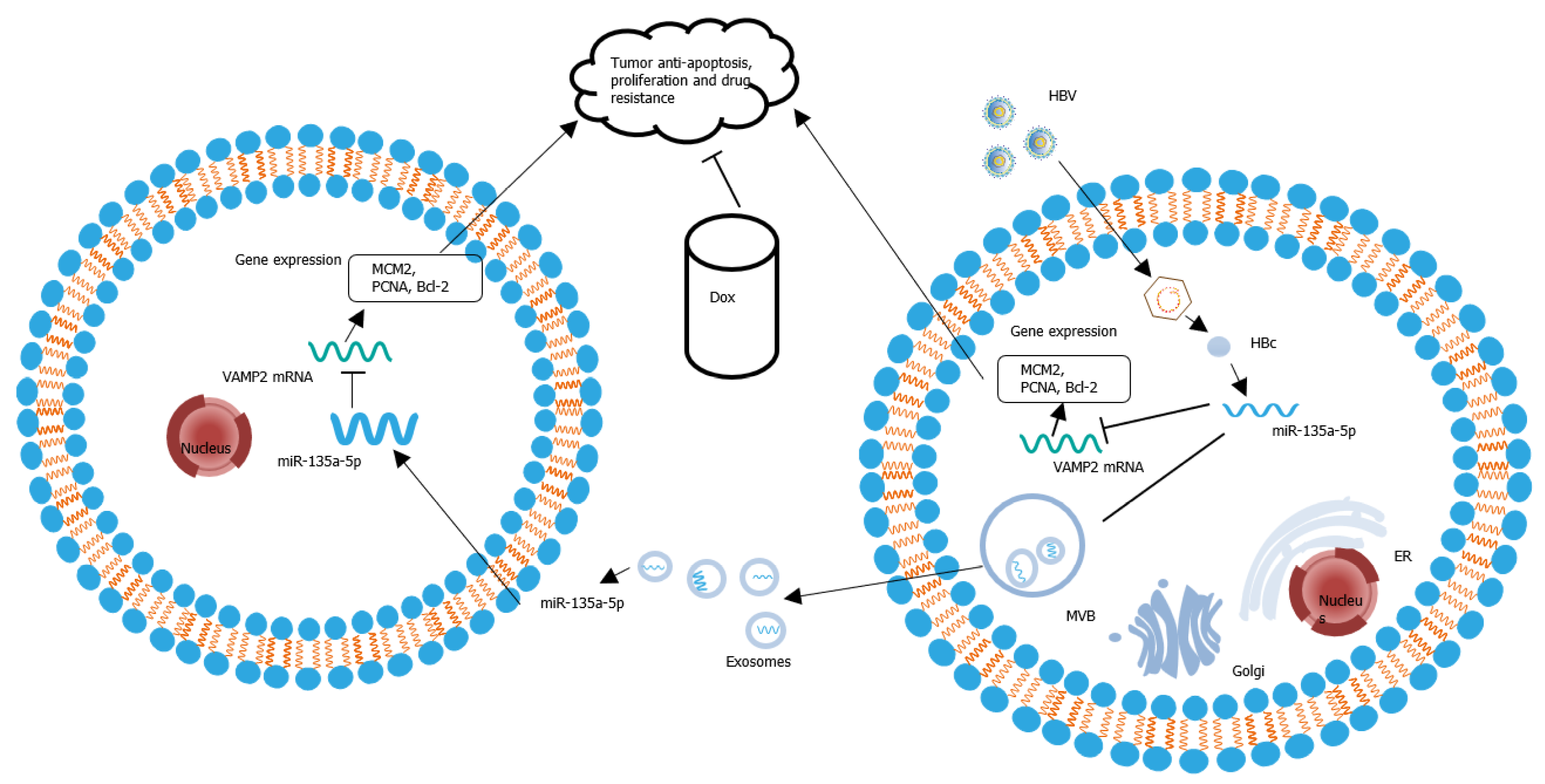
Figure 8 Hepatitis B core antigen promoted tumor anti-apoptosis, proliferation and chemoresistance in hepatocellular carcinoma cells by the miR-135a-5p/vesicle-associated membrane protein 2 axis.
HBV: Hepatitis B virus; HBc: Hepatitis B core antigen; Bcl-2: B-cell lymphoma-2; MCM2: Mini-chromosome maintenance protein-2; PCNA: Proliferating cell nuclear antigen; VAMP2: Vesicle-associated membrane protein 2; Dox: Doxorubicin hydrochloride; MVB: Multivesicular body; ER: Endoplasmic reticulum.
- Citation: Wei XC, Xia YR, Zhou P, Xue X, Ding S, Liu LJ, Zhu F. Hepatitis B core antigen modulates exosomal miR-135a to target vesicle-associated membrane protein 2 promoting chemoresistance in hepatocellular carcinoma. World J Gastroenterol 2021; 27(48): 8302-8322
- URL: https://www.wjgnet.com/1007-9327/full/v27/i48/8302.htm
- DOI: https://dx.doi.org/10.3748/wjg.v27.i48.8302


