Copyright
©The Author(s) 2021.
World J Gastroenterol. Dec 28, 2021; 27(48): 8227-8241
Published online Dec 28, 2021. doi: 10.3748/wjg.v27.i48.8227
Published online Dec 28, 2021. doi: 10.3748/wjg.v27.i48.8227
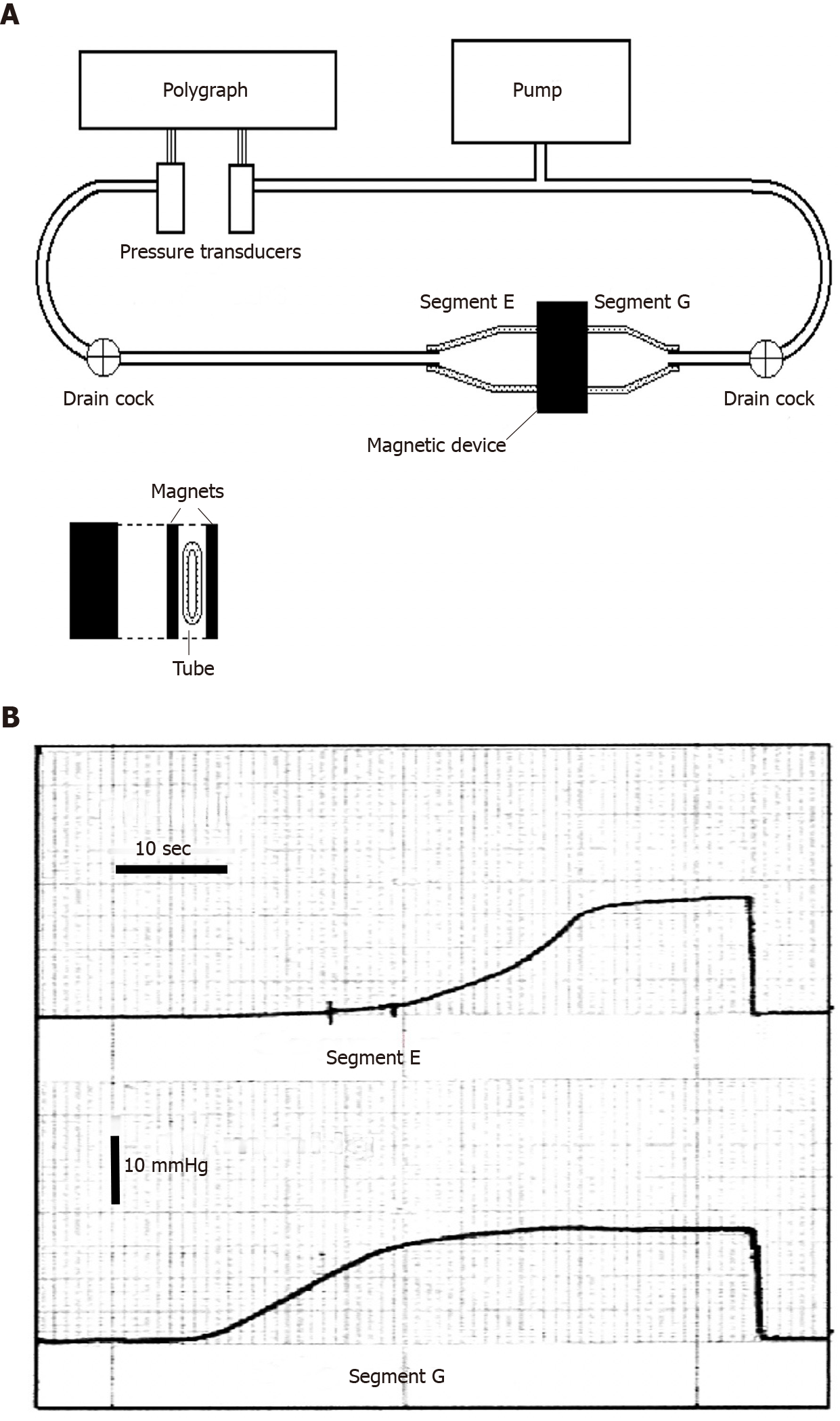
Figure 1 Benchtop experiment to demonstrate the possibility of creating a sphincter with two magnetic plaques.
A: Schematic illustration of the bench model used to study the new anti-reflux device based on magnets. On the right there is a flaccid polyethylene tube 2.8 cm in diameter, mimicking the gastro-esophageal junction. It is squeezed perpendicularly by two rectangular magnets made of plastoferrite (Flexo) 2 cm × 4 cm × 0.5 cm with an attraction force of 0.36 N/cm2, when in contact and 0.16 N/cm2, at a distance of 7 mm. It creates a high pressure zone 2 cm wide, that divides the tube in segment E (esophagus) and G (stomach). The tube is perfused with water by a pump and the pressure variations in each segment are detected with 2 pressure transducers and recorded by a polygraph; B: Intraluminal pressure variations in segment G (bottom) and E (top). The pressure in segment G (stomach) was progressively increased by the pump and when it reached approximately 11.5 mmHg, the magnets, simulating the sphincter, get detached, so that the pressure in segment E (esophagus) starts to increase, mimicking a gastro-esophageal reflux and reaching the level of the segment G. Once the pump stops the pressure falls and the magnets adhere again, closing the passage. Exchanging the letter E for G and G for E, this sequence of events may represent the passage of a bolus through the zone squeezed by the magnets. A-B: Citation: Bortolotti M. A novel anti-reflux device based on magnets. J Biomech 2006; 39: 564-7. Copyright© The Authors 2020. Published by Elsevier. The authors obtained permission for use of the figure from the Elsevier Publishing Group (Supplementary material).
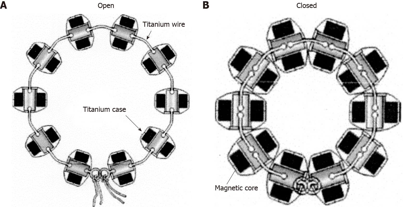
Figure 2 Schematic drawing of the Linx magnetic augmentation device to insert around the abdominal portion of the esophagus in the open and closed position.
A: Open position; B: Closed position. A-B: Citation: Bonavina L, Saino GI, Bona D, Lipham J, Ganz RA, Dunn D, DeMeester T. Magnetic augmentation of the lower esophageal sphincter: results of a feasibility clinical trial. J Gastrointest Surg 2008; 12: 2133-40. Copyright© The Authors 2020. Published by Springer Nature. The authors obtained permission for use of the figure from Springer Nature (Supplementary material).
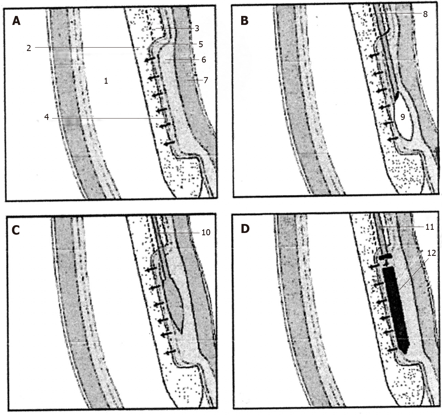
Figure 3 Extremity of the special endoesophageal probe positioned at the LES level in a sequence of operations for the deployment of a magnetic plaque seen in profile.
A: The mucosa of the distal esophagus is sucked onto the perforated wall of the operative chamber; B: The needle injects milliliters of saline solution to create a blister in the submucosa; C: The end of the catheter with a blunted bolt creates a pouch in the submucosa; D: The magnetic plaque (seen in profile) is pushed into the pouch. 1: Esophageal lumen; 2: Delivery probe; 3: Deployment channel; 4: Perforated wall of the aspiration chamber; 5: Mucosal layer; 6: Submucosal layer; 7: Muscular layer; 8: Needle-catheter; 9: Saline solution; 10: Bolt-catheter; 11: Magnetic plaque seen in profile. A-D: Citation: Bortolotti M, Grandis A, Mazzero G. A novel endoesophageal magnetic device to prevent gastroesophageal reflux. Surg Endosc 2009; 4: 885-9. Copyright© The Authors 2020. Published by Springer Nature. The authors obtained permission for use of the figure from Springer Nature (Supplementary material).
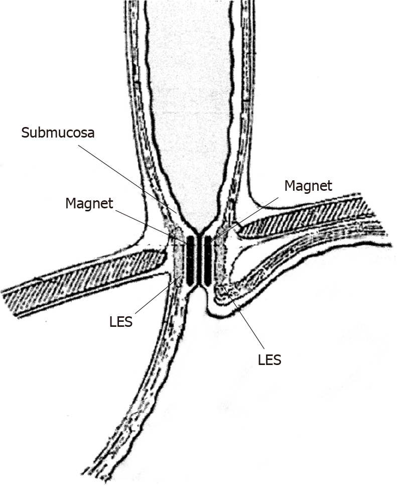
Figure 4 Schematic section following a vertical frontal plane through the lower portion of the esophago-gastric wall showing in profile the two magnetic plaques inserted face to face in the submucosal position at the lower esophageal sphincter level; these attracting each other close the gastro-esophageal junction.
Citation: Bortolotti M, Grandis A, Mazzero G. A novel endoesophageal magnetic device to prevent gastroesophageal reflux. Surg Endosc 2009; 4: 885-9. Copyright© The Authors 2020. Published by Springer Nature. The authors obtained permission for use of the figure from Springer Nature (Supplementary material).
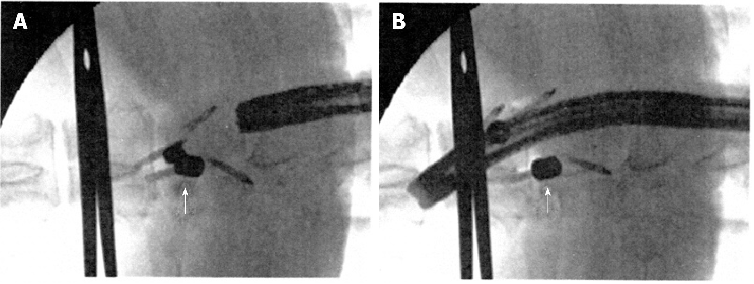
Figure 5 Fluoroscopic view after insertion of the magnets.
A: Magnets in the sub-adventitial space opposing the respective esophageal walls. A surgical clamp indicates the level of the esophago-gastric junction and the arrow indicates the magnets attracted to one another and closing the lumen; B: Magnets separated by the passage of the endoscope. The arrow indicates one of the magnets separated from the other. A-B: Citation: Modified from: Dobashi A, Wu SW, Deters JL, Miller CA, Knipschield MA, Cameron GP, Lu L, Rajan E, Gostout CJ. Endoscopic magnet placement into subadventitial tunnels for augmenting the lower esophageal sphincter using submucosal endoscopy: ex vivo and in vivo study in a porcine model (with video). Gastrointest Endosc 2019; 89: 422-428. Copyright© The Authors 2020. Published by Elsevier. The authors obtained permission for use of the figure from Elsevier (Supplementary material).
- Citation: Bortolotti M. Magnetic challenge against gastroesophageal reflux . World J Gastroenterol 2021; 27(48): 8227-8241
- URL: https://www.wjgnet.com/1007-9327/full/v27/i48/8227.htm
- DOI: https://dx.doi.org/10.3748/wjg.v27.i48.8227


