Copyright
©The Author(s) 2021.
World J Gastroenterol. Aug 28, 2021; 27(32): 5404-5423
Published online Aug 28, 2021. doi: 10.3748/wjg.v27.i32.5404
Published online Aug 28, 2021. doi: 10.3748/wjg.v27.i32.5404
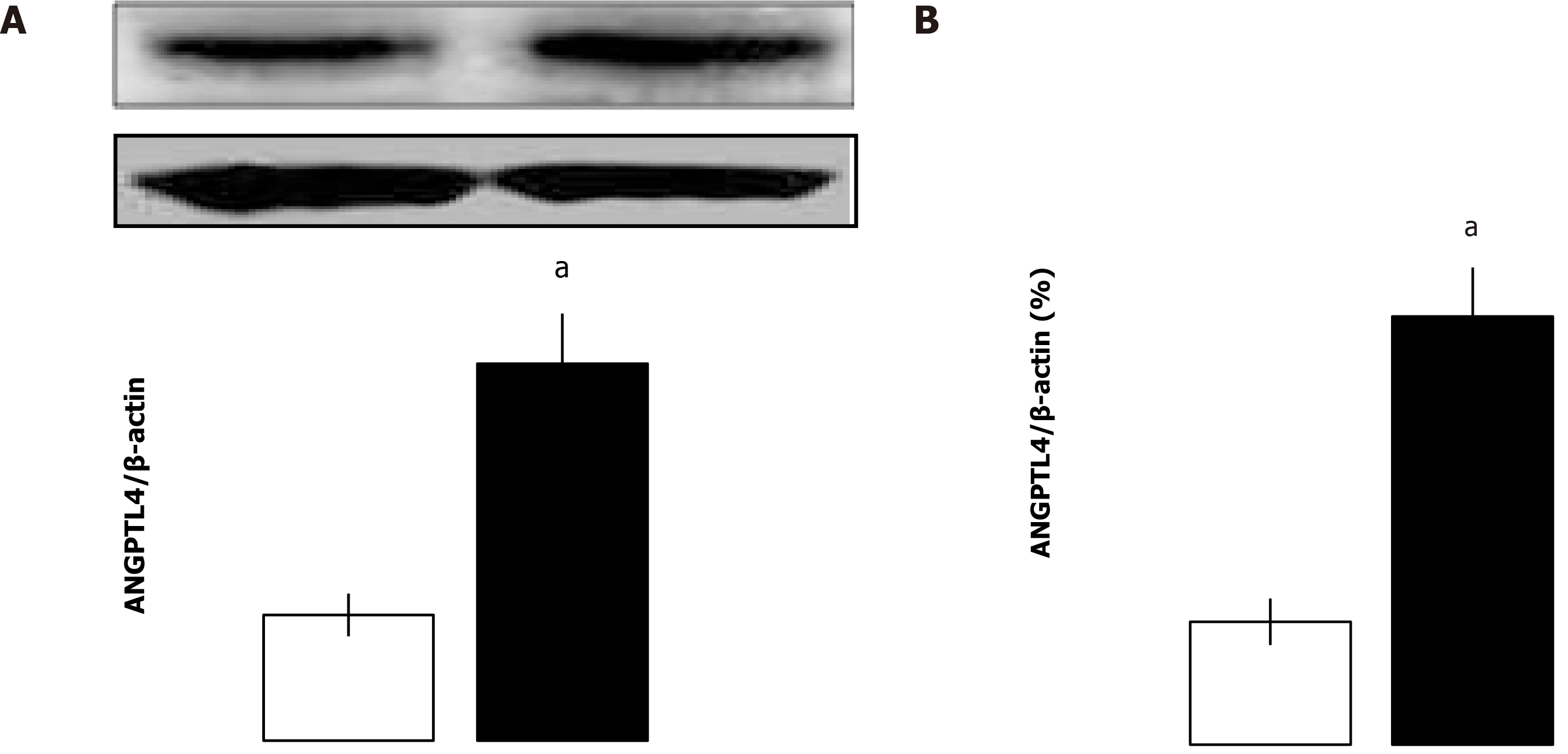
Figure 1 Intestinal angiopoietin-like protein 4 level after intestinal ischemia-reperfusion in different groups (mean ± SD, n = 8).
A: Angiopoietin-like protein 4 protein levels were assessed by western blot; B: Angiopoietin-like protein 4 mRNA levels in the intestine were measured by quantitative real-time polymerase chain reaction. aP < 0.05 vs sham by Student’s t test. ANGPTL4: Angiopoietin-like protein 4.
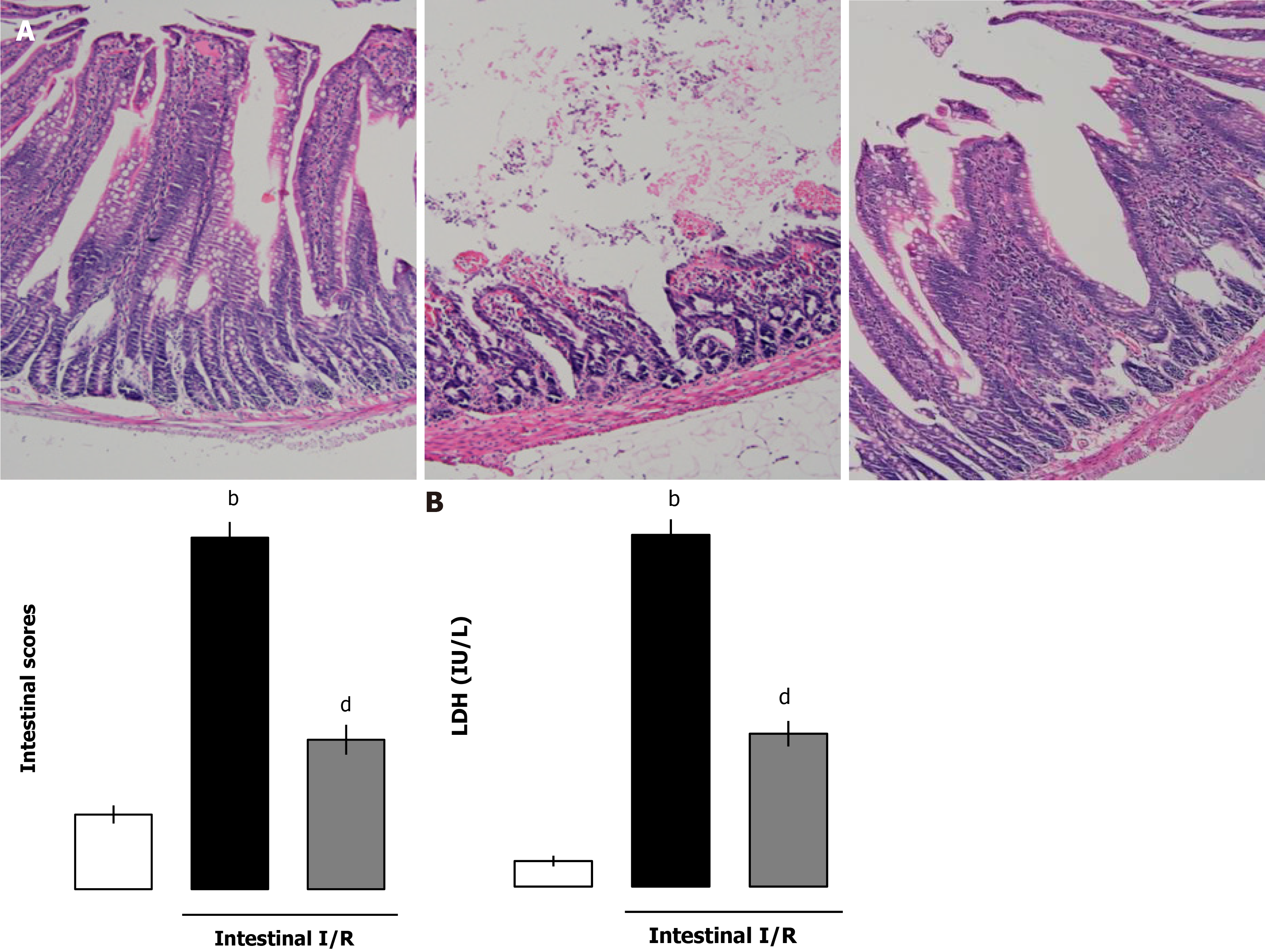
Figure 2 Recombinant human angiopoietin-like protein 4 improved ischemia-reperfusion-induced intestinal histopathologically and serum lactate dehydrogenase.
A and B: Pathologic changes of intestine (scale bar = 100 μm) of (A) tissue and (B) serum lactate dehydrogenase (LDH) in different groups (mean ± SD, n = 8). bP < 0.01 vs sham; dP < 0.01 vs ischemia-reperfusion. I/R: Ischemia-reperfusion.
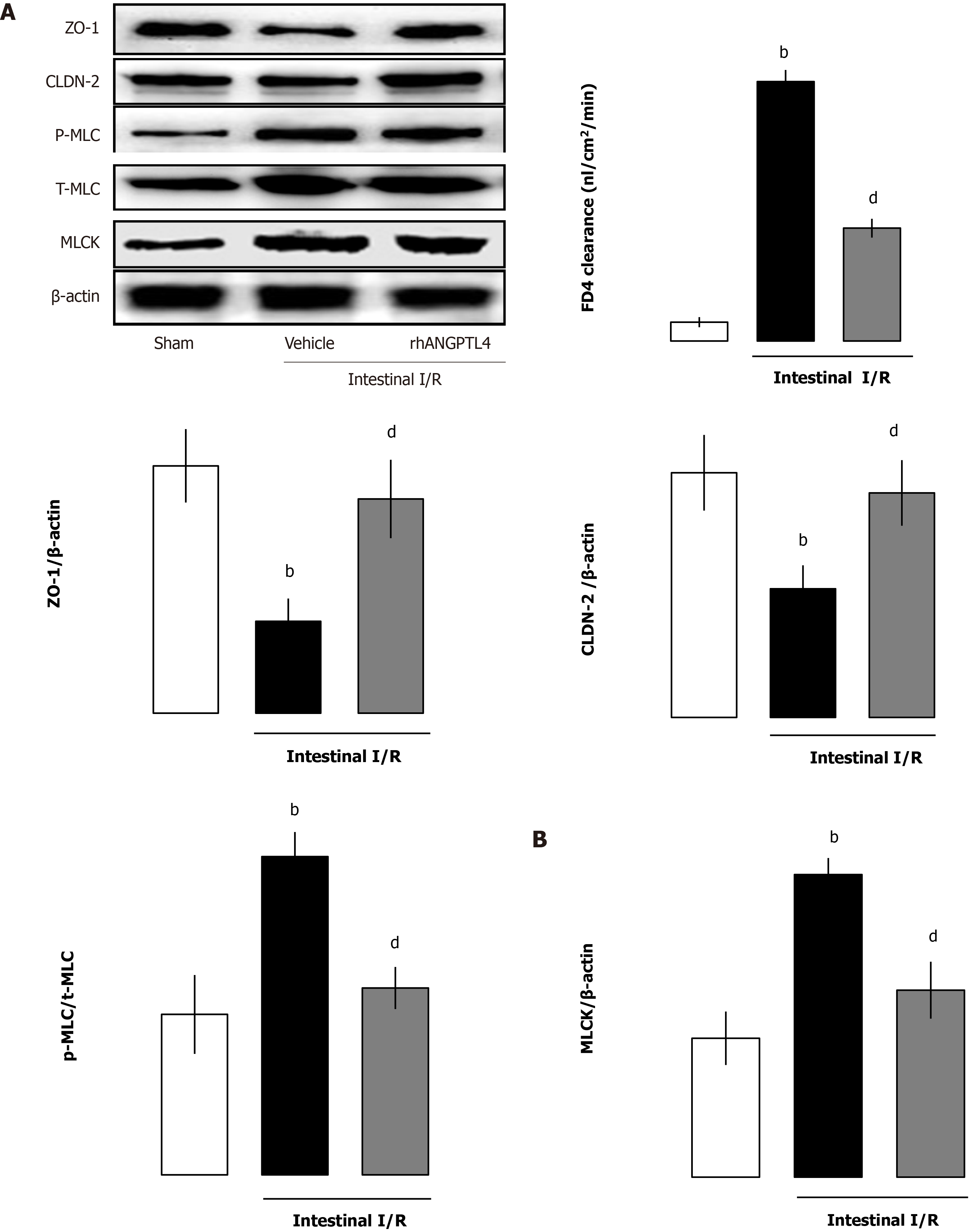
Figure 3 Recombinant human angiopoietin-like protein 4 regulates the intestinal permeability barrier and myosin light chain kinase-tight junction pathway related proteins in vivo.
Intestinal mucosal tissue myosin light chain kinase (MLCK), phosphorylated-myosin light chain (p-MLC)/myosin light chain, zonula occludens-1 (ZO-1) and claudin-2 (CLDN-2) (A) and intestinal mucosal permeability (B) in different groups (mean ± SD, n = 8). bP < 0.01 vs sham; dP < 0.01 vs ischemia-reperfusion (I/R). ANGPTL4: Angiopoietin-like protein 4; t-MLC: Total-myosin light chain.
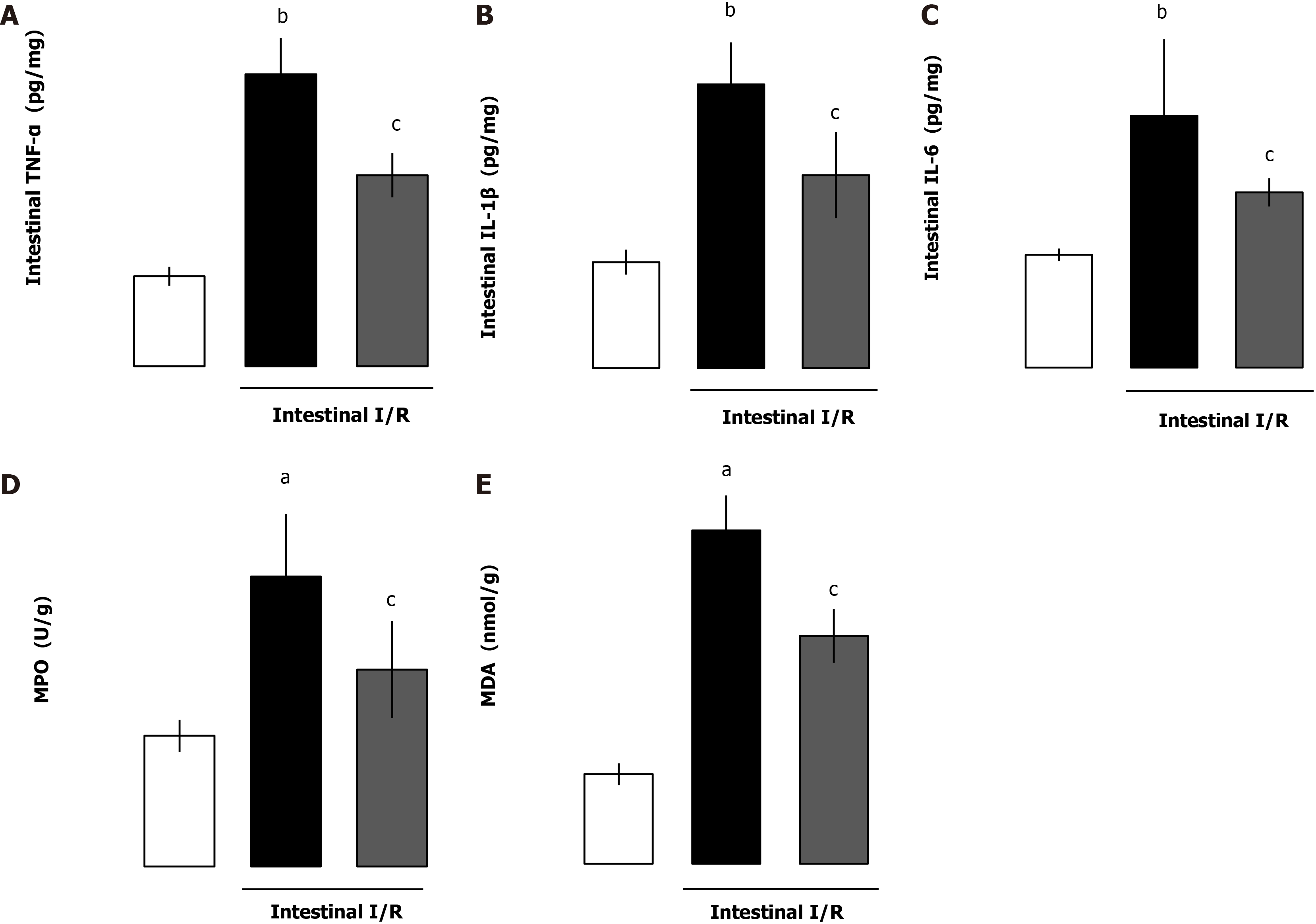
Figure 4 Recombinant human angiopoietin-like protein 4 suppresses the intestinal inflammatory response and oxidation stress after intestinal ischemia-reperfusion.
A-E: Intestinal cytokine tumor necrosis factor-alpha (TNF-α) (A), interleukin (IL)-1β (B), interleukin-6 (C), intestinal myeloperoxidase (MPO) (D) and malondialdehyde (MDA) (E) levels in different groups (mean ± SD, n = 8). aP < 0.05, bP < 0.01 vs sham; cP < 0.05 vs ischemia-reperfusion (I/R).
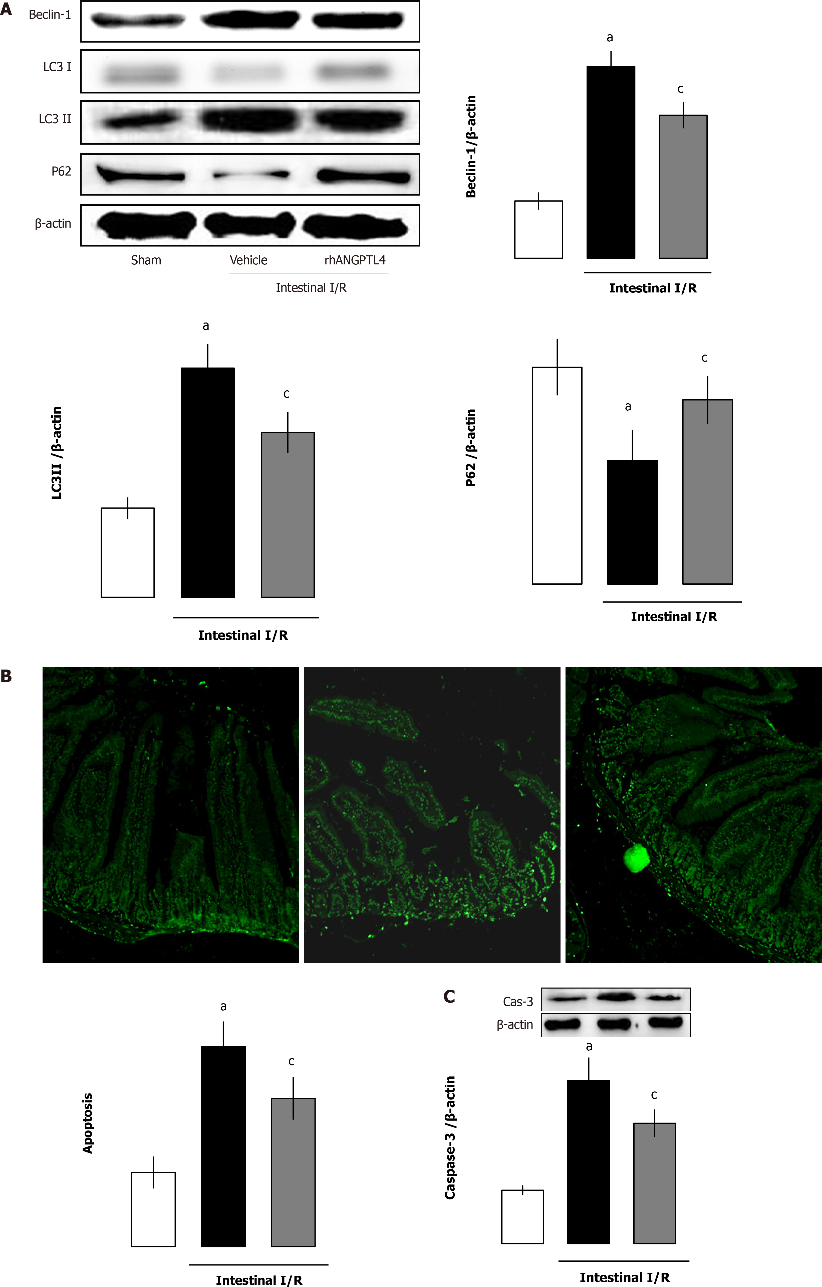
Figure 5 Recombinant human angiopoietin-like protein 4 reduced the increase of intestinal autophagy and apoptosis after ischemia-reperfusion.
A: Intestinal autophagy-related protein beclin-1, microtubule-associated light chain 3 (LC3) I/II and p62 in different groups (mean ± SD, n = 8). aP < 0.05 vs sham; cP < 0.05 vs ischemia-reperfusion (I/R) groups; B and C: Intestinal apoptosis (scale bar = 100 μm) (B) and cleaved caspase-3 (Cas-3) (C) in different groups (mean ± SD, n = 8). aP < 0.05 vs sham; cP < 0.05 vs I/R. ANGPTL4: Angiopoietin-like protein 4.
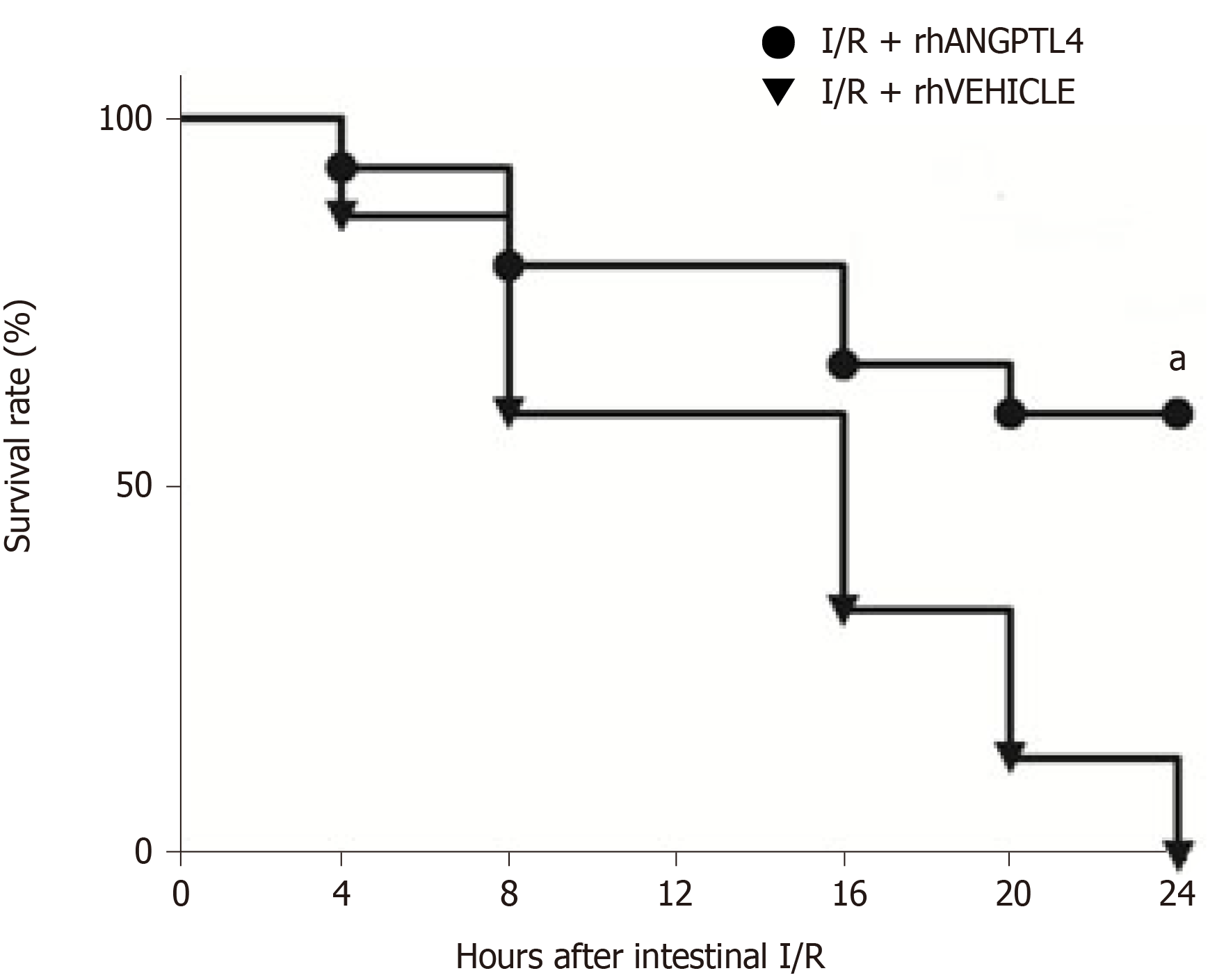
Figure 6 Survival benefit in recombinant human angiopoietin-like protein 4-treated rats after intestinal ischemia-reperfusion injury (n = 15, Kaplan Meyer log-rank test).
Each point in the figure shows the mean survival rate at each time point. aP < 0.05 vs vehicle. I/R: Ischemia-reperfusion; rhANGPTL4: Recombinant human angiopoietin-like protein 4.
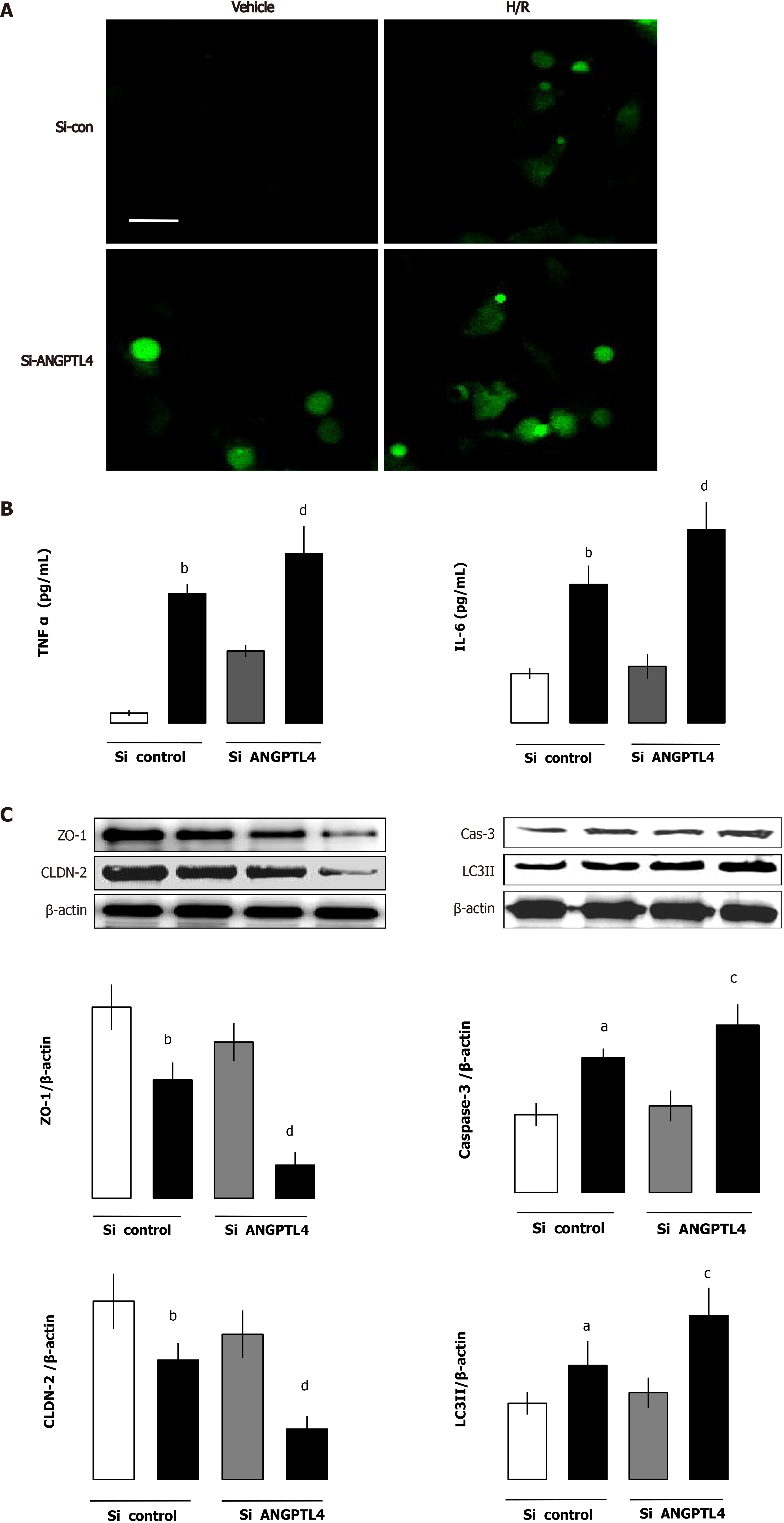
Figure 7 Angiopoietin-like protein 4 deficiency aggravates Caco-2 cells injury challenge by hypoxia/reoxygenation.
A and B: Reactive oxygen species (ROS) levels (scale bar = 12.5 μm) (A) determined by immunofluorescence and tumor necrosis factor-alpha (TNF-α) and interleukin (IL)-6 levels (B) determined by enzyme-linked immunosorbent assay; C: Tight junction zonula occludens-1 (ZO-1) and claudin-2 (CLDN-2), autophagy marker microtubule-associated protein light chain 3 (LC3) II and apoptosis marker caspase-3 were determined using western blot assays (mean ± SD, n = 3). aP < 0.05, bP < 0.01 vs vehicle; cP < 0.05, dP < 0.01 vs vehicle (n = 3 per group). ANGPTL4: Angiopoietin-like protein 4; H/R: Hypoxia/reoxygenation; si: Small interfering RNA.
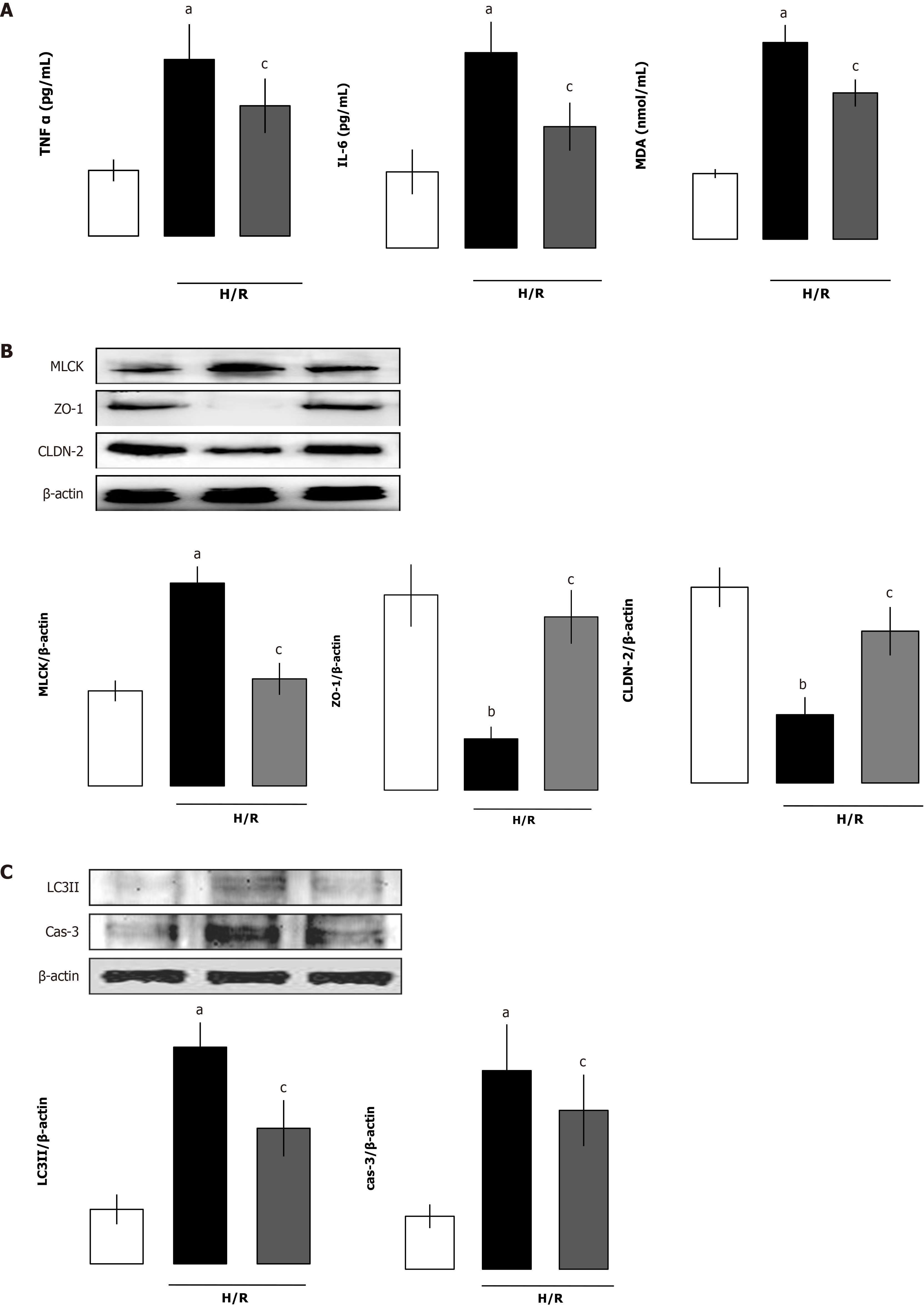
Figure 8 Beneficial effects of recombinant human angiopoietin-like protein 4 on angiopoietin-like protein 4 deficient Caco-2 cells challenged after hypoxia/reoxygenation.
A: Cellular levels of tumor necrosis factor-alpha (TNF-α), interleukin (IL)-6 and malondialdehyde (MDA) levels; B: Myosin light chain kinase (MLCK), zonula occludens-1 (ZO-1) and claudin-2 (CLDN-2) levels; C: Microtubule-associated protein light chain 3II and cleaved caspase-3 levels were measured 4 h after reoxygenation (mean ± SD, n = 3). aP < 0.05, bP < 0.01 vs control; cP < 0.05 vs vehicle. H/R: Hypoxia/reoxygenation.
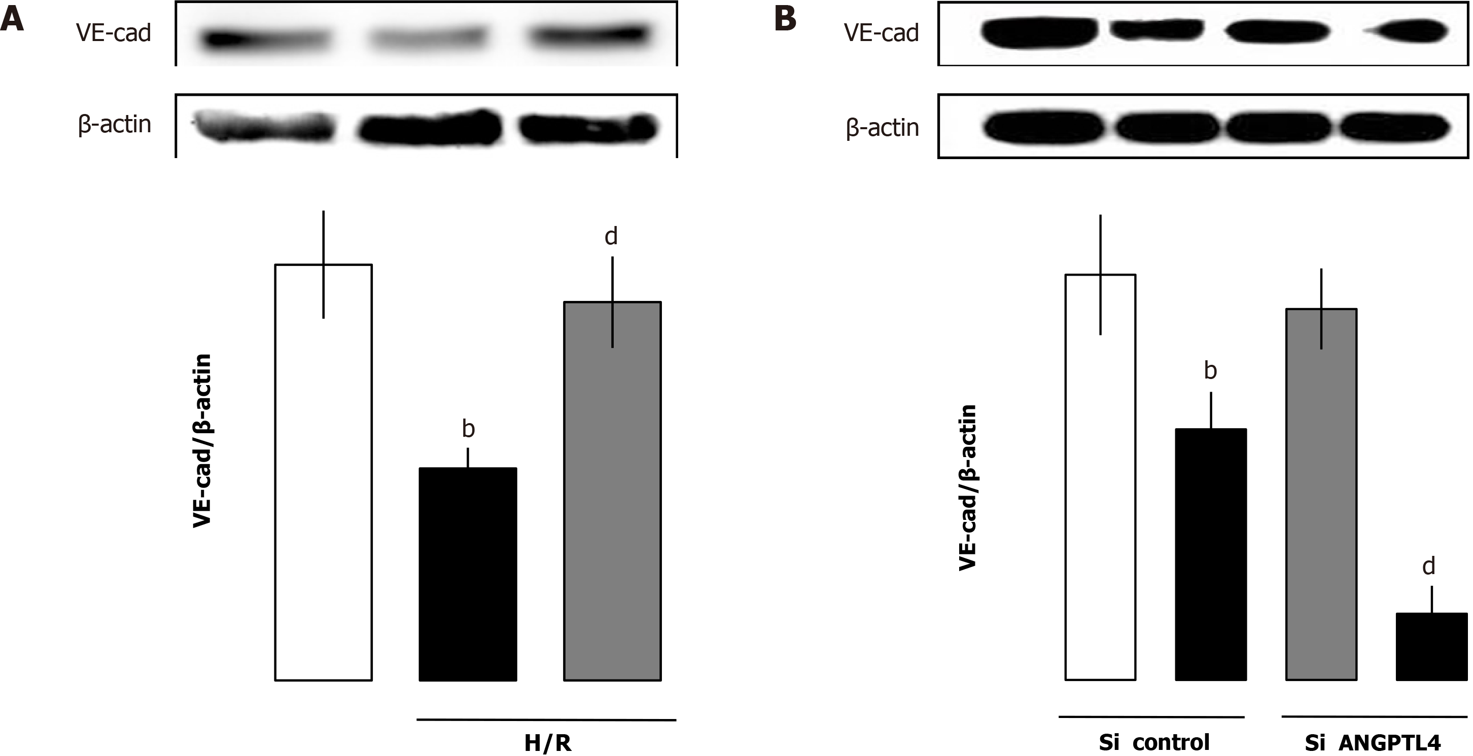
Figure 9 Levels of VE-cadherin after hypoxia/reoxygenation compared with respective vehicle cells.
Recombinant human angiopoietin-like protein 4 restored the VE-cadherin (VE-cad) in human umbilical vein endothelial cells (A) after hypoxia/reoxygenation and VE-cadherin levels in small interfering (si) RNA angiopoietin-like protein 4 control and small interfering RNA angiopoietin-like protein 4 (ANGPLT4) human umbilical vein endothelial cells (B) of different groups (mean ± SD, n = 3). bP < 0.01 vs vehicle; dP < 0.01 vs vehicle. H/R: Hypoxia/reoxygenation.
- Citation: Wang ZY, Lin JY, Feng YR, Liu DS, Zhao XZ, Li T, Li SY, Sun JC, Li SF, Jia WY, Jing HR. Recombinant angiopoietin-like protein 4 attenuates intestinal barrier structure and function injury after ischemia/reperfusion. World J Gastroenterol 2021; 27(32): 5404-5423
- URL: https://www.wjgnet.com/1007-9327/full/v27/i32/5404.htm
- DOI: https://dx.doi.org/10.3748/wjg.v27.i32.5404


