Copyright
©The Author(s) 2021.
World J Gastroenterol. Aug 21, 2021; 27(31): 5126-5151
Published online Aug 21, 2021. doi: 10.3748/wjg.v27.i31.5126
Published online Aug 21, 2021. doi: 10.3748/wjg.v27.i31.5126
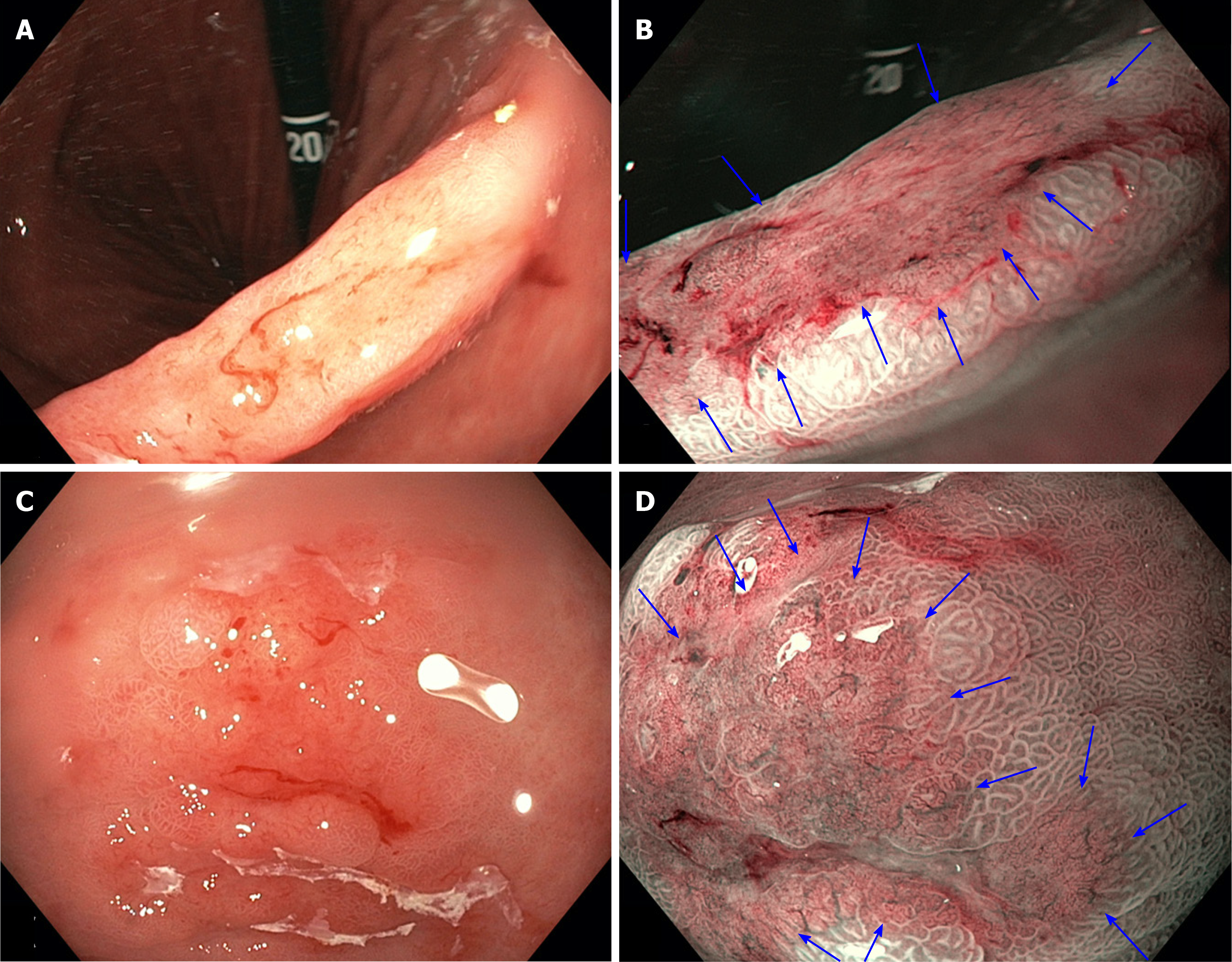
Figure 1 White light endoscopy compared to narrow-band imaging in gastric lesions, demonstrating clear demarcation lines and irregular microvascular/microsurface patterns on narrow-band imaging.
A: Early gastric cancer at the incisura seen on white light endoscopy (WLE); B: The same lesion seen on narrow-band imaging (NBI) (blue arrows); C: Early gastric cancer in the antrum seen on WLE; D: The same lesion seen on NBI (blue arrows).
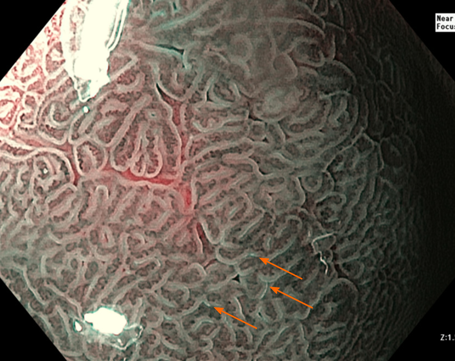
Figure 2 Narrow-band imaging demonstrating the ‘light blue crest’ (orange arrows) consistent with intestinal metaplasia.
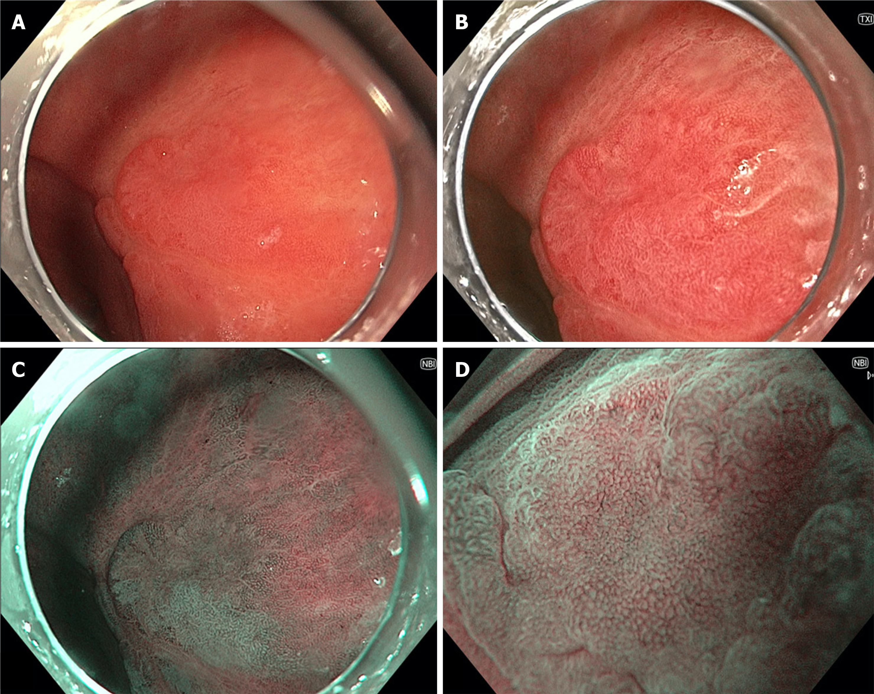
Figure 3 Gastric body lesion with low-grade dysplasia seen on multiple forms of endoscopic imaging.
A: White light endoscopy; B: Texture and colour enhancement imaging; C: Narrow-band imaging (NBI); D: high-magnification NBI.
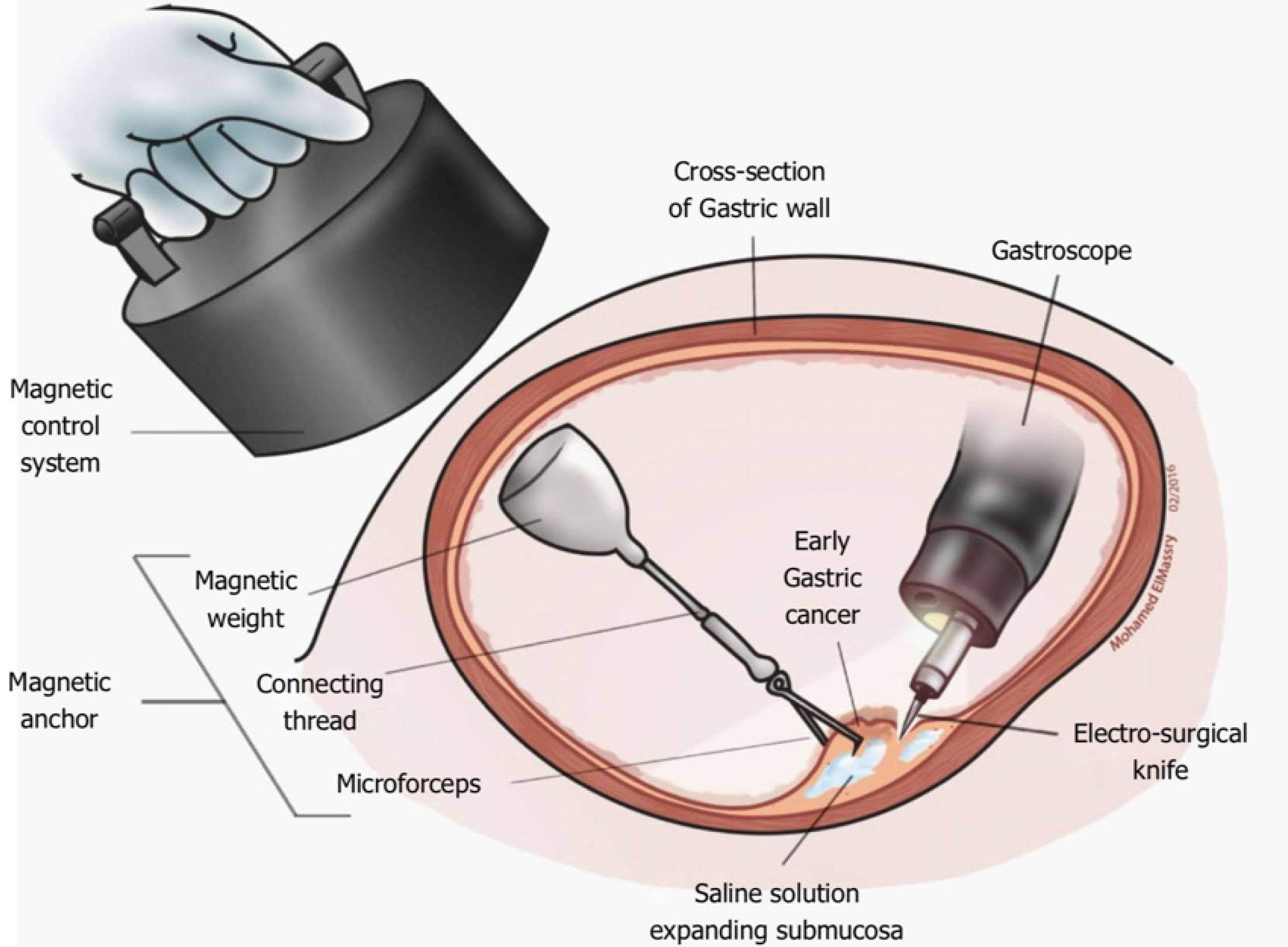
Figure 4 Magnetic anchor-guided endoscopic submucosal dissection[169].
Citation: Mortagy M, Mehta N, Parsi MA, Abe S, Stevens T, Vargo JJ, Saito Y, Bhatt A. Magnetic anchor guidance for endoscopic submucosal dissection and other endoscopic procedures. World J Gastroenterol 2017; 23: 2883-2890. ©The Author(s) 2017. Published by Baishideng Publishing Group Inc.
Figure 5 Endoscopic submucosal dissection using an additional working channel[172].
A: Lesion marked via usual working channel; B: Submucosal injection; C: Near-circumferential incision made; D: Lesion grasped for countertraction via additional working channel while dissection underway. Citation: Knoop RF, Wedi E, Petzold G, Bremer SCB, Amanzada A, Ellenrieder V, Neesse A, Kunsch S. Endoscopic submucosal dissection with an additional working channel (ESD+): a novel technique to improve procedure time and safety of ESD. Surg Endosc 2021; 35: 3506-3512. ©The Author(s) 2021. Published by Springer Open Access Article.
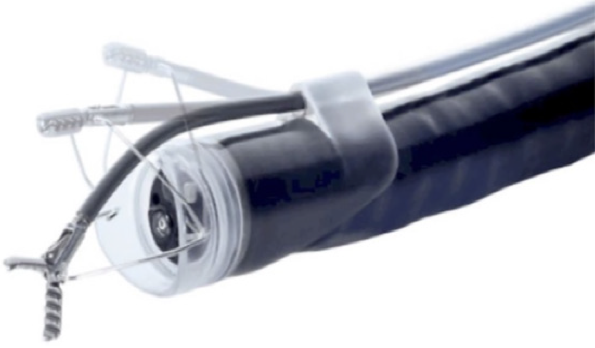
Figure 6 ‘Endo-lifter’ (Olympus-Tokyo, Japan)[192].
Citation: Harlow C, Sivananthan A, Ayaru L, Patel K, Darzi A, Patel N. Endoscopic submucosal dissection: an update on tools and accessories. Ther Adv Gastrointest Endosc 2020; 13: 2631774520957220. ©The Author(s) 2020. Published by Open Access Article.
Figure 7 Spring and loop clip traction[193].
Citation: Nagata M, Fujikawa T, Munakata H. Comparing a conventional and a spring-and-loop with clip traction method of endoscopic submucosal dissection for superficial gastric neoplasms: a randomized controlled trial (with videos). Gastrointest Endosc 2021; 93: 1097-1109. ©The Author(s) 2021. Published by Open Access Article.
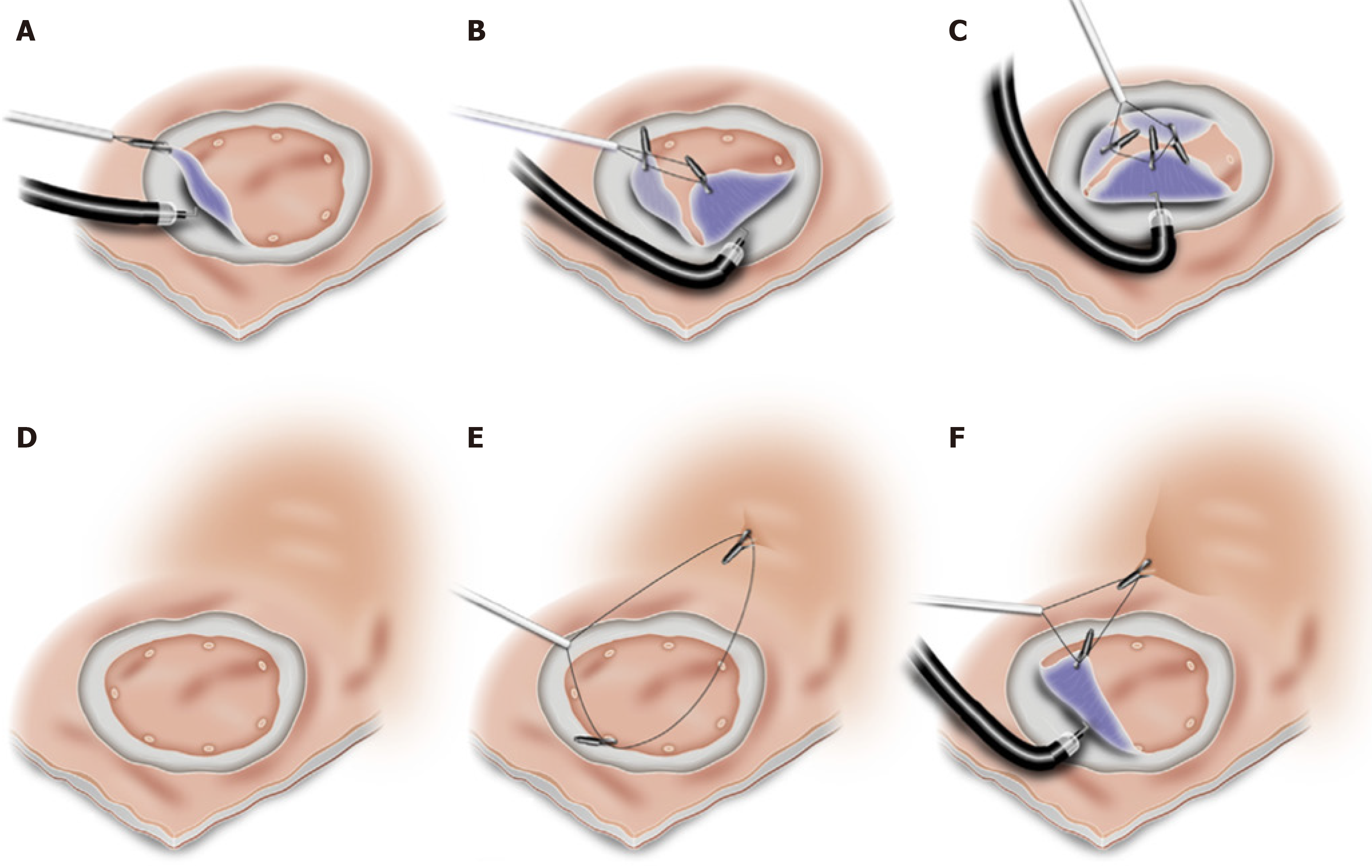
Figure 8 Modified endo-clip and snare[185].
A-C: Multiple clips used to provide multifocal traction; D-F: Clip applied to opposing gastric wall for countertraction. Citation: Zhang Q, Yao X, Wang Z. A modified method of endoclip-and-snare to assist in endoscopic submucosal dissection with mucosal traction in the upper GI tract. VideoGIE 2018; 3: 137-141. ©The Author(s) 2018. Published by Open Access Article.
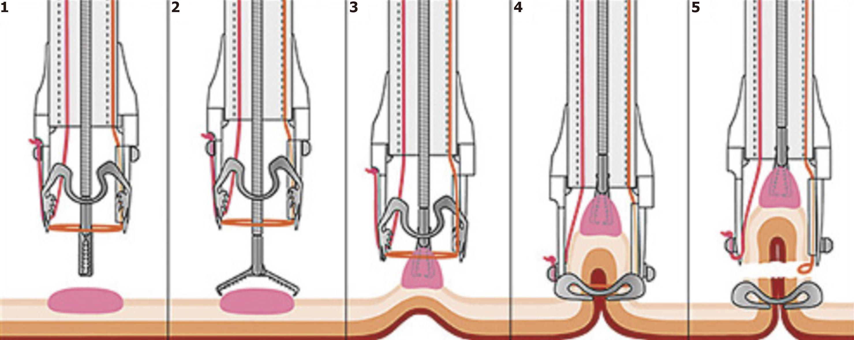
Figure 9 ‘Over-the-scope clip’ (Ovesco, Germany) full-thickness resection[194].
Citation: Mão de-Ferro S, Castela J, Pereira D, Chaves P, Dias Pereira A. Endoscopic Full-Thickness Resection of Colorectal Lesions with the New FTRD System: Single-Center Experience. GE Port J Gastroenterol 2019; 26: 235-241. ©The Author(s) 2019. Published by Open Access Article.
- Citation: Young E, Philpott H, Singh R. Endoscopic diagnosis and treatment of gastric dysplasia and early cancer: Current evidence and what the future may hold. World J Gastroenterol 2021; 27(31): 5126-5151
- URL: https://www.wjgnet.com/1007-9327/full/v27/i31/5126.htm
- DOI: https://dx.doi.org/10.3748/wjg.v27.i31.5126


