Copyright
©The Author(s) 2021.
World J Gastroenterol. Jul 28, 2021; 27(28): 4738-4745
Published online Jul 28, 2021. doi: 10.3748/wjg.v27.i28.4738
Published online Jul 28, 2021. doi: 10.3748/wjg.v27.i28.4738
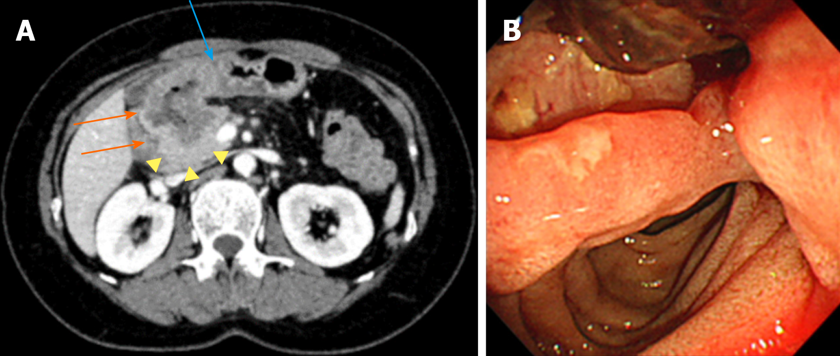
Figure 1 Abdominal computed tomography and gastroduodenoscopy.
Images were obtained before the start of treatment. A: An axial image shows an irregular circumferential mass in the first portion of duodenum (orange arrows), with transmural extension to pylorus (blue arrow) and direct extension to pancreas head (yellow arrow heads); B: During gastroduodenoscopy, mass lesion with ulcer was visible in the first portion of duodenum.
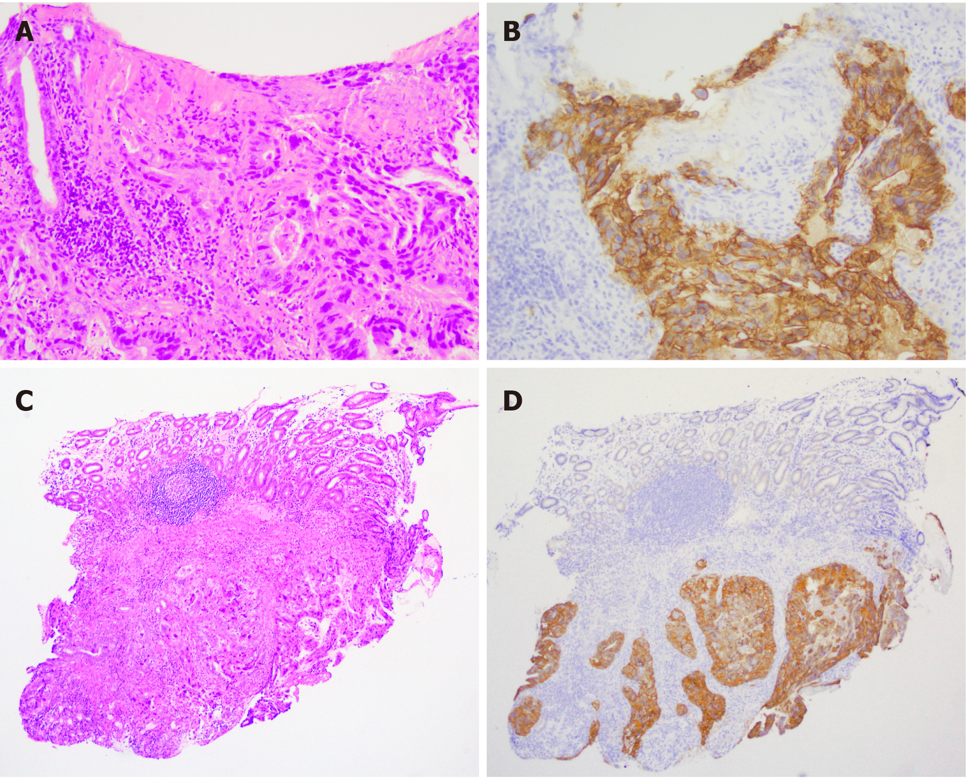
Figure 2 Histological findings of biopsy specimens from duodenal lesion.
A: Adenocarcinoma cells proliferate in the proper mucosal and submucosal layer with ulcer formation (HE staining, × 200); B: Adenocarcinoma cells show strong HER2 membranous expression (× 200); C and D: Adenocarcinoma cells proliferate in the submucosal layer with strong HER2 protein expression (× 54).
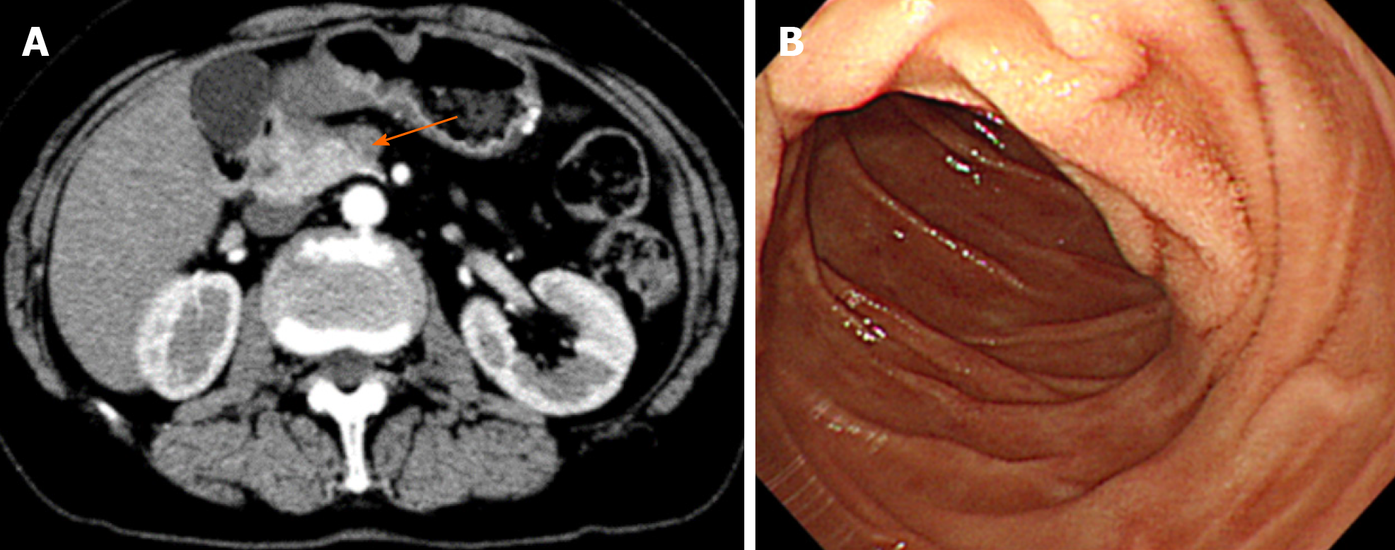
Figure 3 Abdominal computed tomography and gastroduodenoscopy.
Images were obtained after 2 cycles of trastuzumab treatment. A: An axial image shows an irregular mass lesion in the pancreas head (orange arrows), duodenal and pylorus lesion are disappeared; B: During gastroduodenoscopy, mass and ulcer lesion were vanished in the first portion of duodenum.
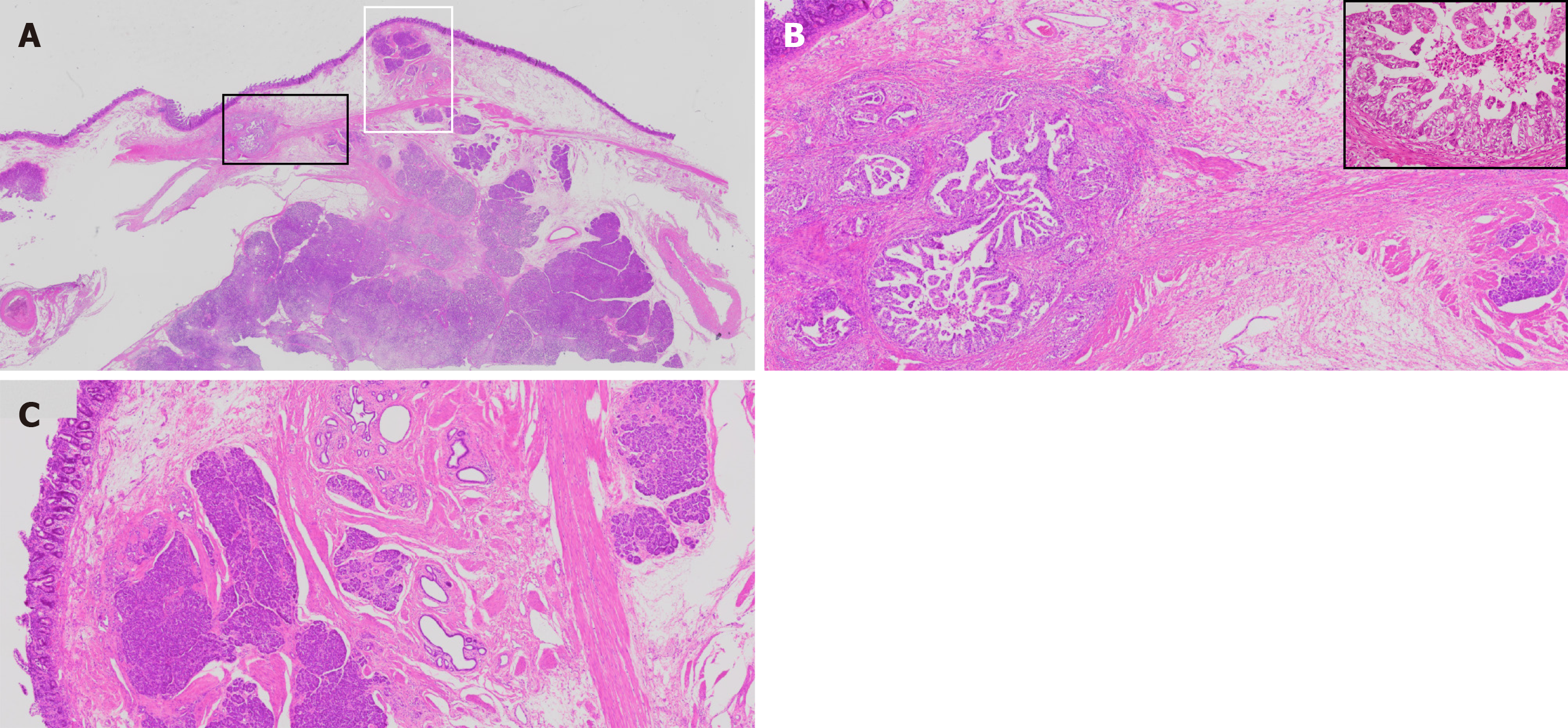
Figure 4 Histological findings of resected duodenum.
A: Heterotopic pancreas tissues are located in the submucosal layer and the laminar muscularis mucosa (HE staining, x 0.08). Adenocarcinoma cells are positioned in closely to the heterotopic pancreas tissue (× 0.08); B: On magnified image of the black square in Panel A, papillary like growth of the adenocarcinoma is present in vicinity of the heterotopic pancreas tissue (HE staining, × 10 and inset × 200); C: On magnified image of the white square in Panel A, submucosal heterotopic pancreas tissue is composed of acinar and duct structure (HE staining, × 10).
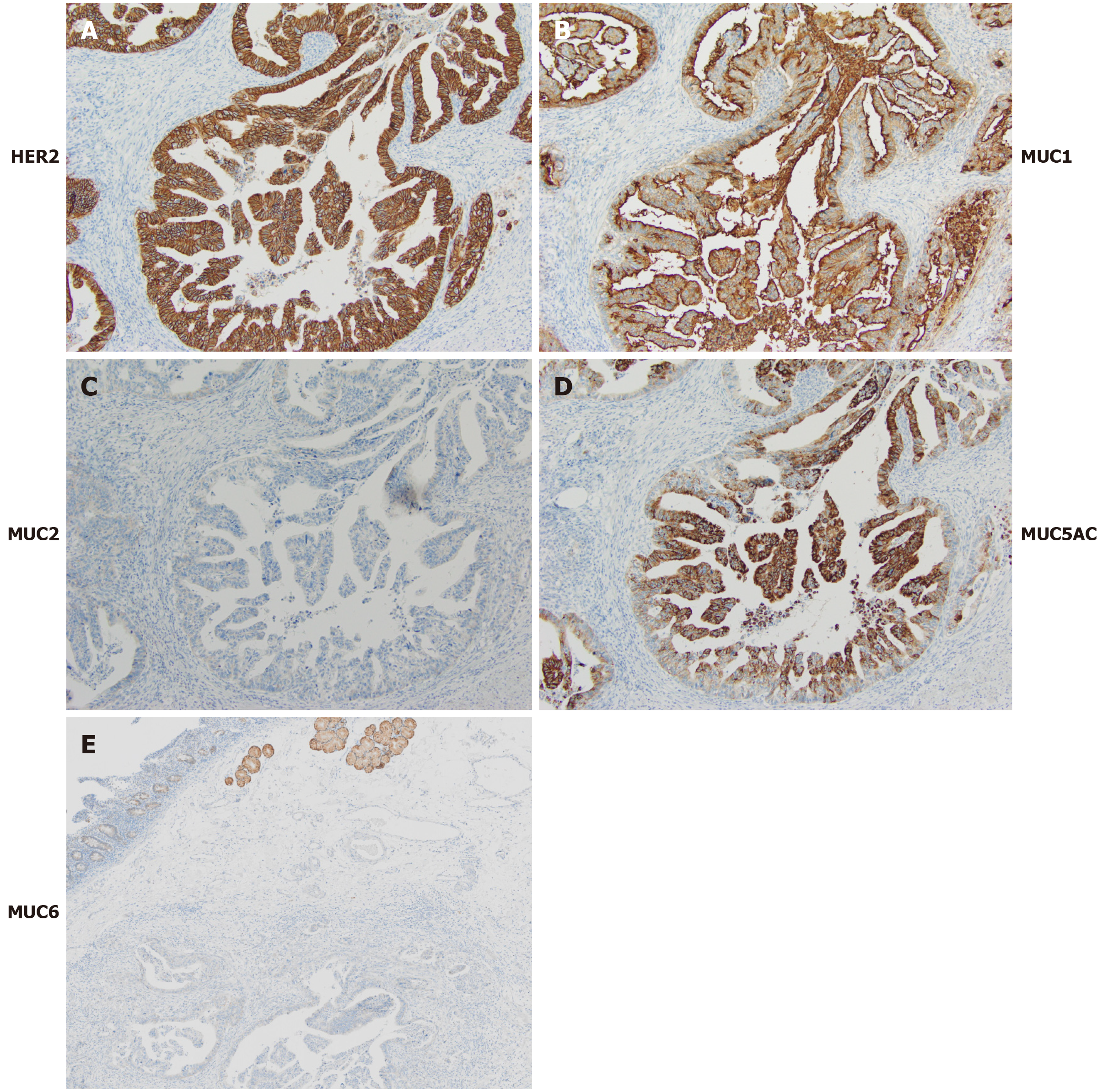
Figure 5 Immunohistochemical staining in adenocarcinoma cells in the submucosal layer.
A: Strong HER2 expression is localized in the cell membrane (× 100); B-E: Adenocarcinoma cells express MUC1 and MUC5AC but no MUC2 or MUC6 protein (B-D: × 100; E: × 3.5).
- Citation: Hirokawa YS, Iwata T, Okugawa Y, Tanaka K, Sakurai H, Watanabe M. HER2-positive adenocarcinoma arising from heterotopic pancreas tissue in the duodenum: A case report. World J Gastroenterol 2021; 27(28): 4738-4745
- URL: https://www.wjgnet.com/1007-9327/full/v27/i28/4738.htm
- DOI: https://dx.doi.org/10.3748/wjg.v27.i28.4738


