Copyright
©The Author(s) 2021.
World J Gastroenterol. May 28, 2021; 27(20): 2531-2544
Published online May 28, 2021. doi: 10.3748/wjg.v27.i20.2531
Published online May 28, 2021. doi: 10.3748/wjg.v27.i20.2531
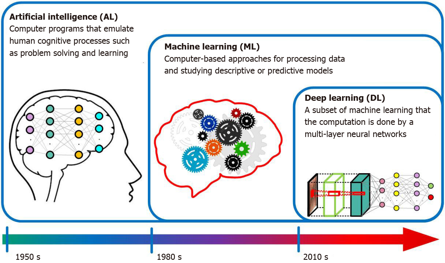
Figure 1
Infographic with icons and timeline for artificial intelligence, machine learning and deep learning.
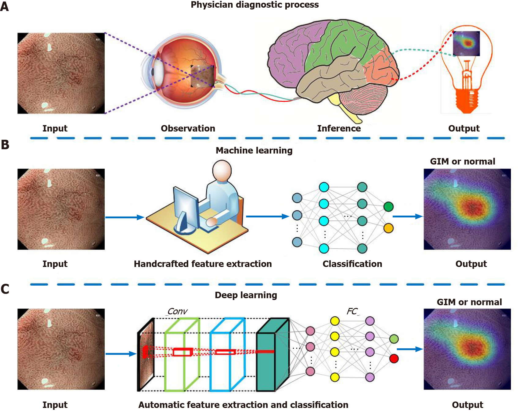
Figure 2 Illustration of the diagnostic process of physician, machine learning and deep learning.
A: Physician diagnostic process; B: Machine learning; C: Deep learning. Conv: Convolutional layer; FC: Fully connected layer; GIM: gastric intestinal metaplasia.
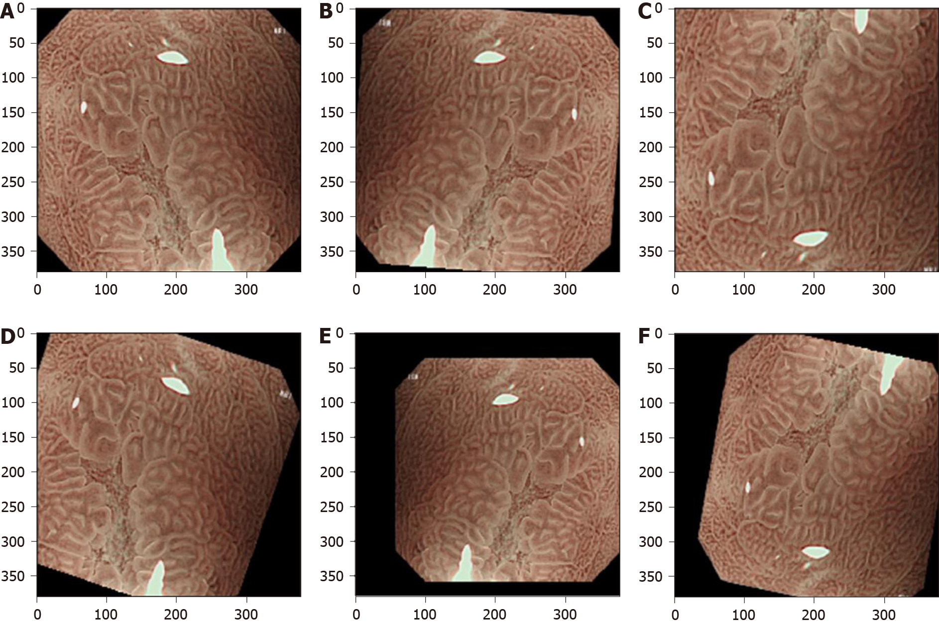
Figure 3 Data augmentation for a typical magnifying narrow band image for training a convolutional neural network model.
This is performed by using a variety of image transformations and their combinations. A: Original image; B: Flip horizontal and random rotation; C: Flip vertical and magnification; D: Random rotation and shift; E: Flip horizontal, minification and shift; F: Flip vertical, rotation and shift.
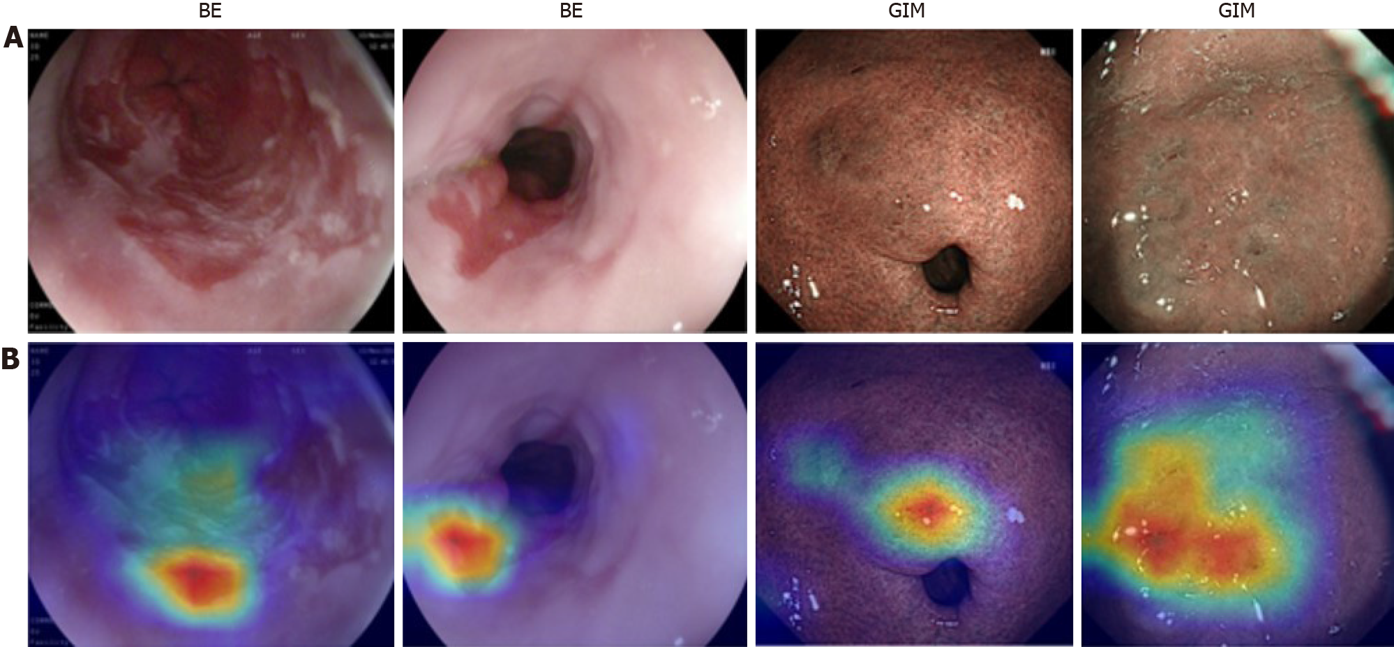
Figure 4 Informative features (partially related to lesions areas) acquired by the convolutional neural networks, where warmer colors mean higher contributions to decision making.
A: Original endoscopic images; B: Corresponding attention. BE: Barrett’s esophagus; GIM: Gastric intestinal metaplasia.
- Citation: Yan T, Wong PK, Qin YY. Deep learning for diagnosis of precancerous lesions in upper gastrointestinal endoscopy: A review. World J Gastroenterol 2021; 27(20): 2531-2544
- URL: https://www.wjgnet.com/1007-9327/full/v27/i20/2531.htm
- DOI: https://dx.doi.org/10.3748/wjg.v27.i20.2531


