Copyright
©The Author(s) 2020.
World J Gastroenterol. Sep 28, 2020; 26(36): 5527-5533
Published online Sep 28, 2020. doi: 10.3748/wjg.v26.i36.5527
Published online Sep 28, 2020. doi: 10.3748/wjg.v26.i36.5527
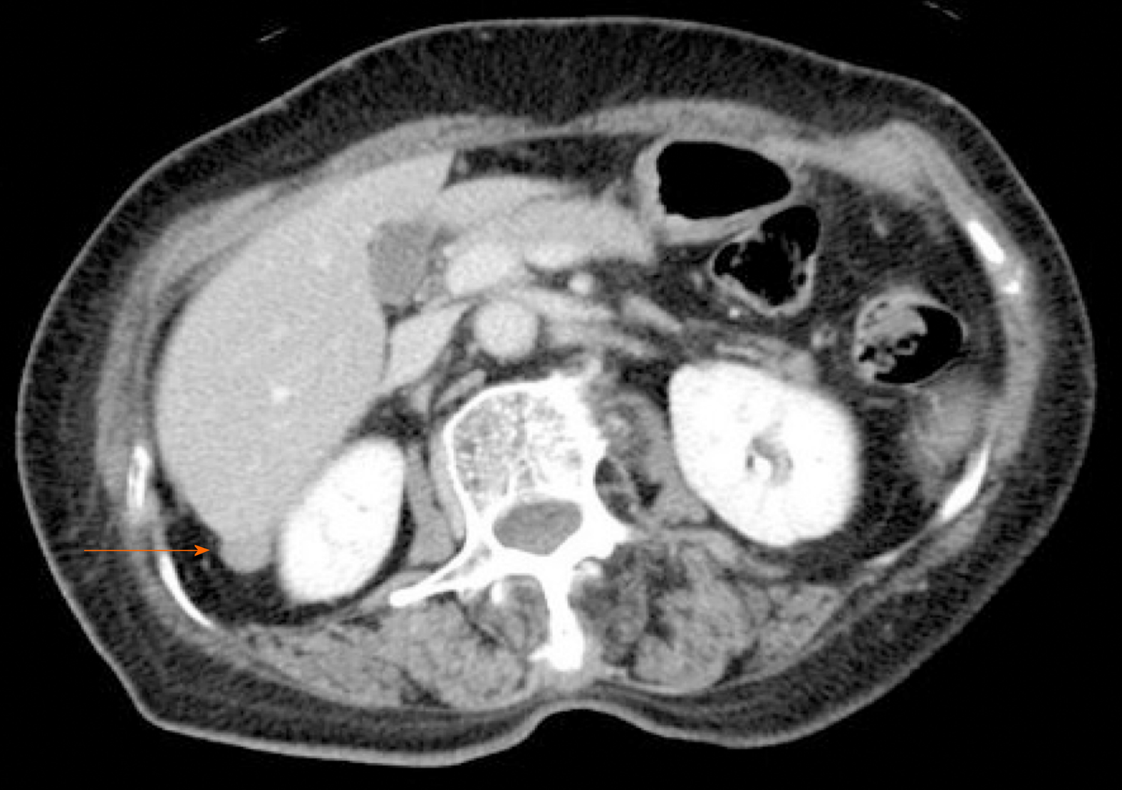
Figure 1 Contrast-enhanced computed tomography.
Contrast-enhanced computed tomography showing a slightly enhanced mass (diameter: 12 mm) located between the dorsal side of the right hepatic lobe and right kidney.
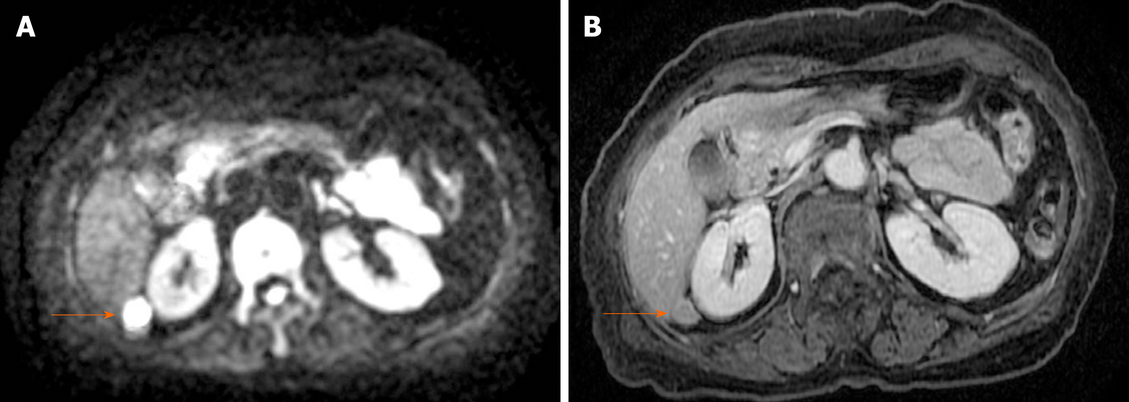
Figure 2 Magnetic resonance imaging.
Diffusion-weighted magnetic resonance imaging showing high signal intensity (A) and a uniform contrast effect (B).
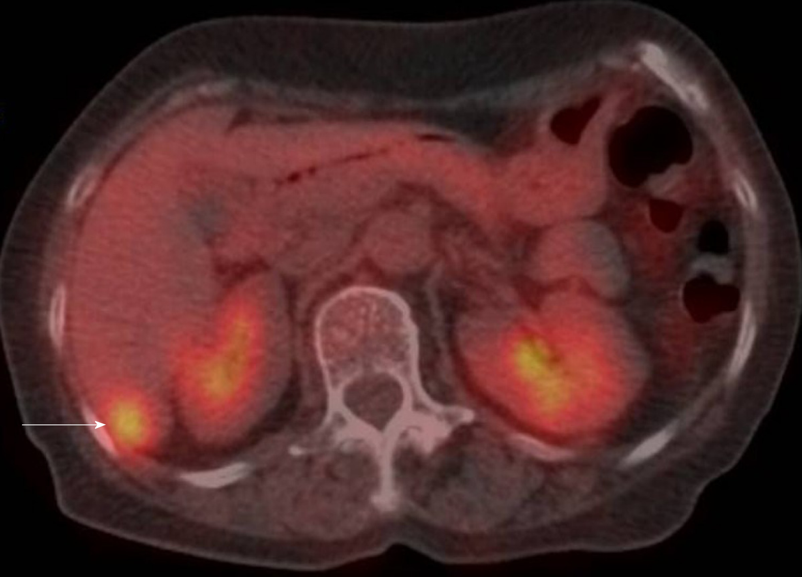
Figure 3 Positron emission tomography/computed tomography.
Positron emission tomography/computed tomography showing a tumor mass with fluorine-18 2-deoxy-2-fluoro-D-glucose accumulation, with a standardized uptake value of 5.5.
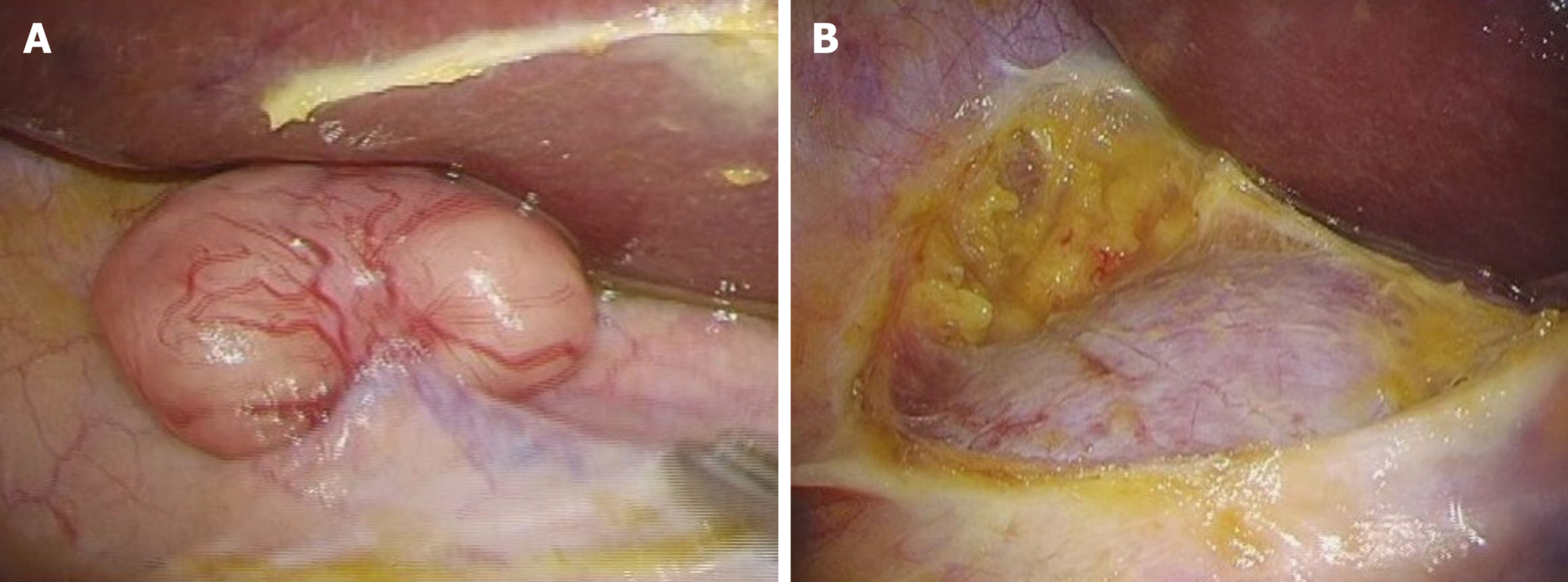
Figure 4 Surgical findings.
A: No tumor due to retroperitoneal or intraperitoneal dissemination is seen; B: We performed laparoscopic tumor resection.
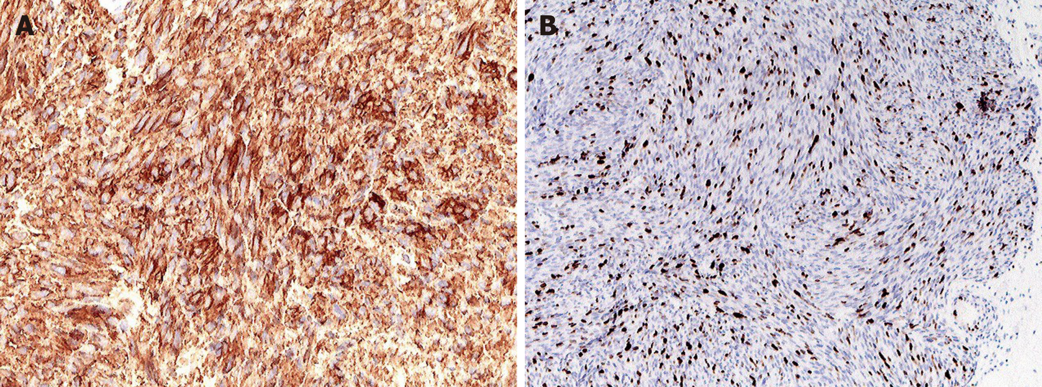
Figure 5 Immunohistochemical staining of the tumor specimen.
A: Immunohistochemical staining of the tumor specimen. Staining showed positivity for c-kit; B: The MIB-1 Labeling index was approximately 15%-20%.
- Citation: Sugiyama Y, Shimbara K, Sasaki M, Kouyama M, Tazaki T, Takahashi S, Nakamitsu A. Solitary peritoneal metastasis of gastrointestinal stromal tumor: A case report. World J Gastroenterol 2020; 26(36): 5527-5533
- URL: https://www.wjgnet.com/1007-9327/full/v26/i36/5527.htm
- DOI: https://dx.doi.org/10.3748/wjg.v26.i36.5527


