Copyright
©The Author(s) 2020.
World J Gastroenterol. Sep 21, 2020; 26(35): 5375-5386
Published online Sep 21, 2020. doi: 10.3748/wjg.v26.i35.5375
Published online Sep 21, 2020. doi: 10.3748/wjg.v26.i35.5375
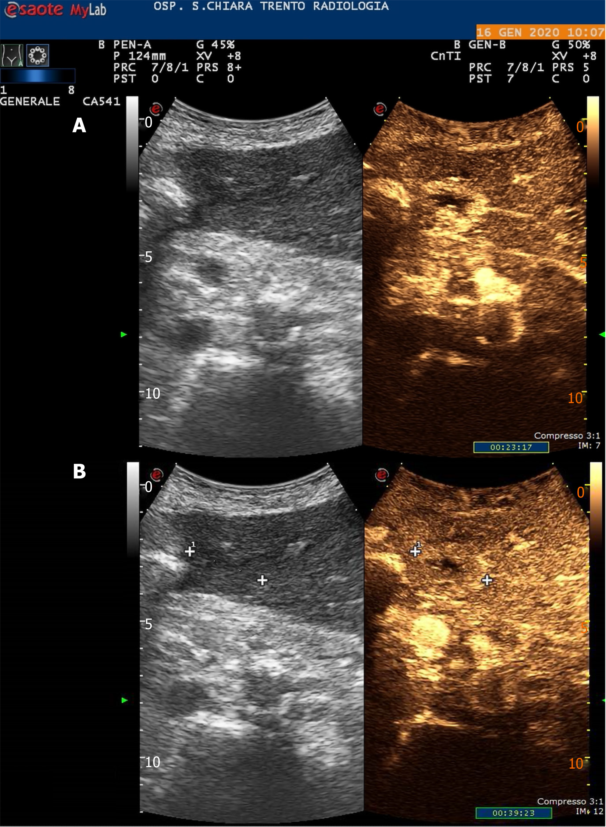
Figure 1 Contrast-enhanced ultrasonography showing the subcapsular 20 mm hepatocellular carcinoma at the 4th liver segment.
A: During wash-in phase; B: During wash-out phase.
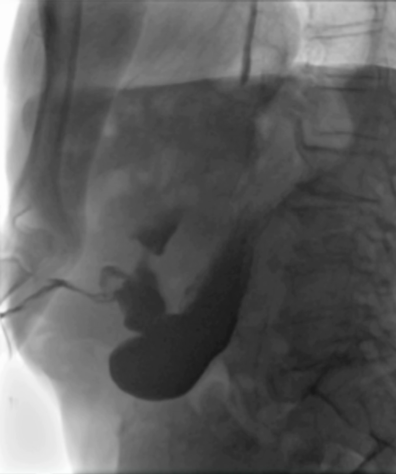
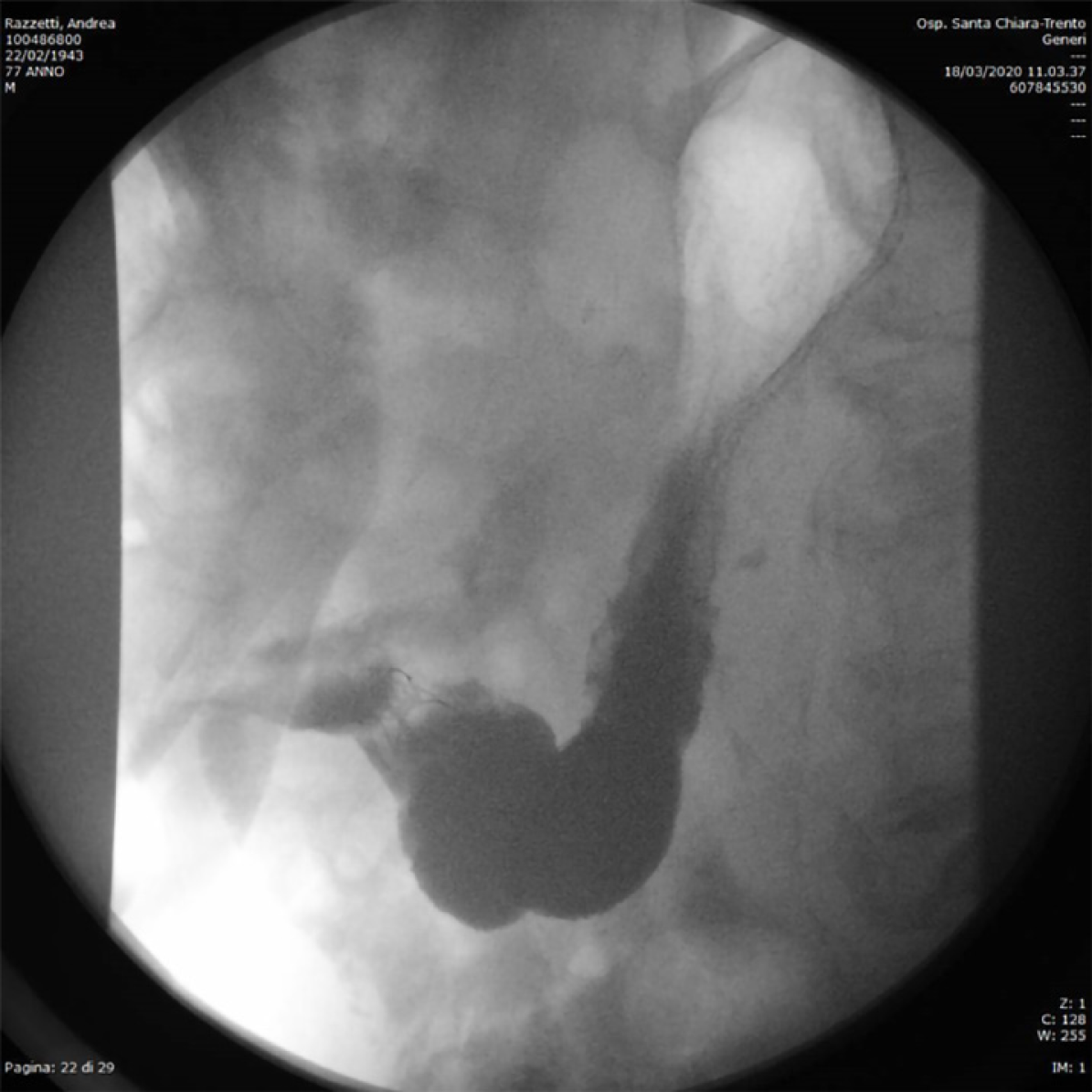
Figure 2 Abdominal film with oral water-soluble contrast agent showing a gastric perforation with a gastro-cutaneous fistulous tract (surgical drain in place).
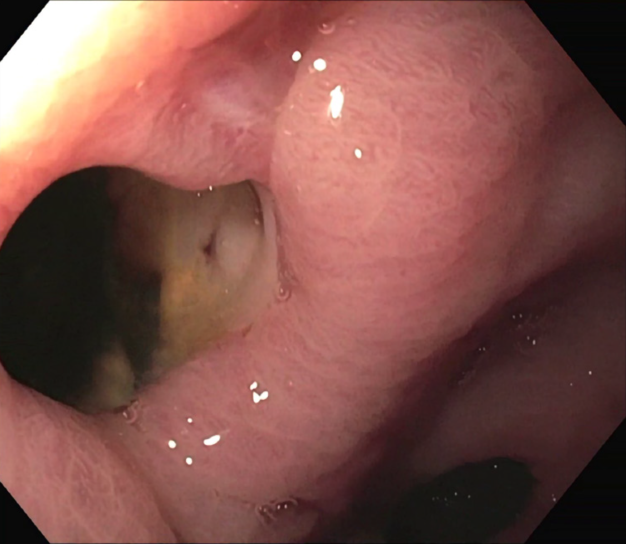
Figure 3 Endoscopic finding of the gastric perforation in communication with a purulent collection.
Pylorus can be seen in the lower part of the picture.
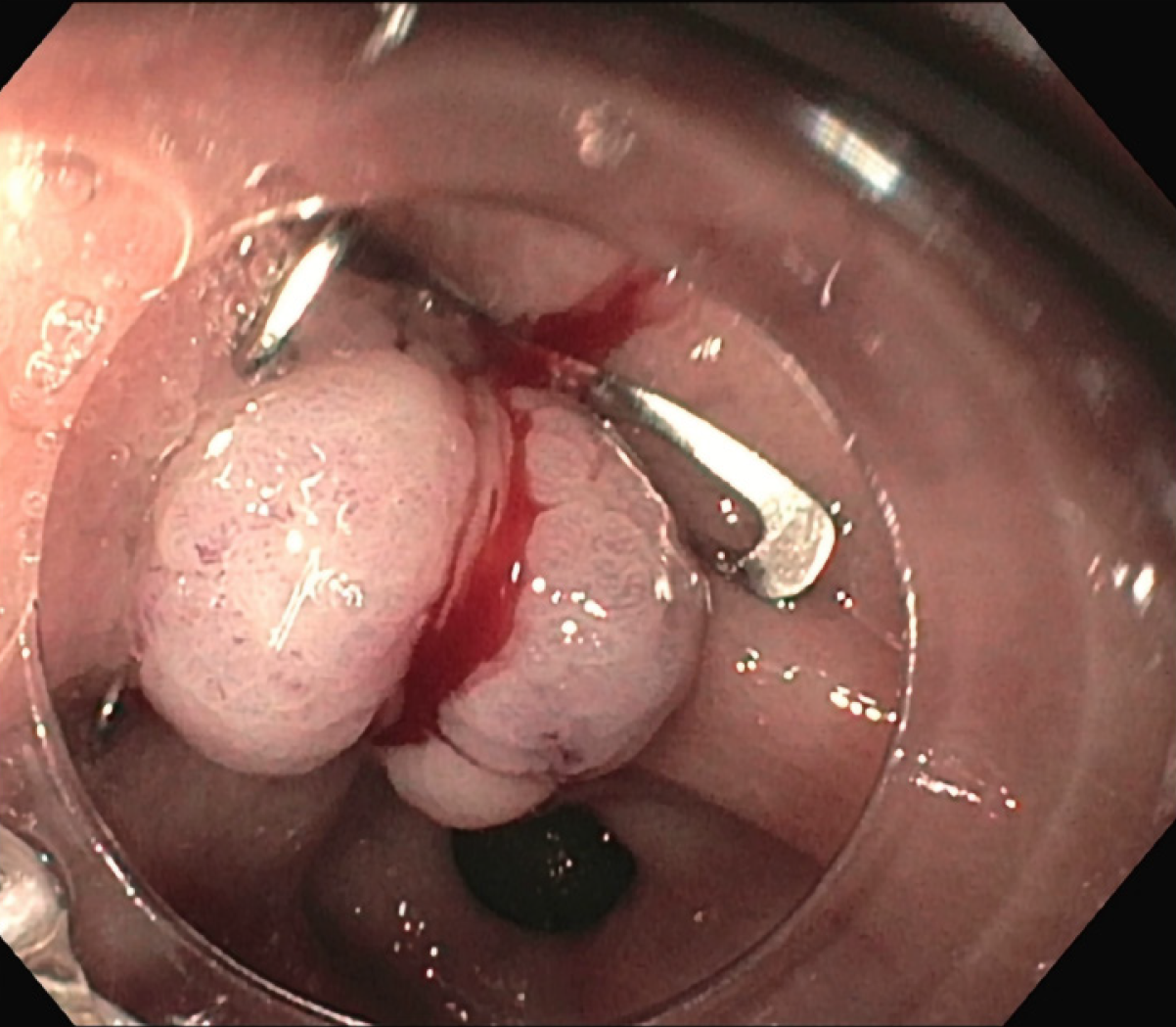
Figure 4 Endoscopic closure of the perforation using an over-the-scope clip.
Figure 5 Abdominal film with oral water-soluble contrast agent showing no active leakage from the stomach.
The over-the-scope clip can be seen in gastric antrum.
- Citation: Rogger TM, Michielan A, Sferrazza S, Pravadelli C, Moser L, Agugiaro F, Vettori G, Seligmann S, Merola E, Maida M, Ciarleglio FA, Brolese A, de Pretis G. Gastrointestinal tract injuries after thermal ablative therapies for hepatocellular carcinoma: A case report and review of the literature. World J Gastroenterol 2020; 26(35): 5375-5386
- URL: https://www.wjgnet.com/1007-9327/full/v26/i35/5375.htm
- DOI: https://dx.doi.org/10.3748/wjg.v26.i35.5375


