Copyright
©The Author(s) 2020.
World J Gastroenterol. Jul 21, 2020; 26(27): 3989-3997
Published online Jul 21, 2020. doi: 10.3748/wjg.v26.i27.3989
Published online Jul 21, 2020. doi: 10.3748/wjg.v26.i27.3989
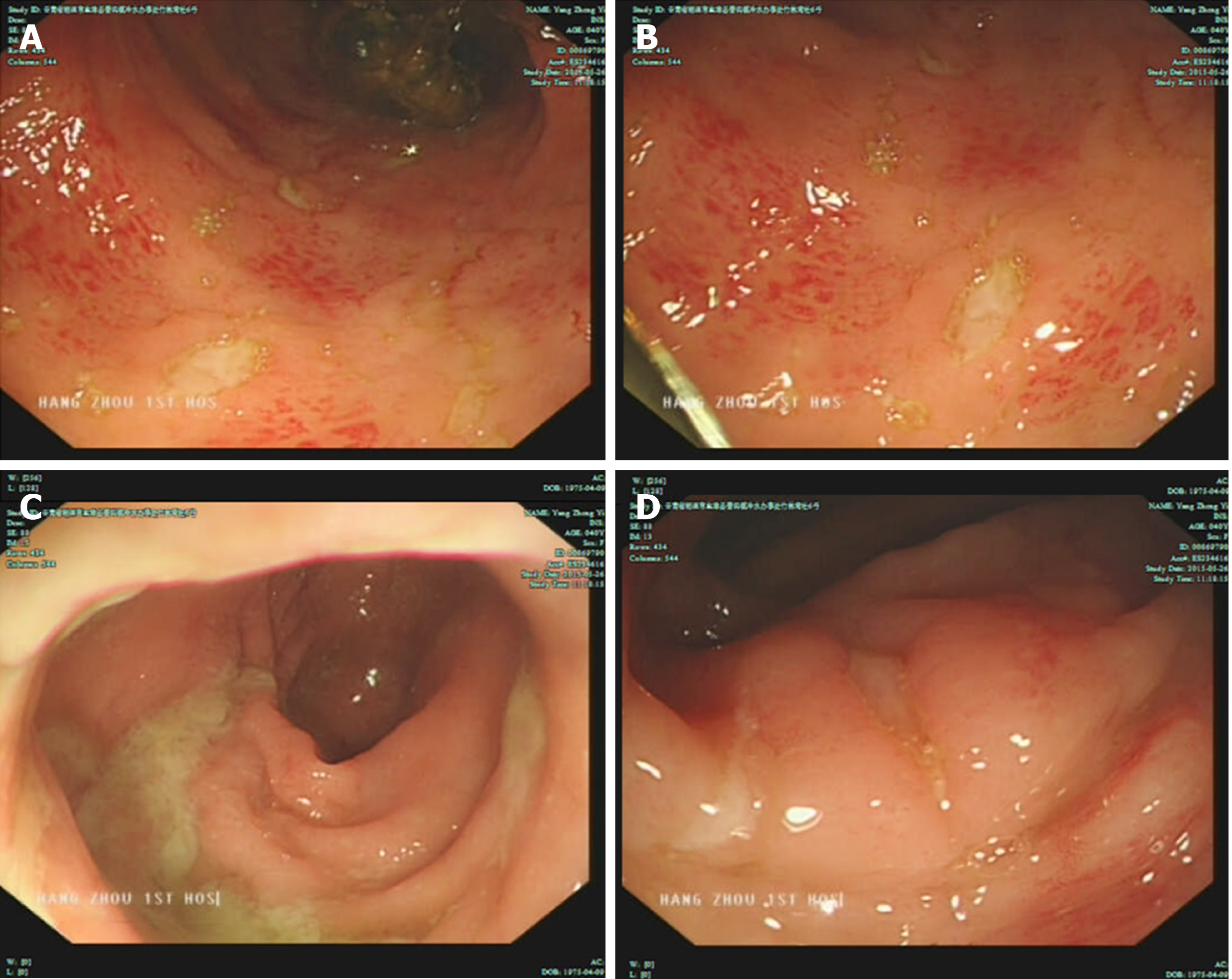
Figure 1 Colonoscopy revealed a large fecal mass located 25 cm from the anus, and it was impossible to continue with the scope.
A: Multiple ulcers of irregular sizes in the colon; B: Ulcers in the colon; C: A large fecal mass in the colon; D: Congestion and edema in the rectum.
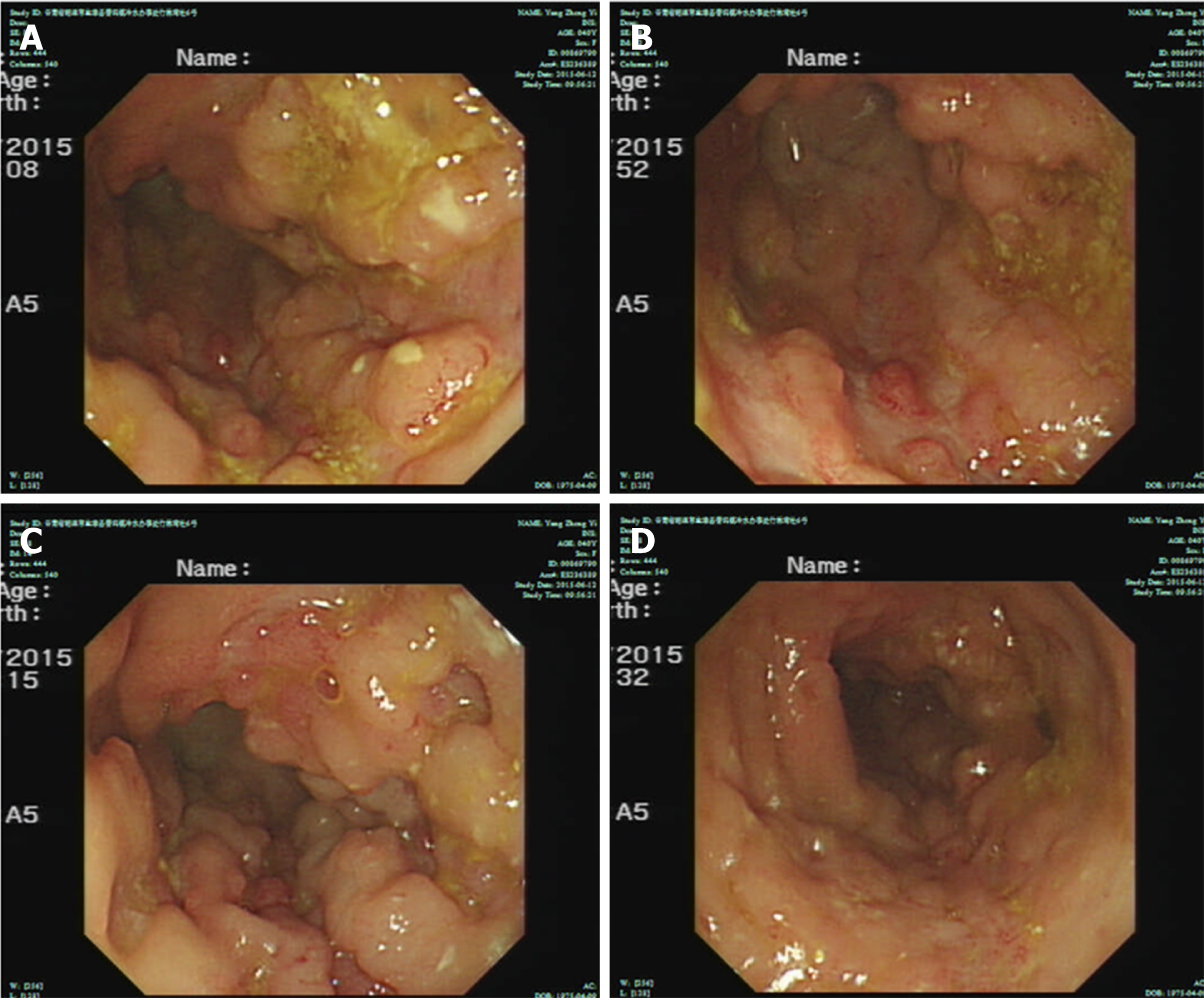
Figure 2 Colonoscopy showed hyperemia and edema of the sigmoid colon mucosa; multiple longitudinal ulcers were seen, with hyperplasia-like changes in the surrounding mucosa.
A: Ulcers and mucosal hyperplasia in the sigmoid; B: Ulcers and mucosal hyperplasia in the sigmoid; C: Ulcers and mucosal hyperplasia in the sigmoid; D: Ulcers and mucosal hyperplasia in the sigmoid.
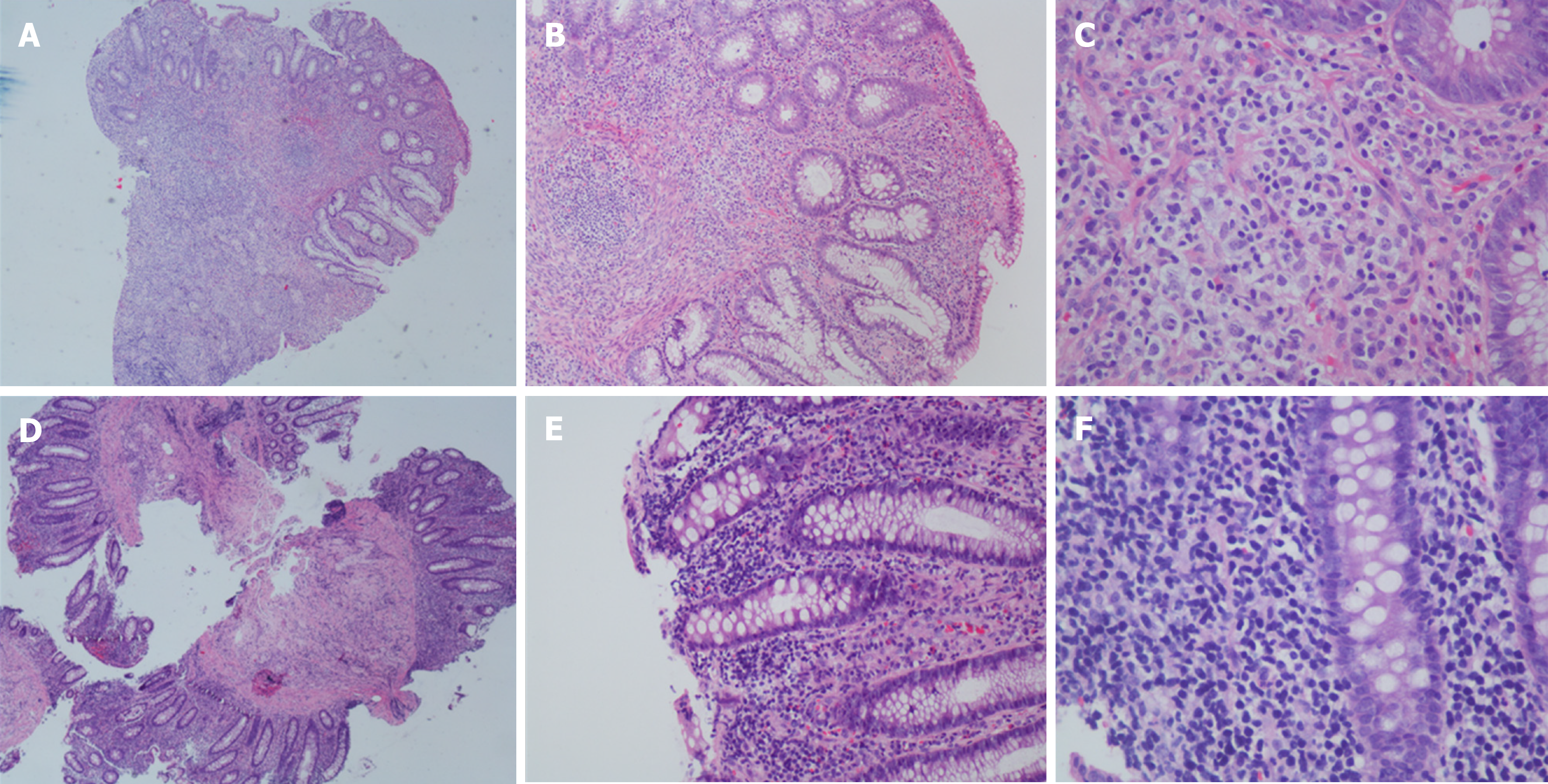
Figure 3 Colonoscopic biopsy findings: intestinal mucosal focal heterotypic lymphocyte infiltration.
A, B and C: Biopsy showed intestinal mucosal focal heterotypic lymphocyte infiltration, B and C were enlargements of a partial area in A; D, E and F: Biopsy showed intestinal mucosal focal heterotypic lymphocyte infiltration, E and F were enlargements of a partial area in D. Hematoxylin and eosin staining.
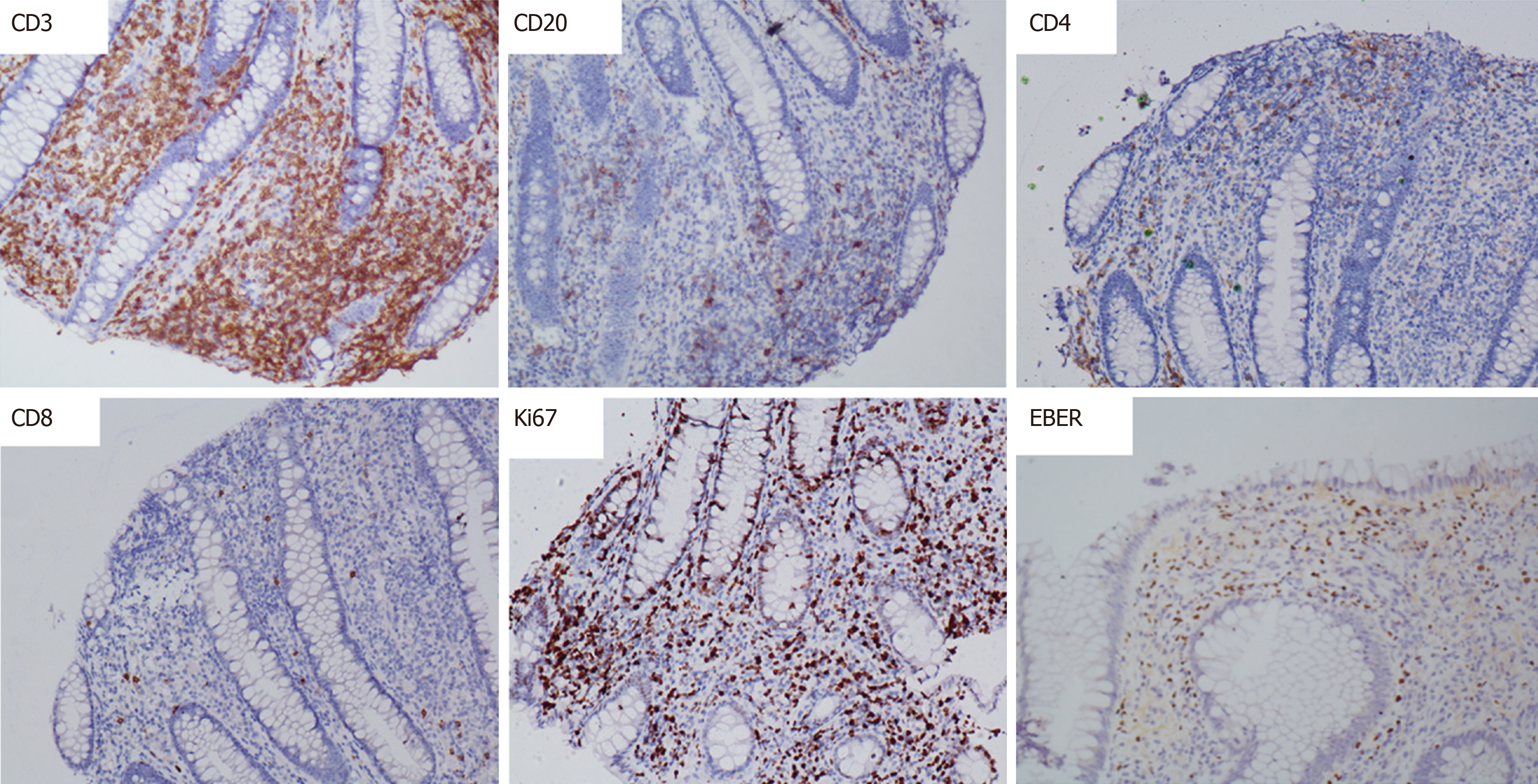
Figure 4 Immunohistochemical results: the expression of CD3 and Epstein-Barr virus encoded RNA was positive.
The expression of CD20, CD4 and CD8 was negative, and Ki-67 was approximately 40%.
- Citation: Li H, Lyu W. Intestinal NK/T cell lymphoma: A case report. World J Gastroenterol 2020; 26(27): 3989-3997
- URL: https://www.wjgnet.com/1007-9327/full/v26/i27/3989.htm
- DOI: https://dx.doi.org/10.3748/wjg.v26.i27.3989


