Copyright
©The Author(s) 2019.
World J Gastroenterol. Feb 21, 2019; 25(7): 744-776
Published online Feb 21, 2019. doi: 10.3748/wjg.v25.i7.744
Published online Feb 21, 2019. doi: 10.3748/wjg.v25.i7.744
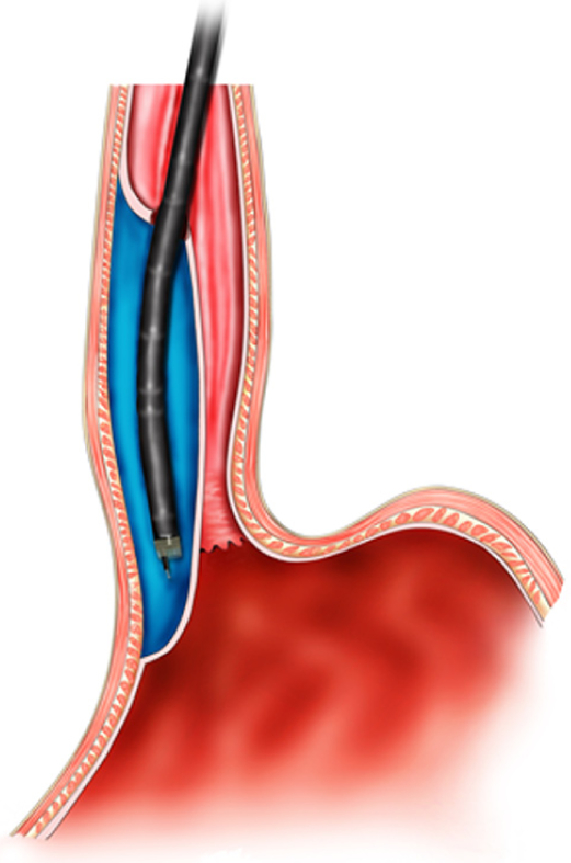
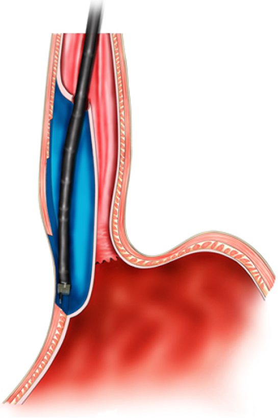
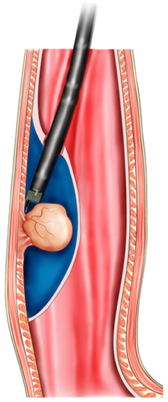
Figure 1 Mechanism of digestive endoscopic tunnel technique, demonstrating a tunnel that is created between the mucosal and muscularis propria layers.
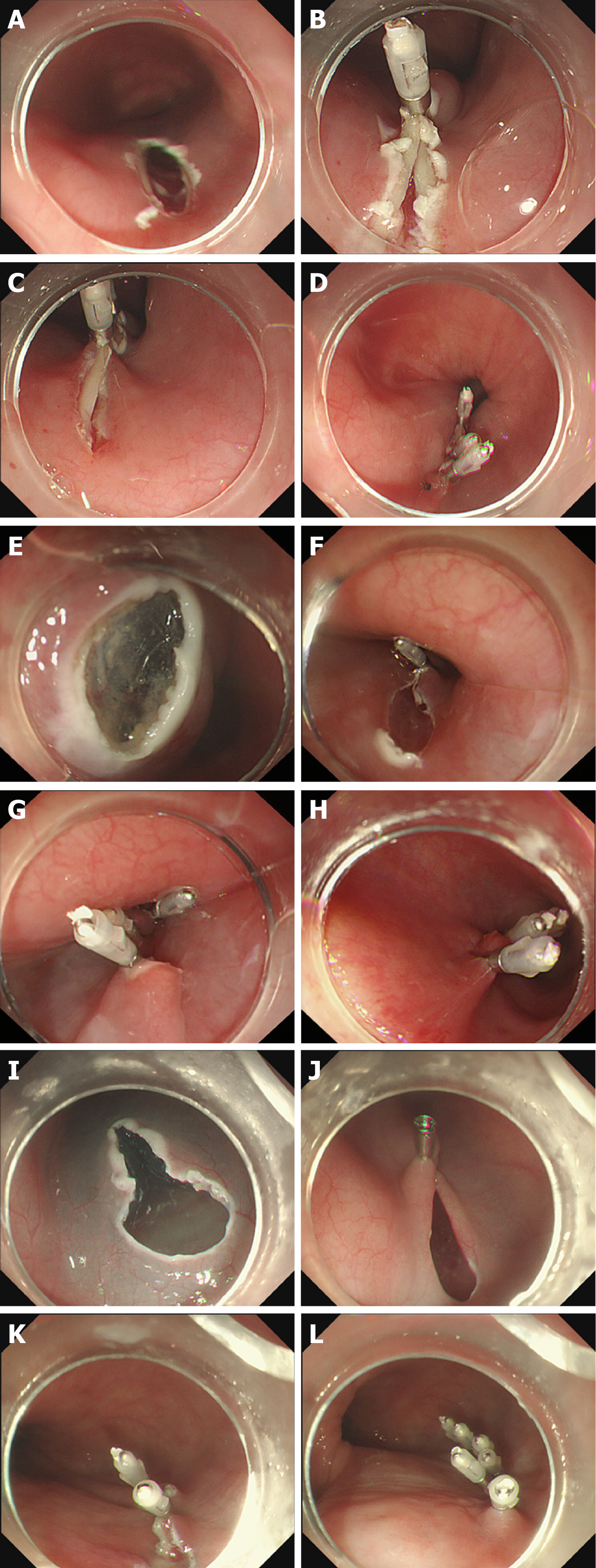
Figure 2 Three methods of tunnel incision and closure.
A and B: Longitudinal incision; C and D: Longitudinal incision closed with titanium clips; E: Transverse incision; F: “Anchoring” of a titanium clip in the middle of the transverse incision; G and H: Longitudinal closure using titanium clips successively; I: Inverted T incision; J-L: Longitudinal closure using titanium clips successively.
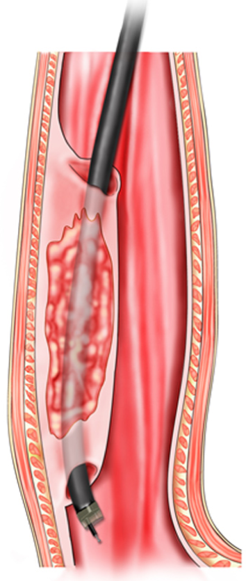
Figure 3 Schema chart of endoscopic submucosal tunnel dissection, demonstrating a tunnel that is created to resect mucosal lesions.
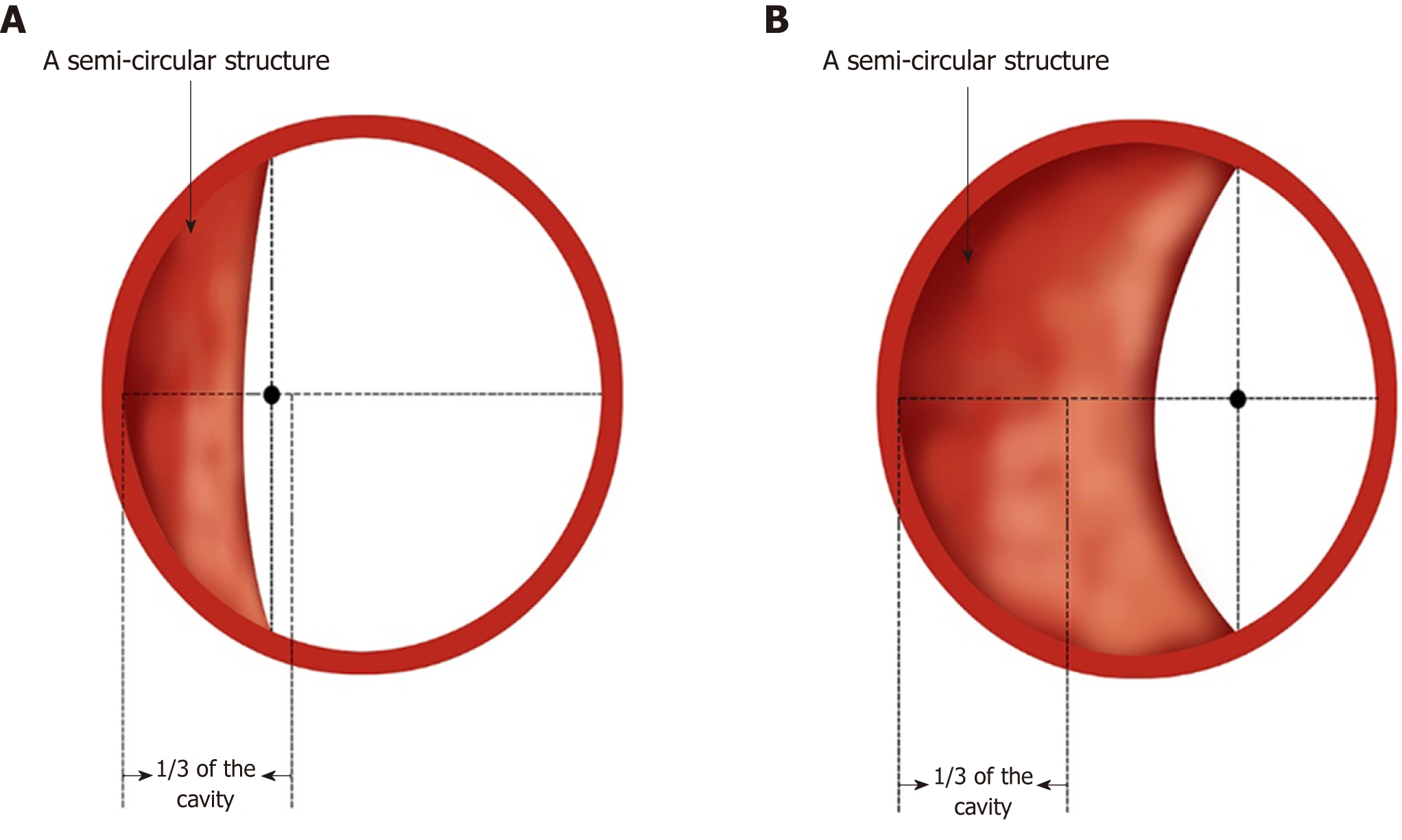
Figure 4 Simulated diagram of endoscopic observations in Ling IIb and Ling IIc.
A: Ling IIb. The arrows indicate 1/3 of the oesophageal cavity, and the semi-annular structure’s midpoint remains within this range; B: Ling IIc. The arrows indicate 1/3 of the oesophageal cavity, and the crescent-like structure's midpoint goes beyond this range.
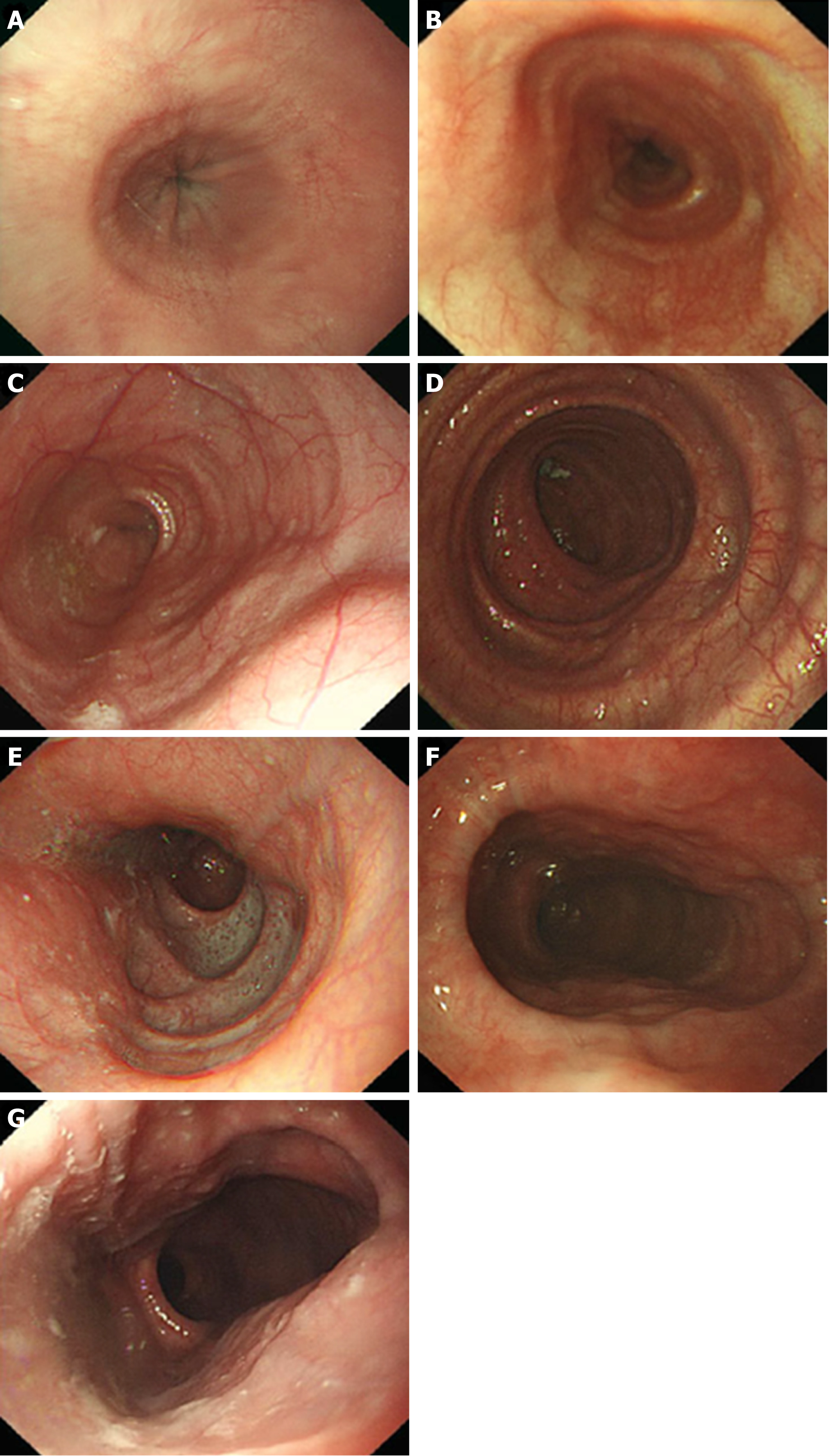
Figure 5 Endoscopic images of the Ling classification of achalasia cardia.
A: Ling I; B: Ling IIa; C: Ling IIb; D: Ling IIc, E: Ling III1; F: Ling IIIr; G: Ling IIIlr.
Figure 6 Correlation between grade A or grade B mucosal inflammation and mild oesophageal submucosal adhesion.
A: Grade A mucosal inflammation; B: Grade B mucosal inflammation; C: Mild oesophageal submucosal adhesion: The fibre filaments are distributed in bundles.
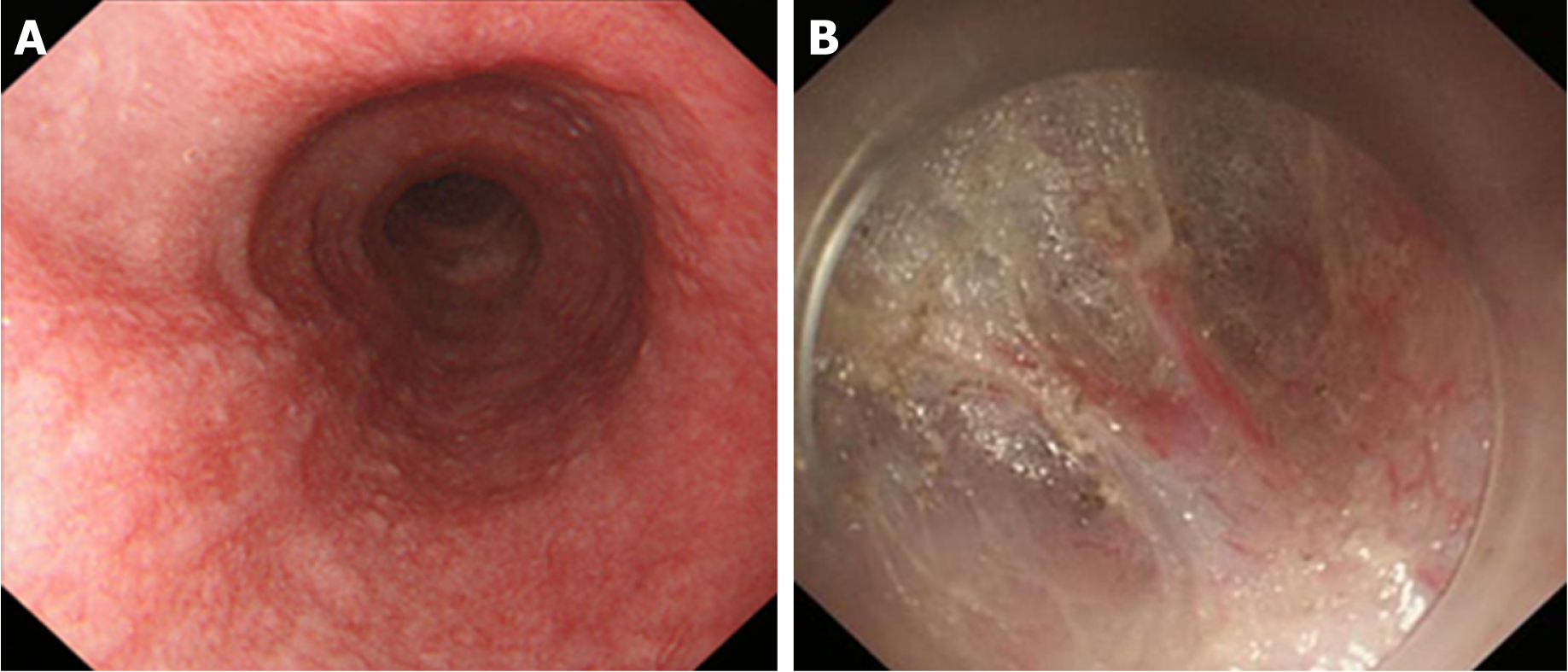
Figure 7 Correlation between Grade C mucosal inflammation and moderate oesophageal submucosal adhesion.
A: Grade C mucosal inflammation; B: Moderate oesophageal submucosal adhesion. The fibres are arranged in disorder, with fusion and decreased transparency.
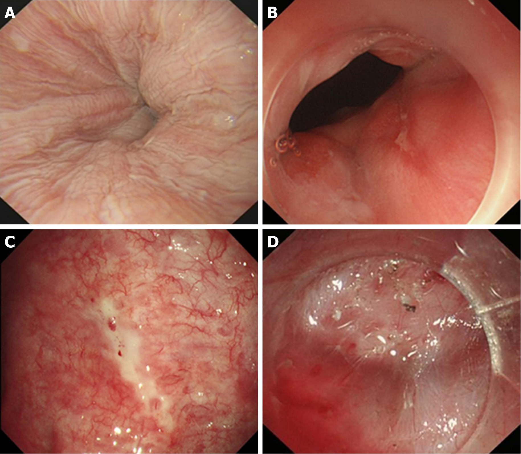
Figure 8 Correlation between Grade D, Grade E, or Grade F mucosal inflammation and severe oesophageal submucosal adhesion.
A: Grade D mucosal inflammation; B: Grade E mucosal inflammation; C: Grade F mucosal inflammation; D: Severe oesophageal submucosal adhesion. The submucosa and muscularis propria are completely adherent.
Figure 9 Anatomical landmark in the tunnel from the lower oesophagus to the cardia.
A: Grid-like blood vessels in the cardia; B: Crescent-like structure visible at the proximal cardia; C: Ampulla-like structure appearing after entering the crescent-like structure; D: Branching vessels with bulky vascular roots in the ampulla-like structure; E: Tunnel below the cardia, showing a steep downward form; F: Stubby and multi-branched vessels below the cardia; G: Beadlike vessels below the cardia.
Figure 10 Schema chart of per-oral endoscopic myotomy, demonstrating a tunnel that is created to incise the muscularis propria.
Figure 11 Endoscope crosses the “ridge” via short-tunnel per-oral endoscopic myotomy.
A: Type Ling IIc oesophagus. The arrow indicates a “ridge” structure formed by the crescent-like structure; B: The short-tunnel entry incision established on a relatively flat oesophageal wall at the oral side of the “ridge”; C: It is easy to cross the “ridge” within the tunnel.
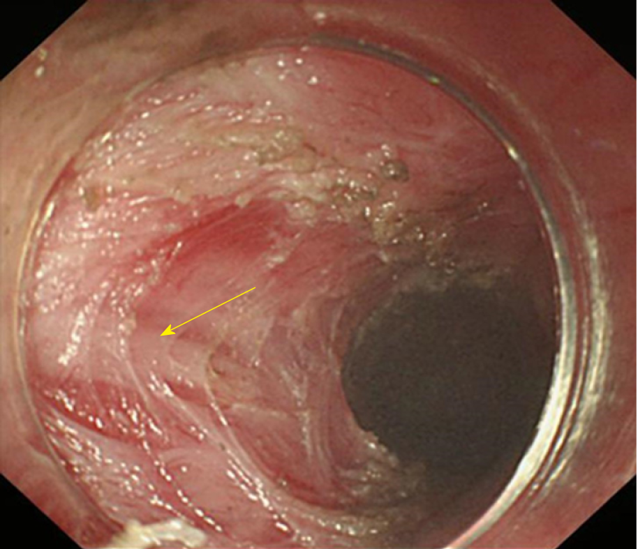
Figure 12 Inner circular muscle myotomy.
The arrow shows the well-retained longitudinal muscle.
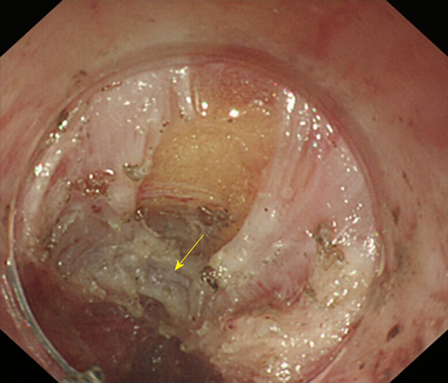
Figure 13 Full-thickness myotomy.
The arrow shows the tunica adventitia of the oesophagus.
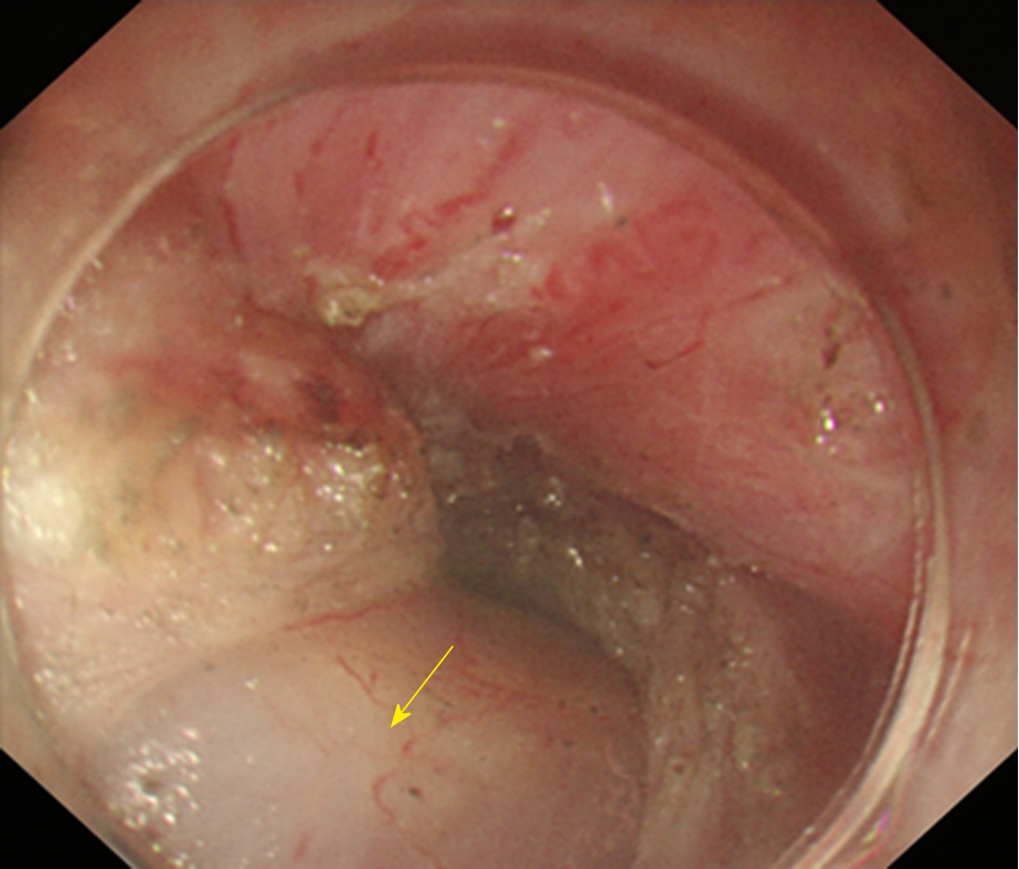
Figure 14 Glasses-style myotomy.
The arrow shows the muscles remaining at the cardia.
Figure 15 Circular muscle myotomy + balloon plasty.
A: The width of the incision should be 1/3 of the circumferential oesophagus; B: Balloon-dilation in the oesophagus; C: The width of the incision after expansion should be 2/3 of the circumferential oesophagus.
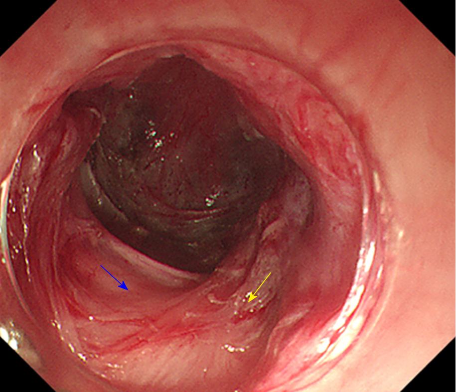
Figure 16 Progressive full-thickness myotomy.
The yellow arrow shows the incision into the inner circular muscles from superficial to deep; the blue arrow shows the full incision into the muscularis propria.
Figure 17 Schema chart of per-oral endoscopic myotomy, demonstrating a tunnel that is created to resect the lesion from the muscularis propria.
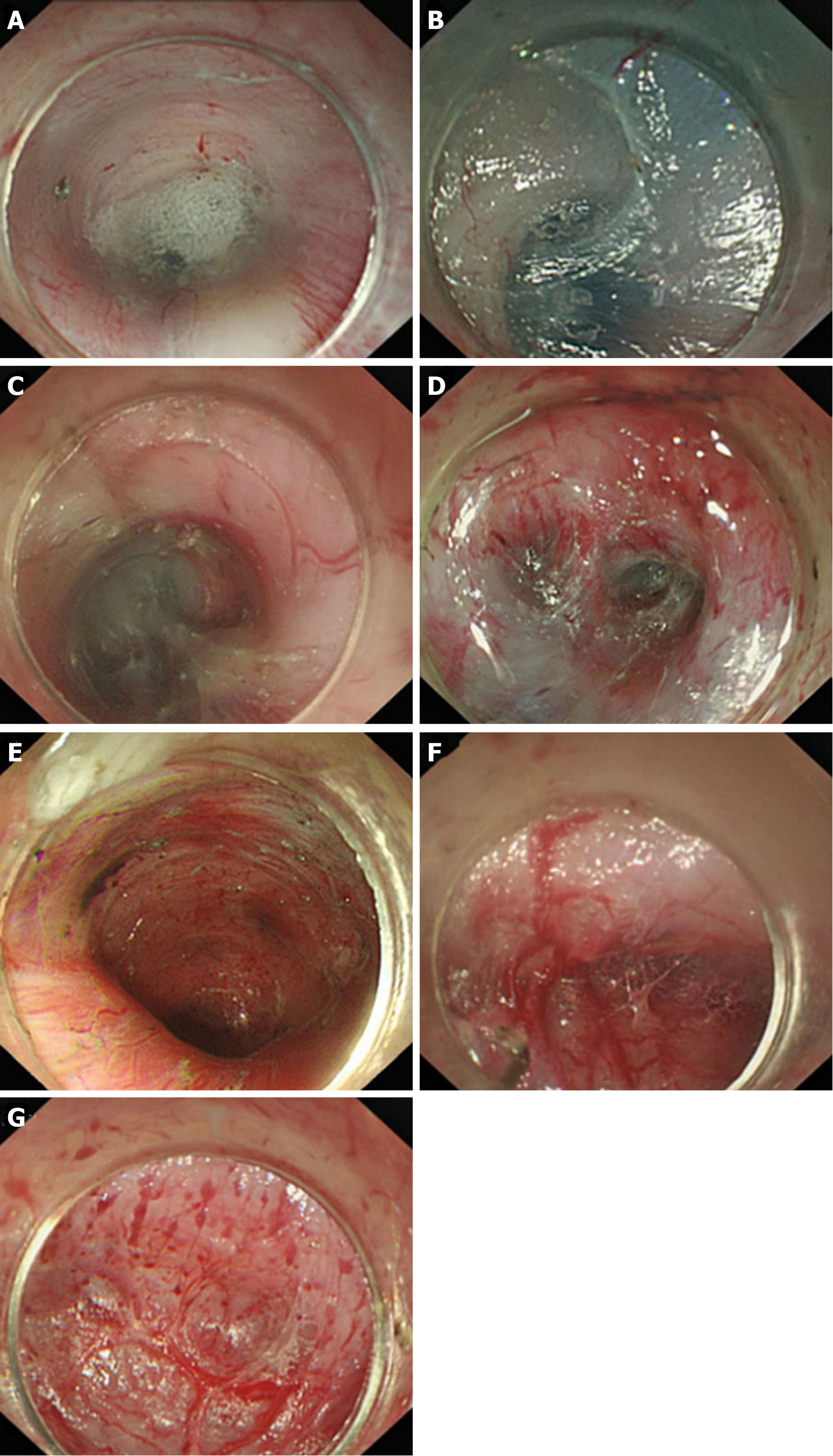
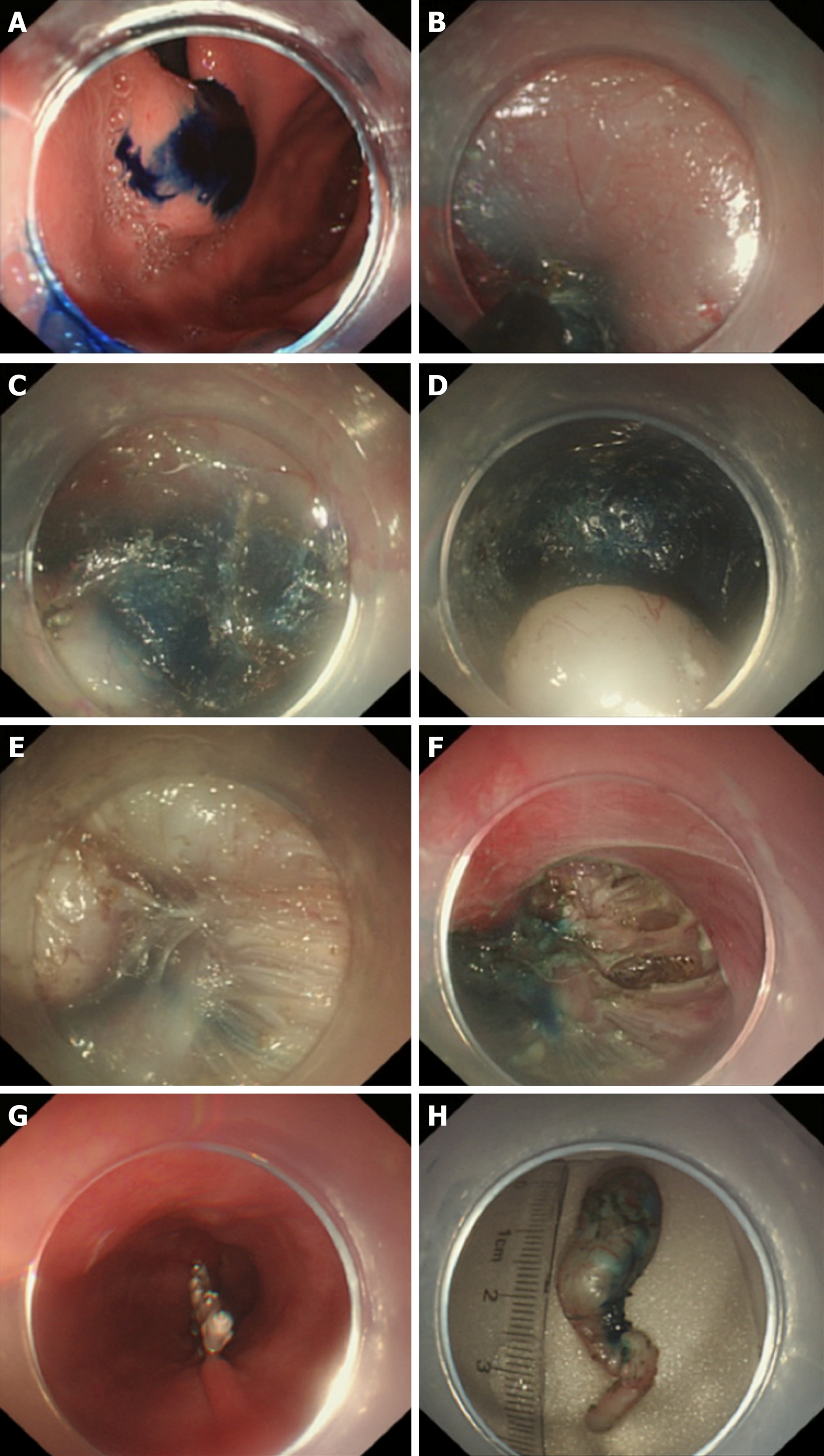
Figure 18 Key steps of submucosal tunnelling endoscopic resection for a cardial submucosal tumour.
A: Injection of methylene blue into the lesion incision site for marking and positioning; B: Establishment of a tunnel; C: Finding of the marking and positioning with methylene blue in the tunnel; D: Exposure of the lesion; E: Resection of the lesion; F: Wound following the resection of the tumour; G: Closure of the mucosal incision; H: The resected specimen.
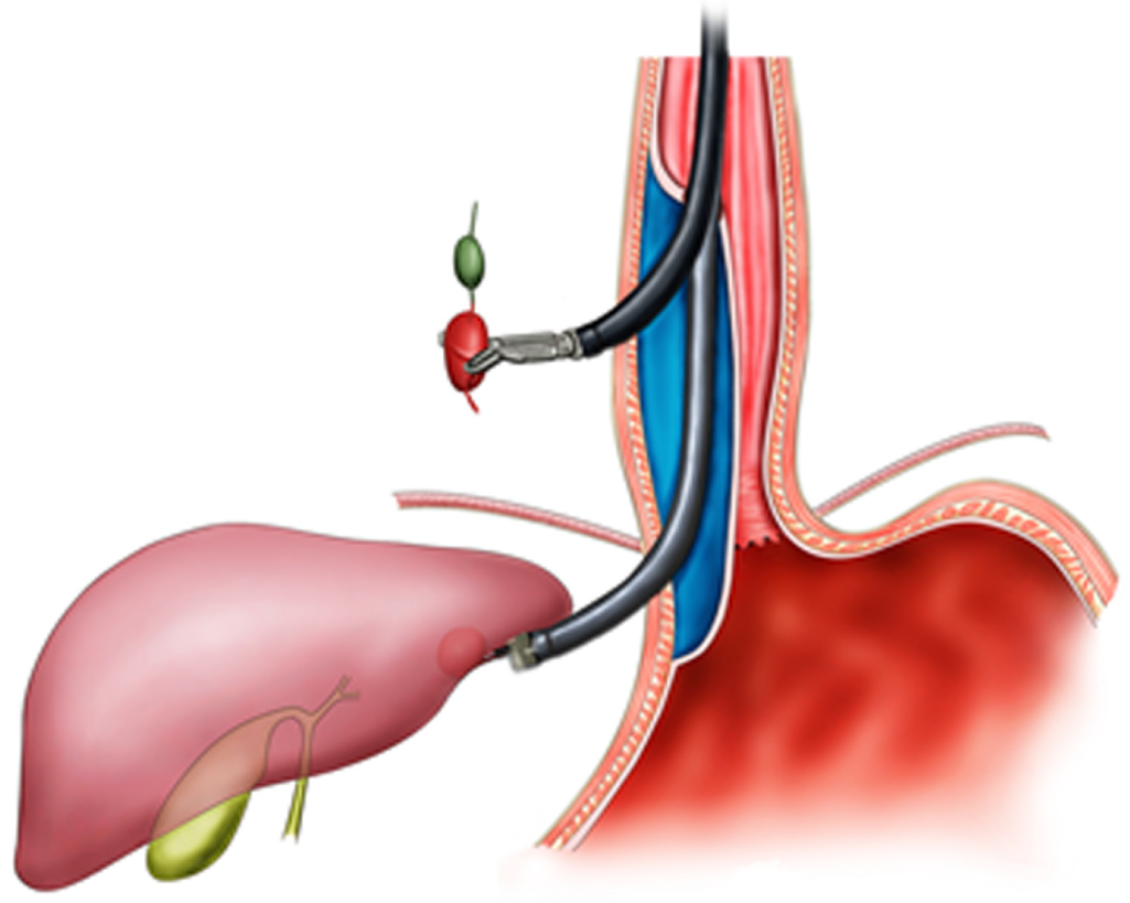
Figure 19 Schema chart of digestive endoscopic tunnel technique on the external digestive tract wall, demonstrating a tunnel that is created to resect a node outside the digestive tract.
- Citation: Chai NL, Li HK, Linghu EQ, Li ZS, Zhang ST, Bao Y, Chen WG, Chiu PW, Dang T, Gong W, Han ST, Hao JY, He SX, Hu B, Hu B, Huang XJ, Huang YH, Jin ZD, Khashab MA, Lau J, Li P, Li R, Liu DL, Liu HF, Liu J, Liu XG, Liu ZG, Ma YC, Peng GY, Rong L, Sha WH, Sharma P, Sheng JQ, Shi SS, Seo DW, Sun SY, Wang GQ, Wang W, Wu Q, Xu H, Xu MD, Yang AM, Yao F, Yu HG, Zhou PH, Zhang B, Zhang XF, Zhai YQ. Consensus on the digestive endoscopic tunnel technique. World J Gastroenterol 2019; 25(7): 744-776
- URL: https://www.wjgnet.com/1007-9327/full/v25/i7/744.htm
- DOI: https://dx.doi.org/10.3748/wjg.v25.i7.744


