Copyright
©The Author(s) 2019.
World J Gastroenterol. Oct 28, 2019; 25(40): 6107-6115
Published online Oct 28, 2019. doi: 10.3748/wjg.v25.i40.6107
Published online Oct 28, 2019. doi: 10.3748/wjg.v25.i40.6107
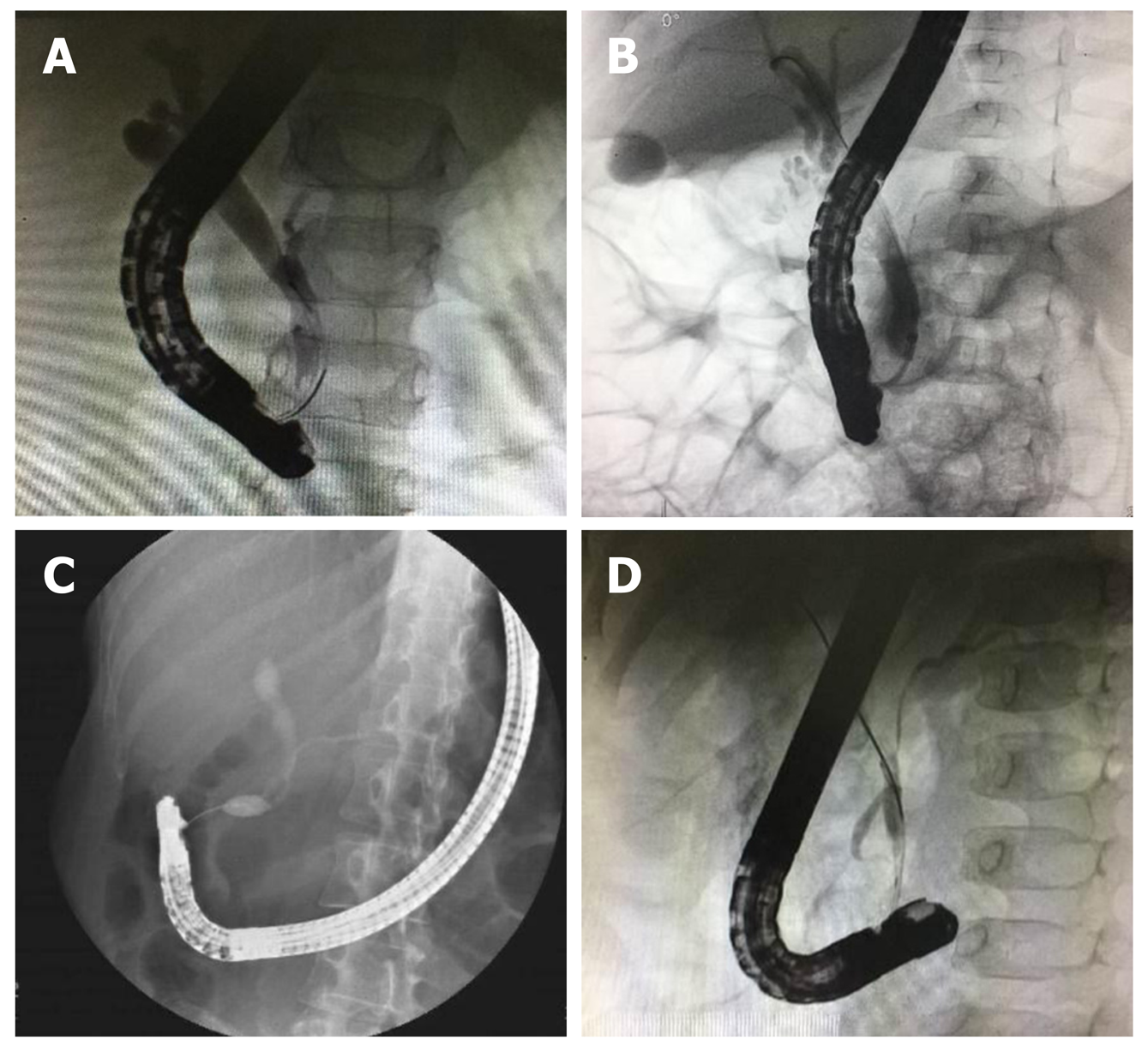
Figure 1 Fluoroscopy images of four pancreaticobiliary maljunction types.
A: Type A (stenotic type). The stenotic segment of the distal common bile duct joins the common channel, with the dilatation of the common bile duct; B: Type B (non-stenotic type). The distal common bile duct without any stenotic segment smoothly joins the common channel; C: Type C (dilated channel type). The common channel is dilated, the narrow segment of the distal common bile duct joins the common channel, and abrupt dilatation of the common channel is seen; D: Type D (complex type). Complicated union of the pancreaticobiliary ductal system occurs as follows: Pancreaticobiliary maljunction is associated with annular pancreas, pancreatic divisum, or other duct system complications.
- Citation: Zeng JQ, Deng ZH, Yang KH, Zhang TA, Wang WY, Ji JM, Hu YB, Xu CD, Gong B. Endoscopic retrograde cholangiopancreatography in children with symptomatic pancreaticobiliary maljunction: A retrospective multicenter study. World J Gastroenterol 2019; 25(40): 6107-6115
- URL: https://www.wjgnet.com/1007-9327/full/v25/i40/6107.htm
- DOI: https://dx.doi.org/10.3748/wjg.v25.i40.6107


