Copyright
©The Author(s) 2019.
World J Gastroenterol. Oct 21, 2019; 25(39): 5926-5935
Published online Oct 21, 2019. doi: 10.3748/wjg.v25.i39.5926
Published online Oct 21, 2019. doi: 10.3748/wjg.v25.i39.5926
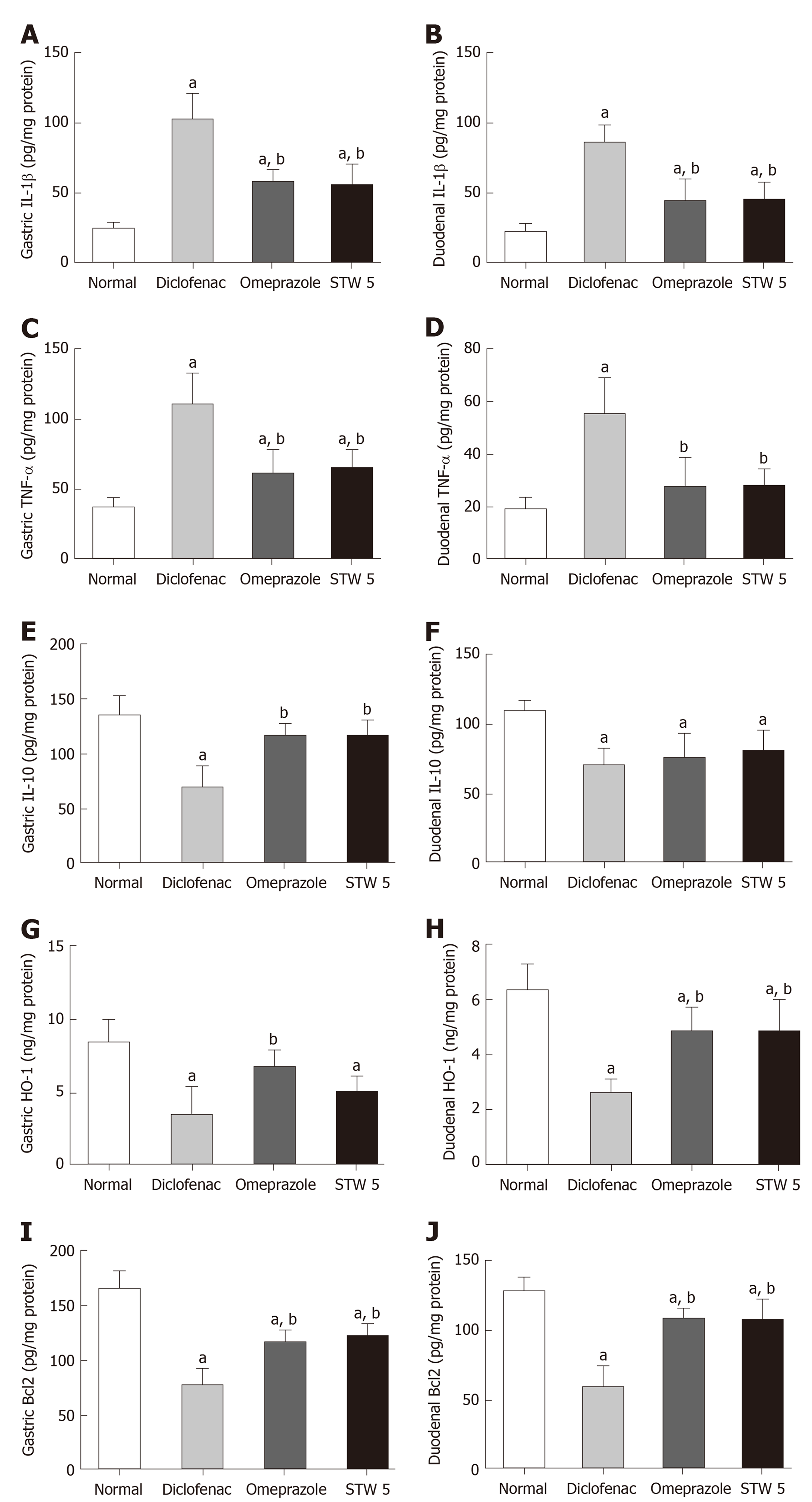
Figure 1 Changes in interleukin 1β, tumor necrosis factor-α, interleukin 10, heme oxygenase-1, and B-cell lymphoma 2 concentration in gastro-intestinal lesions induced by diclofenac in rats after treatment with omeprazole (20 mg/kg) or STW 5 (5 mL/kg).
A: Changes in interleukin 1β concentration in gastric lesions; B: Changes in interleukin 1β concentration in duodenal lesions; C: Changes in tumor necrosis factor-α concentration in gastric lesions; D: Changes in tumor necrosis factor-α concentration in duodenal lesions; E: Changes in interleukin 10 concentration in gastric lesions; F: Changes in interleukin 10 concentration in duodenal lesions; G: Changes in heme oxygenase-1 concentration in gastric lesions; H: Changes in heme oxygenase-1 concentration in duodenal lesions; I: Changes in B-cell lymphoma 2 concentration in gastric lesions; J: Changes in B-cell lymphoma 2 concentration in duodenal lesions. Data are expressed as means ± SD. The significance of the difference between means was tested by ANOVA followed by Tukey Kramer multiple comparisons test. aP < 0.05 vs normal; bP < 0.01 vs diclofenac-treated group, n = 7. IL-1β: Interleukin 1β; IL-10: Interleukin 10; TNF-α: Tumor necrosis factor-α; HO-1: Heme oxygenase-1; Bcl2: B-cell lymphoma 2.
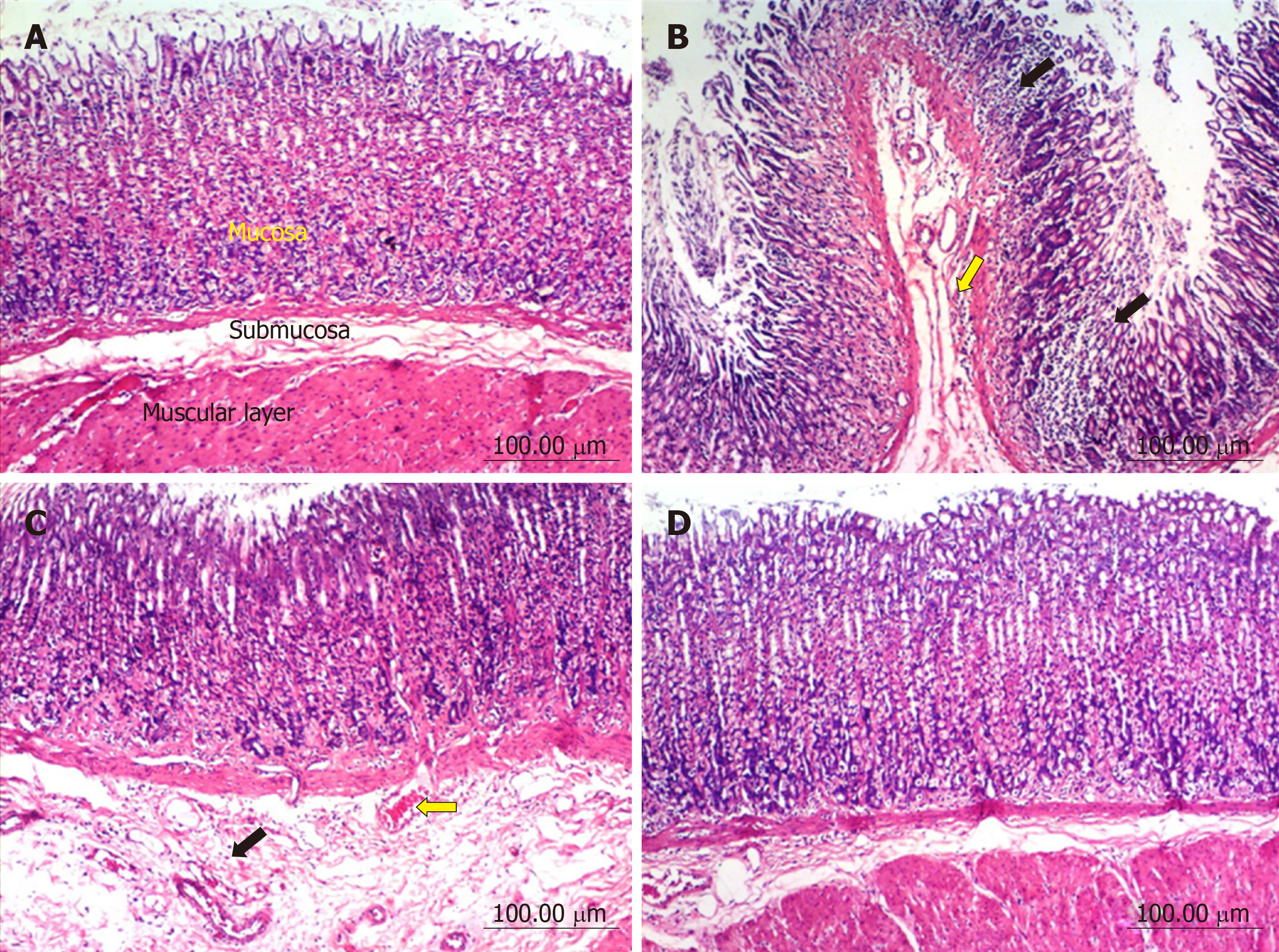
Figure 2 Histopathological changes in rat stomach.
A: Untreated controls showing a normal histological structure of gastric layers (mucosa, submucosa and musculosa); B: Diclofenac treated rats, showing multiple focal necrosis of gastric mucosa associated with mononuclear cell infiltration (black arrows), sub-mucosal edema and inflammatory cells infiltration (yellow arrow); C: Diclofenac together with omeprazole treated rats, showing sub-mucosal edema (black arrow) and congested sub-mucosal blood vessel (yellow arrow); D: Diclofenac together with STW 5 treated rats showing no histopathological alterations.
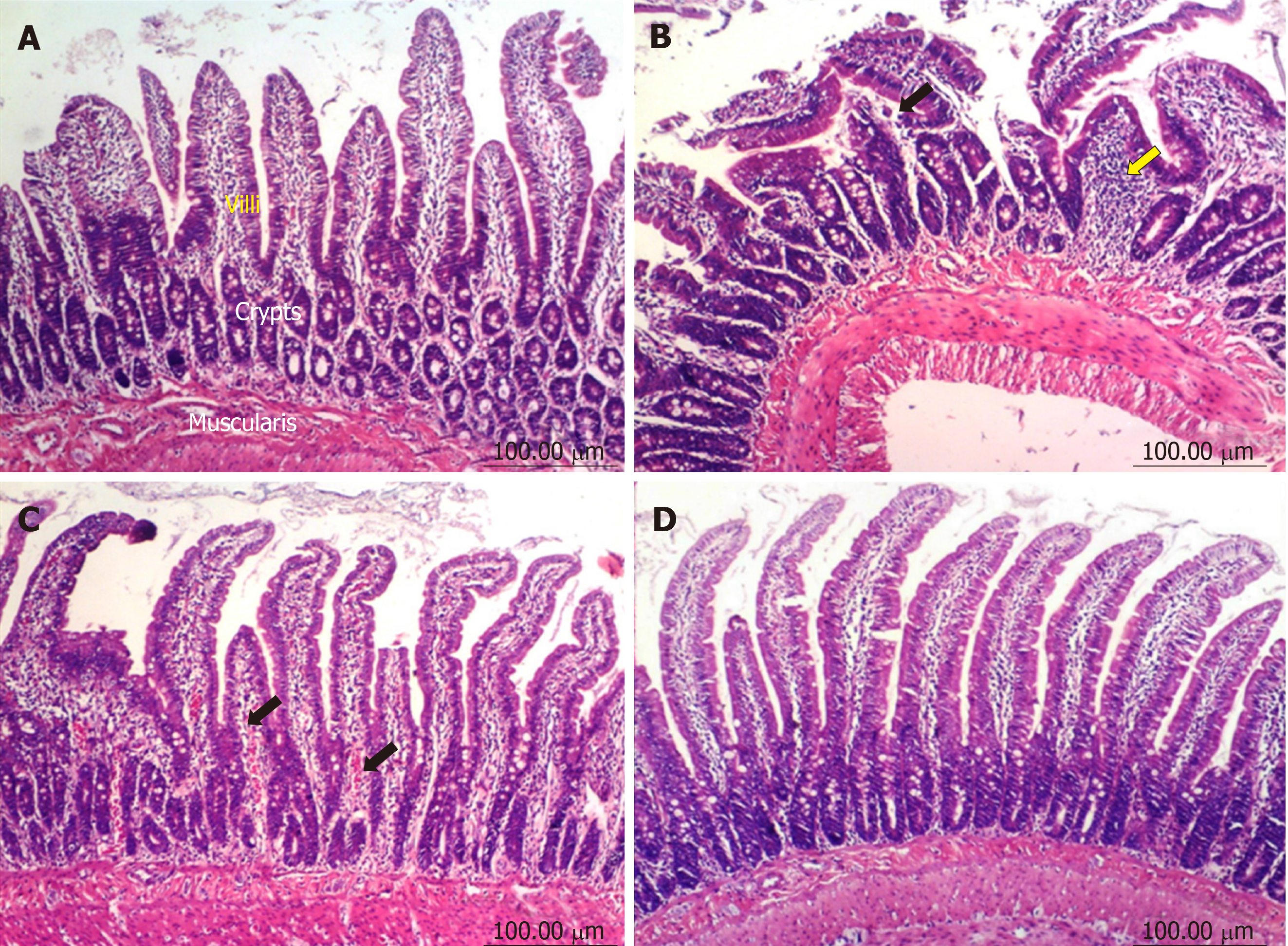
Figure 3 Histopathological changes in rat duodenum.
A: Untreated controls showing a normal histological structure of villi, crypts and muscularis layer; B: Diclofenac treated rats showing focal mucosal necrosis (black arrow) associated with focal inflammatory cells infiltration in lamina propria (yellow arrow); C: Diclofenac together with omeprazole treated rats showing congestion of blood vessels in the lamina propria (black arrows); D: Diclofenac together with STW 5 treated rats showing no histopathological alterations.
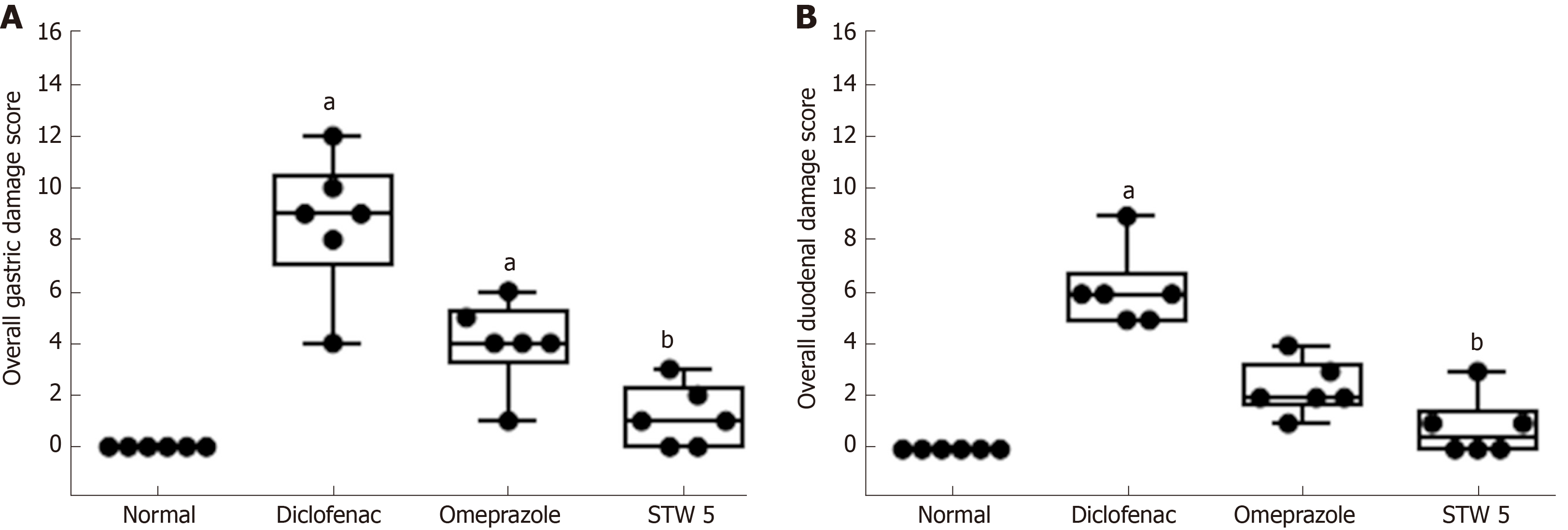
Figure 4 Changes in overall damage score in gastro-intestinal lesions induced by diclofenac in rats after treatment with omeprazole (20 mg/kg) or STW 5 (5 mL/kg).
A: Gastric lesions; B: Duodenal lesions. Data are expressed as box plots of the median of six animals. aP < 0.05 vs normal; bP < 0.01 vs diclofenac-treated group.
- Citation: Khayyal MT, Wadie W, Abd El-Haleim EA, Ahmed KA, Kelber O, Ammar RM, Abdel-Aziz H. STW 5 is effective against nonsteroidal anti-inflammatory drugs induced gastro-duodenal lesions in rats. World J Gastroenterol 2019; 25(39): 5926-5935
- URL: https://www.wjgnet.com/1007-9327/full/v25/i39/5926.htm
- DOI: https://dx.doi.org/10.3748/wjg.v25.i39.5926


