Copyright
©The Author(s) 2019.
World J Gastroenterol. Jul 14, 2019; 25(26): 3380-3391
Published online Jul 14, 2019. doi: 10.3748/wjg.v25.i26.3380
Published online Jul 14, 2019. doi: 10.3748/wjg.v25.i26.3380
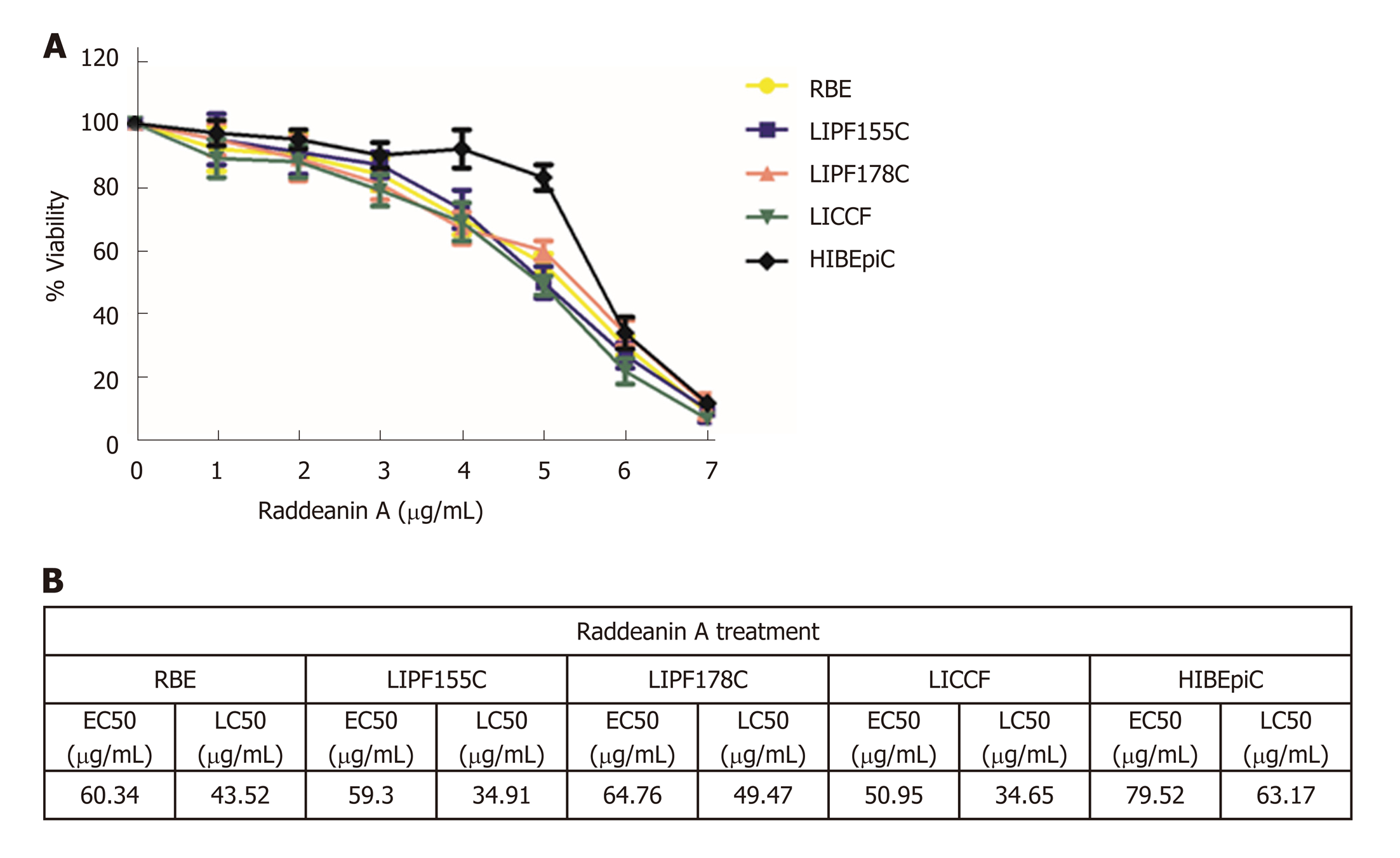
Figure 1 Effects of raddeanin A on cholangiocarcinoma cell lines (n = 3 independent experiments).
A: Increasing concentrations of raddeanin A (RA) were administered to cholangiocarcinoma cell lines for 24 h before being analyzed by the ATPlite assay. The percentage cell viability normalized to control is shown and data are expressed as the mean ± standard deviation; B: The half-maximal effective concentration and the half-maximal lethal concentration values for cholangiocarcinoma cell lines treated with RA. RA: Raddeanin A; EC50: Half-maximal effective concentration; LC50: Half-maximal lethal concentration.
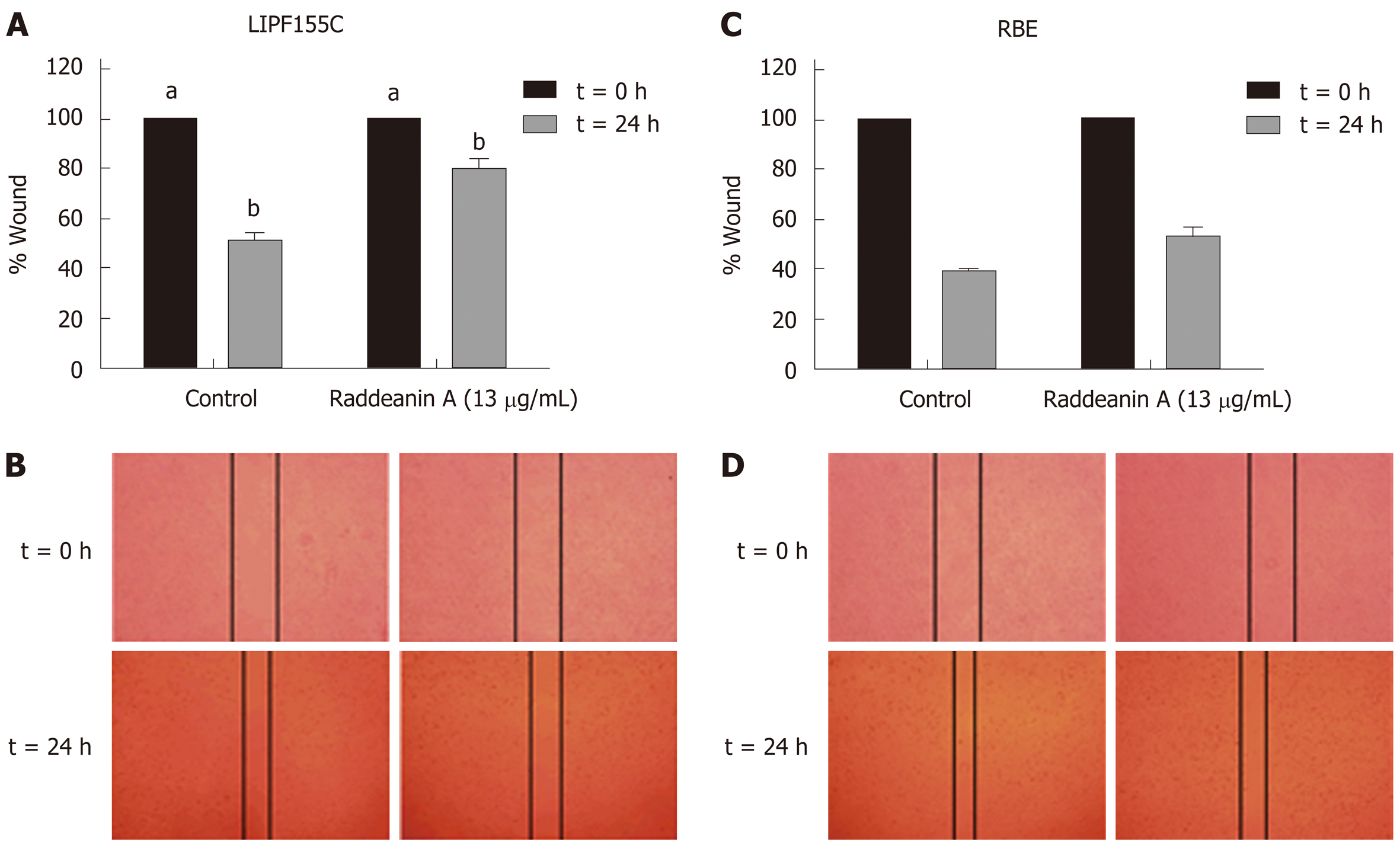
Figure 2 Wound healing assay of RBE and LIPF155C cell lines treated with raddeanin A (13 µg/mL) (n = 3 independent experiments).
A and C: The percentage of wound width normalized to the controls is shown and data are expressed as the mean ± standard deviation. Unique letters shared by the bars suggest significant differences between groups and P < 0.05 was regarded to be statistically significant; B and D: Representative histograms for the percentage of wound width.
Figure 3 Transwell migration and clonogenic assays of RBE and LIPF155C cell lines treated with raddeanin (13 µg/mL) (n = 3 independent experiments).
A: The percentage of migration cells normalized to the controls are shown and data are expressed as the mean ± standard deviation. Unique letters shared by the bars suggest significant differences between groups and P < 0.05 was regarded to be statistically significant; B: Representative histograms for cell colony formation.
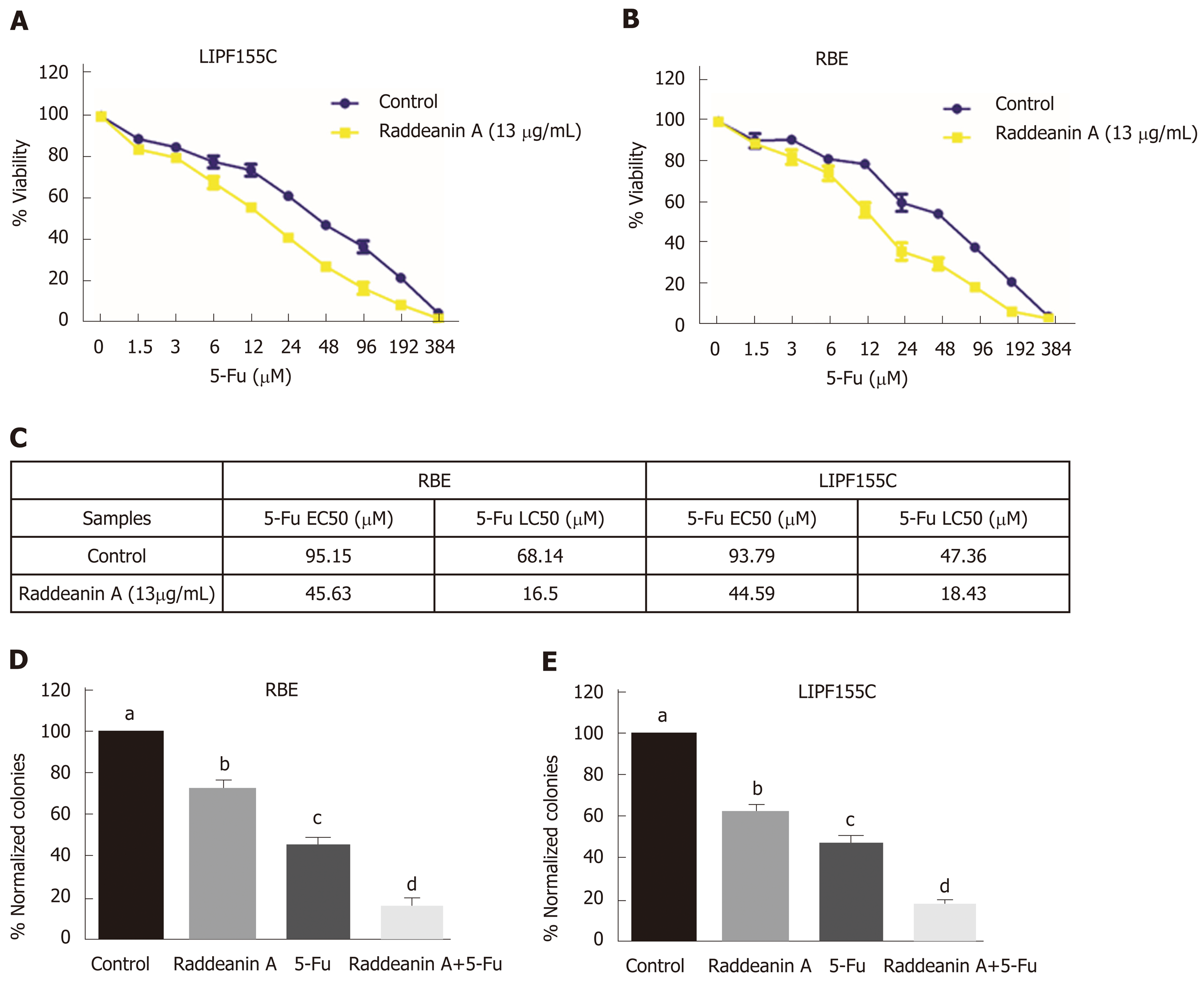
Figure 4 Effects of raddeanin A on 5-fluorouracil effectiveness in RBE and LIPF155C cell lines.
A and B: RBE and LIPF155C cell lines treated with either control or raddeanin A (13 μg/mL) in combination with increasing doses of 5-Fu; C: The half-maximal effective concentration (EC50) and half-maximal lethal concentration values for RBE and LIPF155C cell lines; D and E: The cell colony formation ability in RBE and LIPF155C cell lines. Unique letters shared by the bars suggest significant differences between groups and P < 0.05 was regarded to be statistically significant. 5-Fu: 5-fluorouracil.
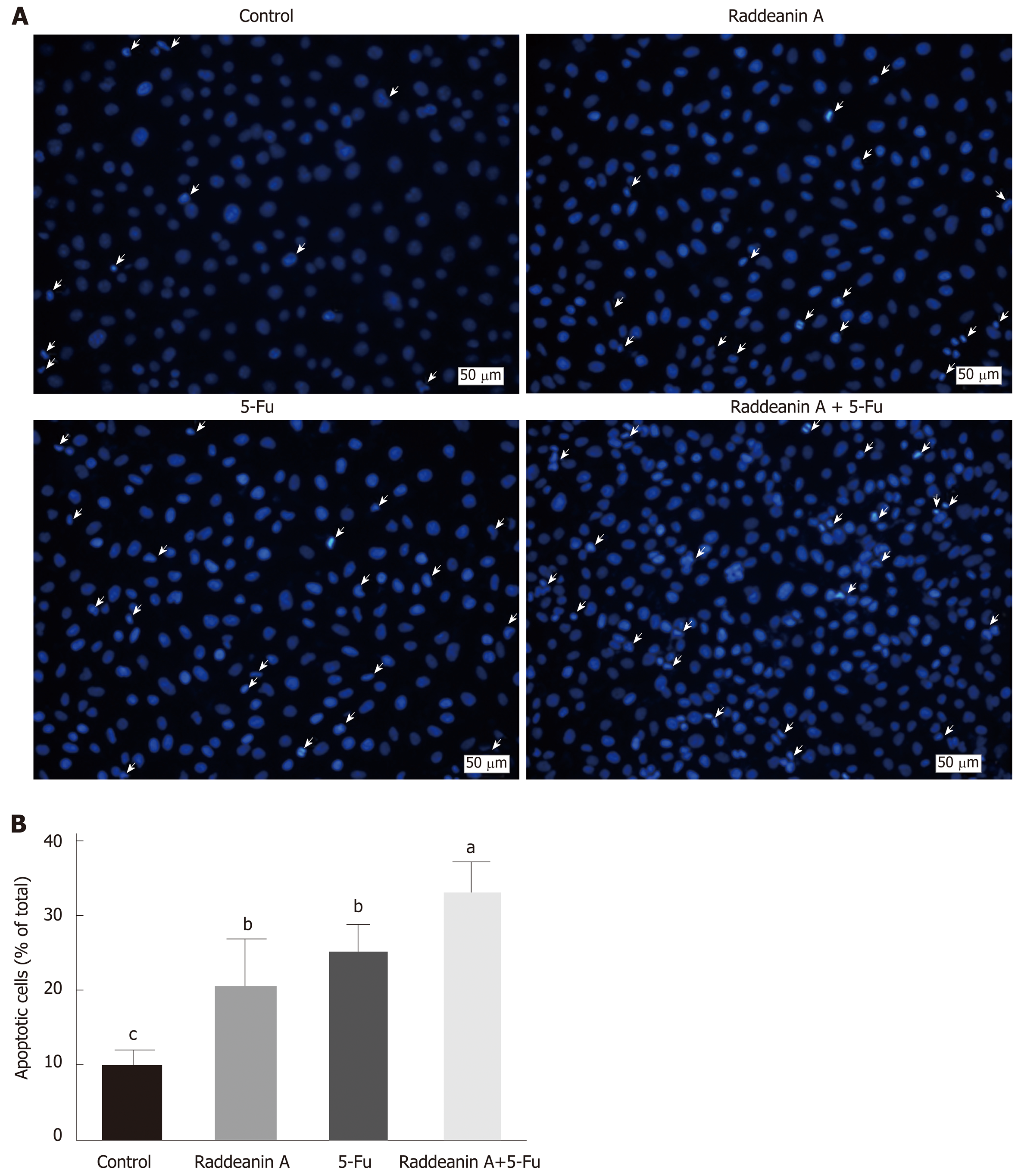
Figure 5 Effects of raddeanin A on 5-fluorouracil cell line RBE/5-Fu.
A: Representative graphs for apoptotic RBE/5-Fu cells treated with 13 μg/mL raddeanin A; B: The percentage of apoptotic cells is shown and data are expressed as the mean ± standard deviation. Unique letters shared by the bars suggest significant differences between groups and P < 0.05 was regarded to be statistically significant. 5-Fu: 5-fluorouracil.
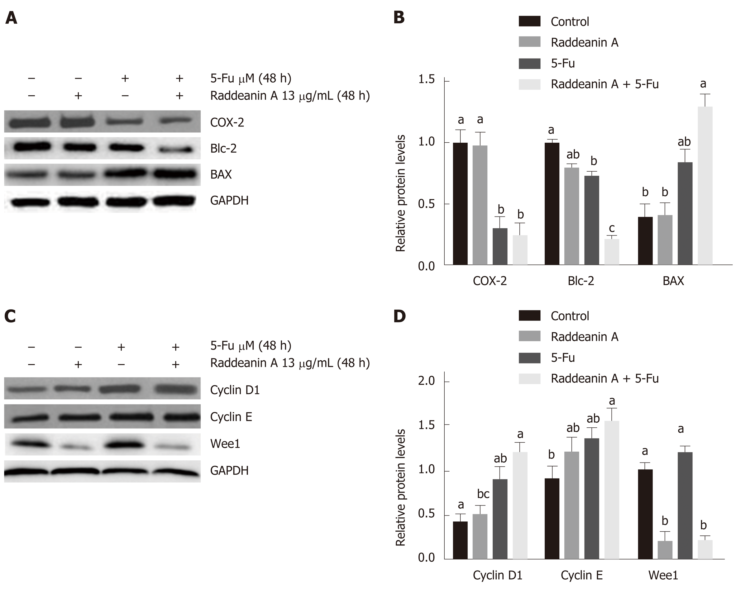
Figure 6 Effects of raddeanin A on cell cycle- and apoptosis-related protein expression in RBE cell line treated with 13 μg/mL raddeanin A for 48 h.
A and B: Apoptosis-related protein expression detected by Western blot; C and D: Cell cycle-related protein expression detected by Western blot. Data are expressed as the mean ± standard deviation. Unique letters shared by the bars suggest significant differences between groups and P < 0.05 was regarded to be statistically significant. RA: Raddeanin A; 5-Fu: 5-fluorouracil; COX-2: Cyclooxygenase-2.
- Citation: Guo SS, Wang Y, Fan QX. Raddeanin A promotes apoptosis and ameliorates 5-fluorouracil resistance in cholangiocarcinoma cells. World J Gastroenterol 2019; 25(26): 3380-3391
- URL: https://www.wjgnet.com/1007-9327/full/v25/i26/3380.htm
- DOI: https://dx.doi.org/10.3748/wjg.v25.i26.3380


