Copyright
©The Author(s) 2019.
World J Gastroenterol. Jul 7, 2019; 25(25): 3108-3115
Published online Jul 7, 2019. doi: 10.3748/wjg.v25.i25.3108
Published online Jul 7, 2019. doi: 10.3748/wjg.v25.i25.3108
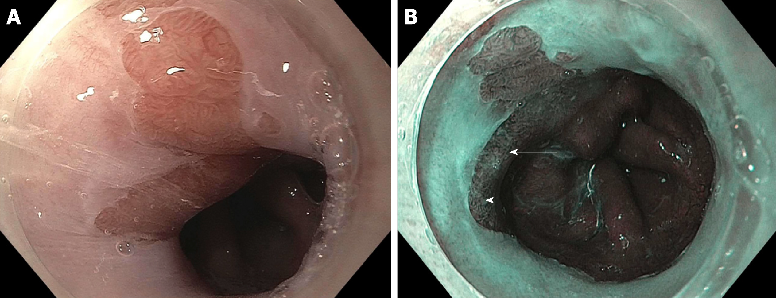
Figure 1 A patient with Barrett’s and high-grade dyspalsia with narrow band imaging.
A: A segment of Barrett’s esophagus on high definition white light endoscopy (HDWLE); B: narrow band imaging (NBI) from a patient with prior long segment disease post two sessions of endoscopic resection, 4 sessions of radiofrequency ablation, and one session of cryotherapy. The HDWLE did not show any features concerning for dysplasia. The NBI shows an area of disrupted vessels (upper white arrow, lower white arrow) concerning for dysplasia.
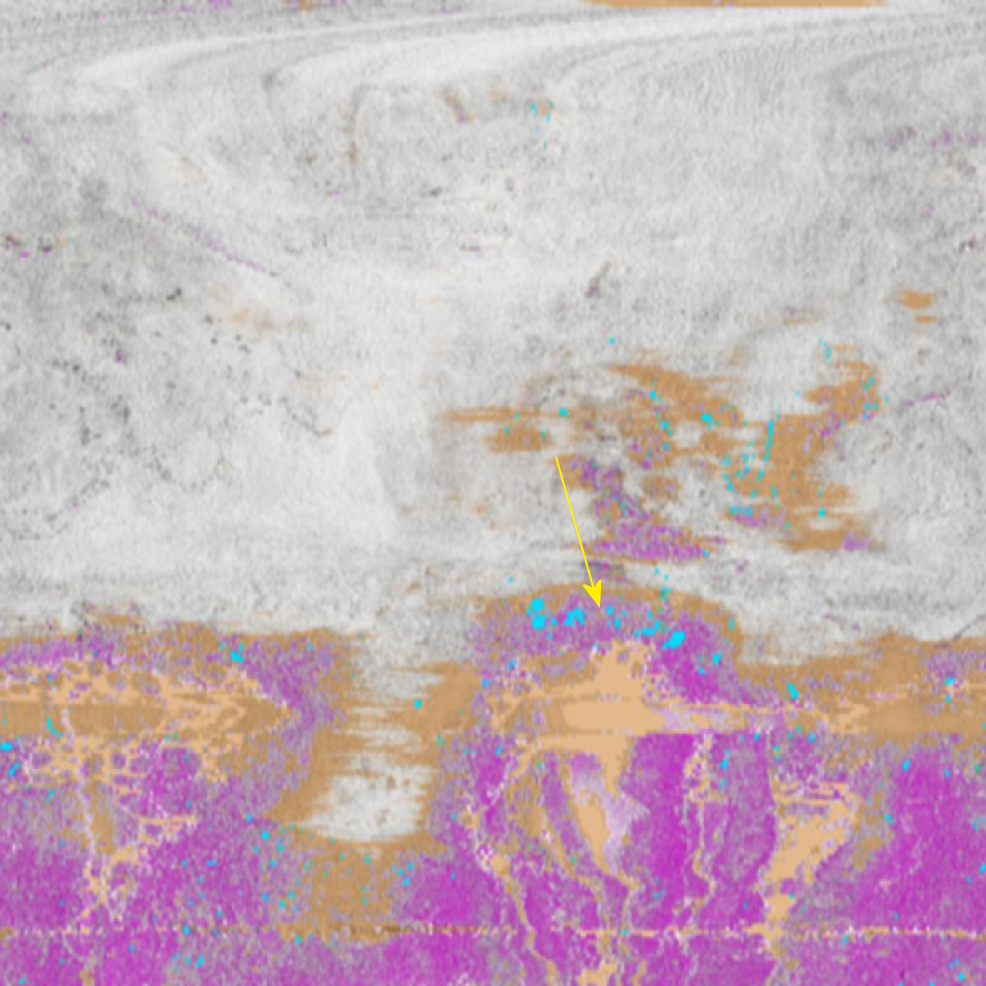
Figure 2 Volumetric laser endomicroscopy with artifical intelligence from the same patient as in Figure 1 with an en-face view showing an area of overlap (yellow arrow) between three features of dysplasia (orange is lack of layering, blue is glandular structures, and pink is a hyper-reflective surface).
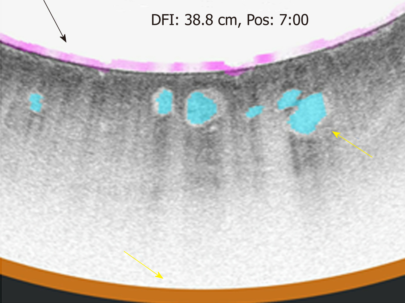
Figure 3 Volumetric laser endomicroscopy from the same patient showing cross-sectional view of the area of overlap (yellow arrow 5.
73” 5.41”) between three features of dysplasia (orange is lack of layering, blue is glandular structures, and pink is a hyper-reflective surface).
Figure 4 Volumetric laser endomicroscopy with artificial intelligence showing an up close snap shot of the abnormal area of overlap between three features of dysplasia (orange is lack of layering, blue is glandular structures, and pink is a hyper-reflective surface).
Figure 5 Endoscopy view of the targeted laser marks (upper yellow arrow, lower yellow arrow) placed using volumetric laser endomicroscopy.
This corresponds to the same area highlighted by narrow band imaging. The pathology showed high-grade dysplasia.
- Citation: Cerrone SA, Trindade AJ. Advanced imaging in surveillance of Barrett’s esophagus: Is the juice worth the squeeze? World J Gastroenterol 2019; 25(25): 3108-3115
- URL: https://www.wjgnet.com/1007-9327/full/v25/i25/3108.htm
- DOI: https://dx.doi.org/10.3748/wjg.v25.i25.3108


