Copyright
©The Author(s) 2019.
World J Gastroenterol. Jan 14, 2019; 25(2): 245-257
Published online Jan 14, 2019. doi: 10.3748/wjg.v25.i2.245
Published online Jan 14, 2019. doi: 10.3748/wjg.v25.i2.245
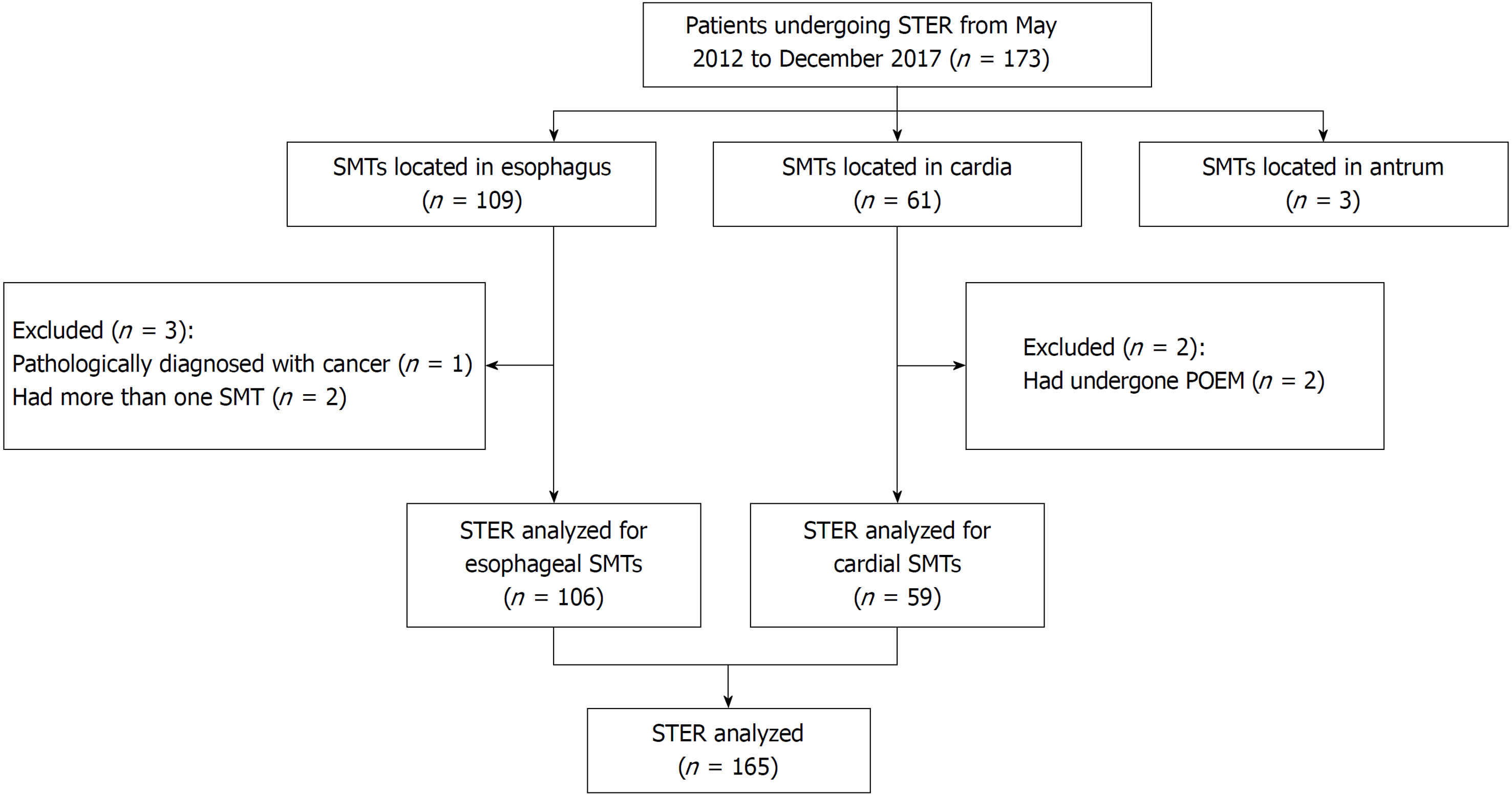
Figure 1 Study flowchart.
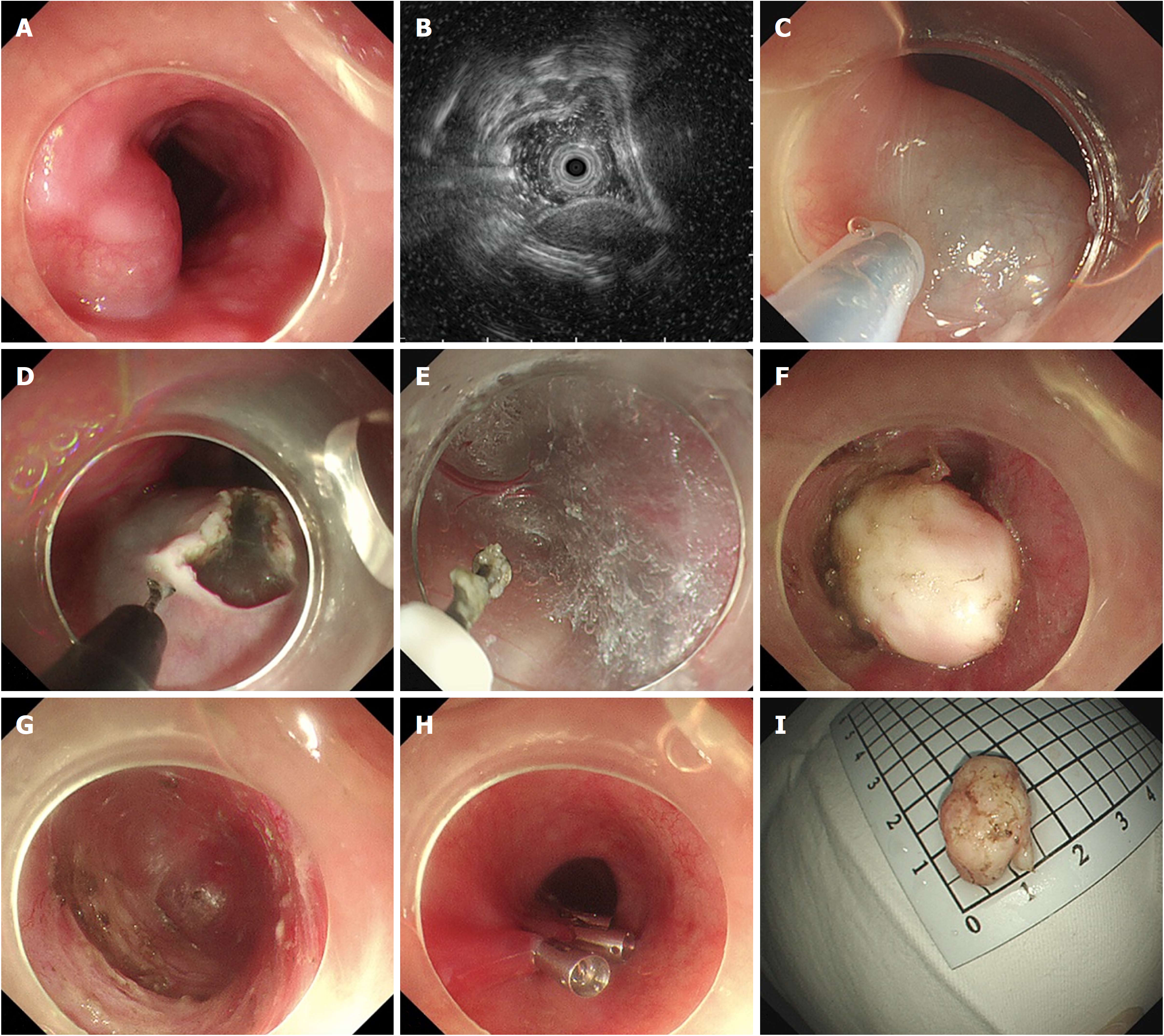
Figure 2 Submucosal tunneling endoscopic resection for a submucosal tumor originating from the muscularis propria layer in the esophagus.
A: Endoscopic view of a submucosal tumor (SMT) in the esophagus; B: Endoscopic ultrasound view of the same SMT, showing that lesion originates from the muscularis propria (MP) layer; C: Submucosal injection at 5 cm proximal to the SMT; D: An inverted T mucosal incision; E: Establishment of a submucosal tunnel between the mucosal and MP layers; F: Exposure of the SMT; G: The tunnel after the resection of the tumor; H: Closure of the mucosal incision site with clips; I: The resected specimen.
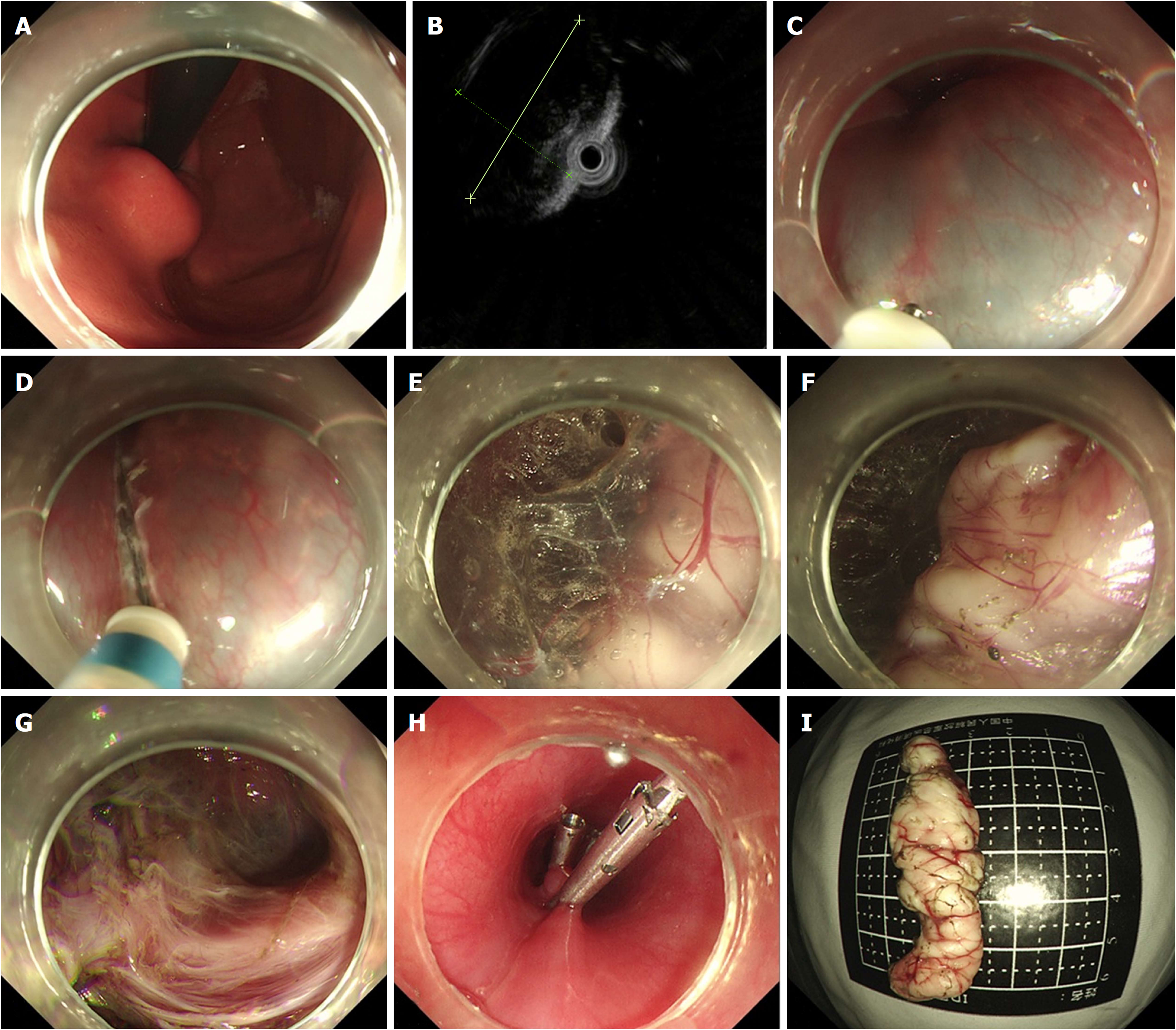
Figure 3 Submucosal tunneling endoscopic resection for a submucosal tumor originating from the muscularis propria layer in the cardia.
A: Endoscopic view of a submucosal tumor (SMT) in the cardia; B: Endoscopic ultrasound view of the same SMT; C: Submucosal injection at 5 cm proximal to the SMT; D: A longitudinal mucosal incision; E: Establishment of a submucosal tunnel between the mucosal and muscularis propria layers; F: Exposure of the SMT; G: The tunnel after the resection of the tumor; H: Closure of the mucosal incision site with clips; I: The resected specimen.
- Citation: Du C, Chai NL, Ling-Hu EQ, Li ZJ, Li LS, Zou JL, Jiang L, Lu ZS, Meng JY, Tang P. Submucosal tunneling endoscopic resection: An effective and safe therapy for upper gastrointestinal submucosal tumors originating from the muscularis propria layer. World J Gastroenterol 2019; 25(2): 245-257
- URL: https://www.wjgnet.com/1007-9327/full/v25/i2/245.htm
- DOI: https://dx.doi.org/10.3748/wjg.v25.i2.245


