Copyright
©The Author(s) 2018.
World J Gastroenterol. Sep 14, 2018; 24(34): 3958-3964
Published online Sep 14, 2018. doi: 10.3748/wjg.v24.i34.3958
Published online Sep 14, 2018. doi: 10.3748/wjg.v24.i34.3958
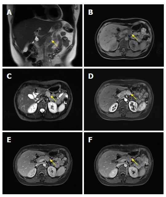
Figure 1 Magnetic resonance imaging of the lesion (yellow arrow) in Case 1.
A: Coronal T2-weighted image sequence; B: Transverse T1-weighted fat-suppression sequence; C: Transverse T2-weighted fat-suppression sequence; D: Transverse T1-weighted fat-suppression sequence (early arterial phase); E: Transverse T1-weighted fat-suppression sequence (late arterial phase); F: Transverse T1-weighted fat-suppression sequence (delayed phase).
Figure 2 Surgery of the retroperitoneal peripancreatic unicentric Castleman disease in Case 1.
A: The tumor was close to the lower side of the pancreas with a clear margin and a volume of 3 cm × 2 cm × 2 cm; B: Image of the free tumor; C: The tumor was completely excised.
Figure 3 Pathological examinations of the tumor in Case 1.
A: The tumor was 3 cm × 2 cm × 2 cm in size. It had a smooth surface, complete envelope, and was white to tan in appearance; B: Pathological section analysis of UCD (HE, × 100); C: Pathological section analysis of UCD (HE, × 200); D: Pathological section analysis of UCD (HE, × 400). HE: Hematoxylin and eosin; UCD: Unicentric Castleman disease.
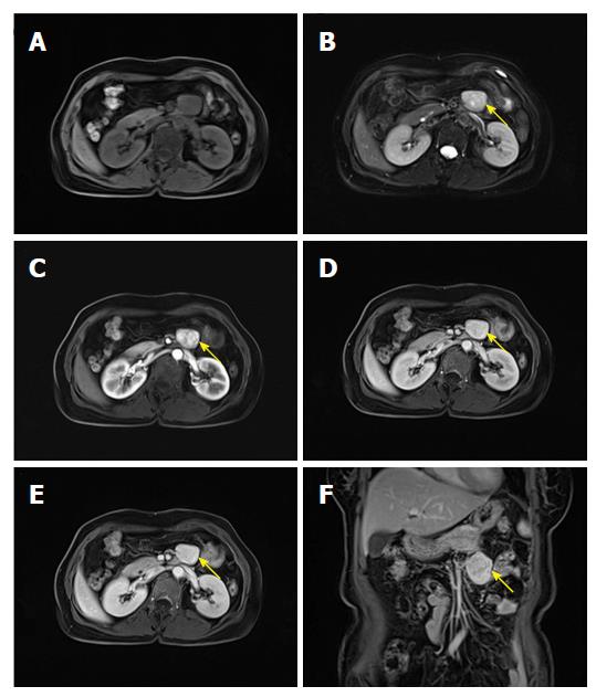
Figure 4 Magnetic resonance imaging of the lesion (yellow arrow) in Case 2.
A: Transverse T1-weighted fat-suppression sequence; B: Transverse T2-weighted fat-suppression sequence; C: Transverse T1-weighted fat-suppression sequence (arterial phase); D: Transverse T1-weighted fat-suppression sequence (venous phase); E: Transverse T1-weighted fat-suppression sequence (delayed phase); F: Coronal T1-weighted fat-suppression sequence (delayed phase).
Figure 5 Surgery of the retroperitoneal peripancreatic unicentric Castleman disease in Case 2.
A: The lesion was located below and behind the transverse mesocolon with a volume of 3.5 cm × 3 cm × 3 cm. Intraoperative ultrasonography indicated that the tumor had an abundant blood supply, and its right side was close to the inferior mesenteric artery and vein; B: Image of the free tumor; C: The tumor was completely excised.
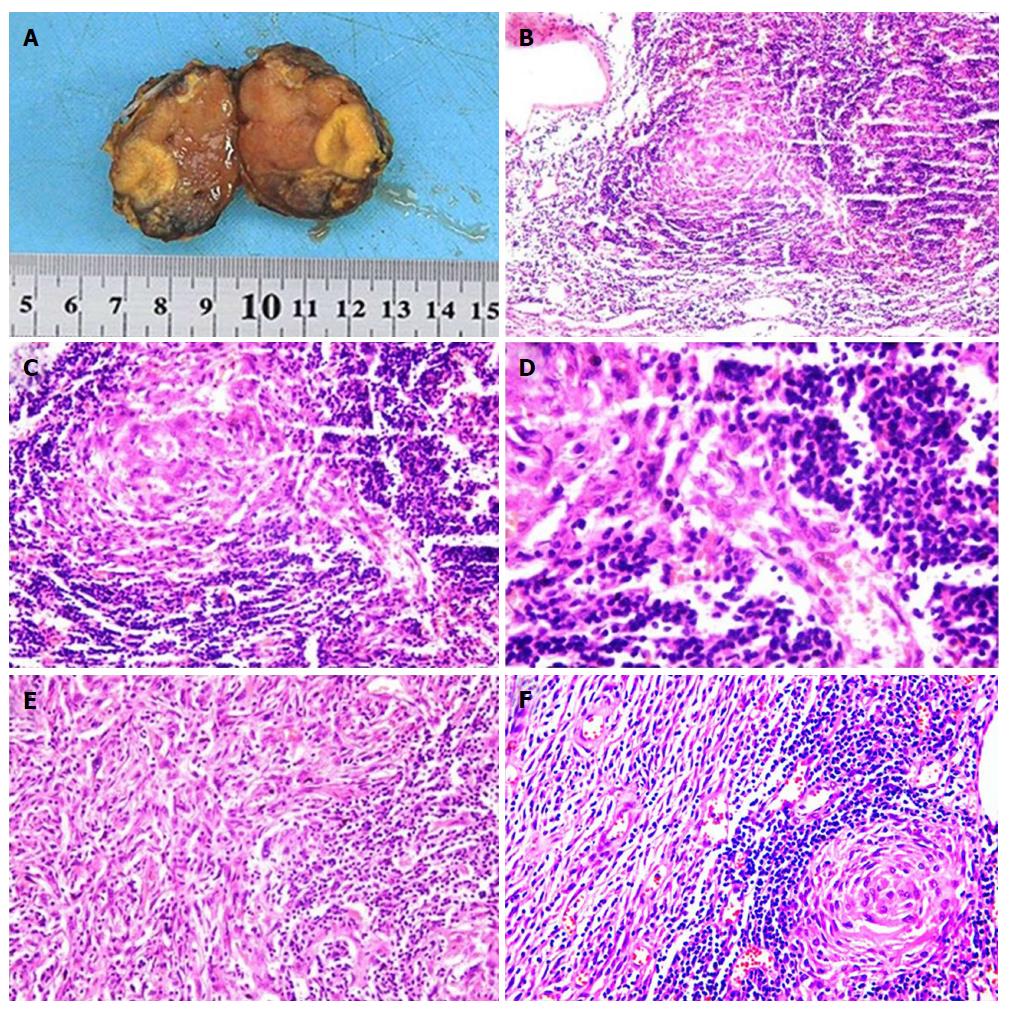
Figure 6 Pathological examinations of the tumor in Case 2.
A: The tumor was 3.5 cm × 3 cm × 3 cm in size. It had a smooth surface, complete envelope, and was white to tan in appearance; B: Pathological section analysis of UCD (HE, × 100); C: Pathological section analysis of UCD (HE, × 200); D: Pathological section analysis of UCD (HE, × 400); E: Pathological section analysis of FDCT (HE, × 200); F: Pathological section analysis of UCD together with FDCT (HE, × 200). HE: Hematoxylin and eosin; UCD: Unicentric Castleman disease; FDCT: Follicular dendritic cell tumor.
- Citation: Cheng JL, Cui J, Wang Y, Xu ZZ, Liu F, Liang SB, Tian H. Unicentric Castleman disease presenting as a retroperitoneal peripancreatic mass: A report of two cases and review of literature. World J Gastroenterol 2018; 24(34): 3958-3964
- URL: https://www.wjgnet.com/1007-9327/full/v24/i34/3958.htm
- DOI: https://dx.doi.org/10.3748/wjg.v24.i34.3958


