Copyright
©The Author(s) 2018.
World J Gastroenterol. Aug 7, 2018; 24(29): 3250-3259
Published online Aug 7, 2018. doi: 10.3748/wjg.v24.i29.3250
Published online Aug 7, 2018. doi: 10.3748/wjg.v24.i29.3250
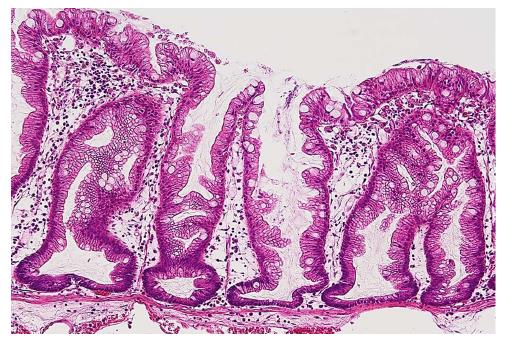
Figure 1 Typical histology of a sessile serrated adenoma/polyp.
Crypts with a serrated architecture include those that are irregularly dilated, branch irregularly, and are horizontally arranged (basal).
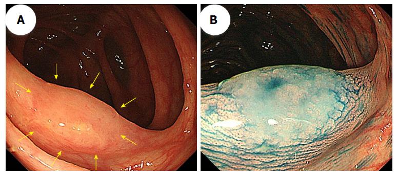
Figure 2 A sessile serrated adenoma/polyp in the transverse colon that measured 13 mm.
A: An image from conventional colonoscopy showing the lesion’s location (arrows); B: An image from chromoendoscopy following indigo carmine dye spraying.
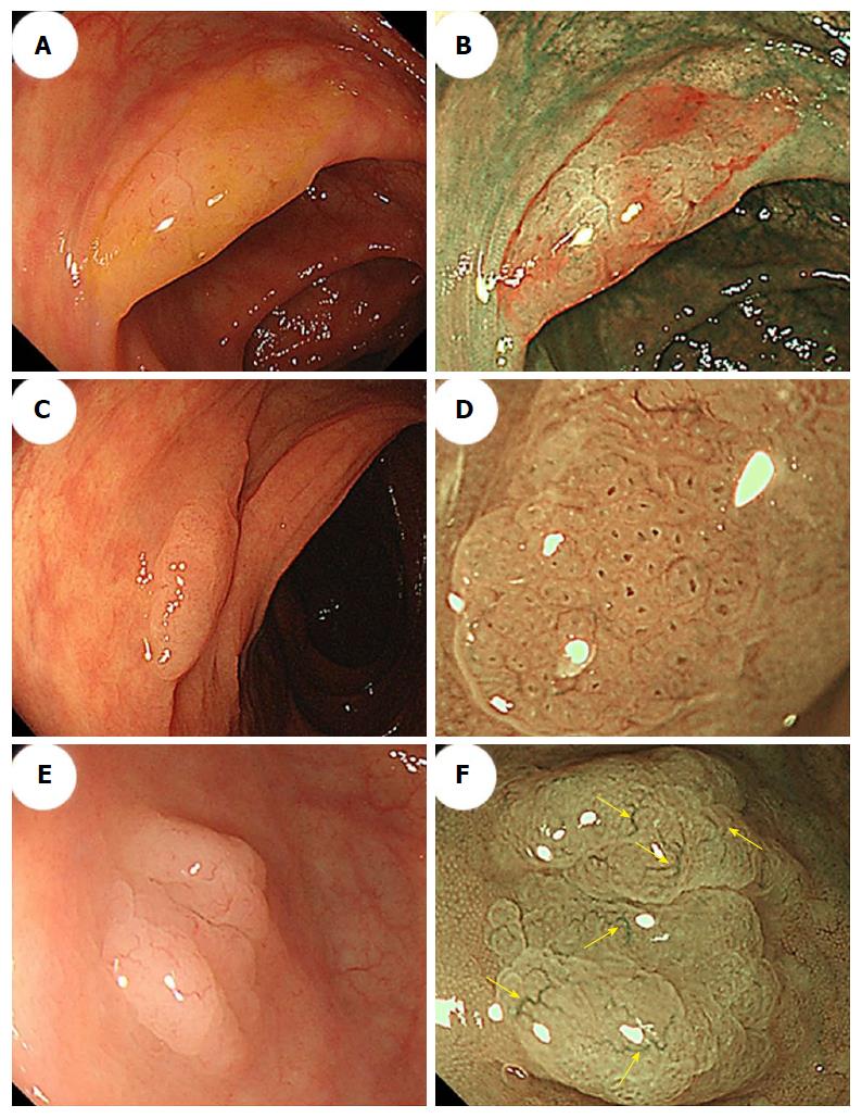
Figure 3 Morphologic characteristics of sessile serrated adenoma/polyps.
A: Conventional endoscopy revealed a flat-elevated lesion with a 20-mm diameter that was covered with a mucus cap in the transverse colon. B: Narrow-band imaging (NBI) showed that the SSA/P in (A) was covered with a mucus cap that appeared intensely red. C: Conventional endoscopy showed a flat-elevated lesion with a 14-mm diameter in the ascending colon. D: Magnifying NBI of the SSA/P in (C) revealed dark spots inside the crypts in part of the lesion. E: A conventional endoscopic image shows a flat-elevated pale colored lesion with a 10-mm diameter in the cecum. F: Magnifying NBI of the SSA/P in (E) revealed varicose microvascular vessels (arrows) in part of the lesion. SSA/P: Sessile serrated adenoma/polyp.
Figure 4 Conventional colonoscopic image (A) and a chromoendoscopic image (B) following indigo carmine dye spraying show an 18-mm sessile serrated adenoma/polyp with a mucus cap that was in the transverse colon (arrows).
C: Magnifying chromoendoscopy using crystal violet staining identified a type II-open pit pattern in the lesion.
Figure 5 Endoscopic images of a sessile serrated adenoma/polyp with high-grade cytologic dysplasia in a representative case.
A-C: A conventional endoscopic view using white-light imaging. A: An endoscopic image shows a pale-color, flat-elevated lesion covered with mucus at the ascending colon (arrows). B: The lesion is covered with mucus cap. C: After washing the target lesion to sufficiently remove mucus, a flat-elevated lesion that had a 13-mm diameter and a dome-shaped double elevation can be clearly seen. The dome-shaped area is slightly red-colored. D and E: Magnifying chromoendoscopic views using crystal violet staining. D: A type II-open pit pattern is partly evident in the edge of the lesion (arrows). E: Type VI-mild pit pattern consisting of areas with irregular pits can be observed at the dome-shaped area. We endoscopically diagnosed the lesion as an SSA/P with cytologic dysplasia, and achieved an en bloc resection by performing an endoscopic mucosal resection. F-H: Histopathologic findings with hematoxylin-eosin staining of the resected specimen. G: Crypts with a serrated architecture exhibit irregularly dilated crypts, irregularly branching crypts, and horizontally arranged basal crypts, corresponding to SSA/P. H: A high-power view shows conventional adenomatous high-grade dysplasia with cytological atypia and architectural dysplasia in the dome-shaped area. The lesion was pathologically consistent with an SSA/P with high-grade cytologic dysplasia. SSA/P: Sessile serrated adenoma/polyp.
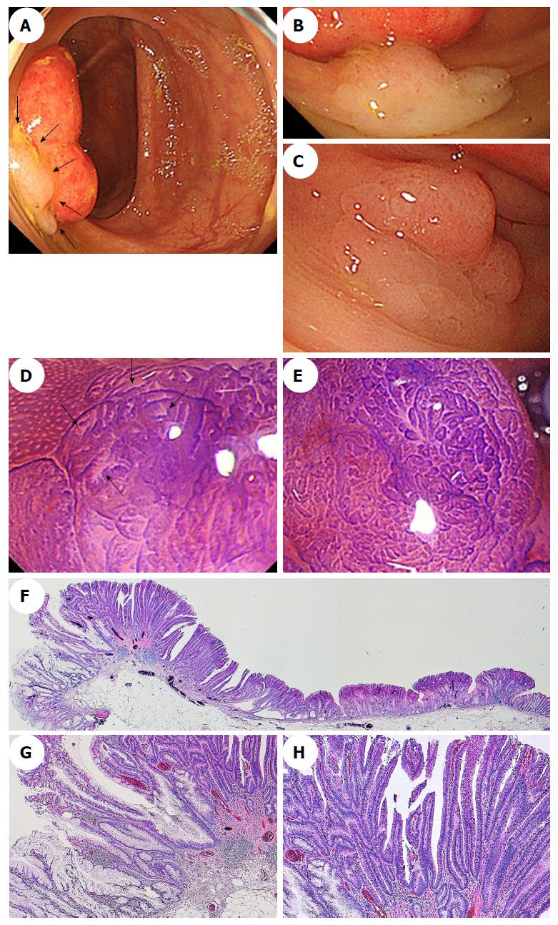
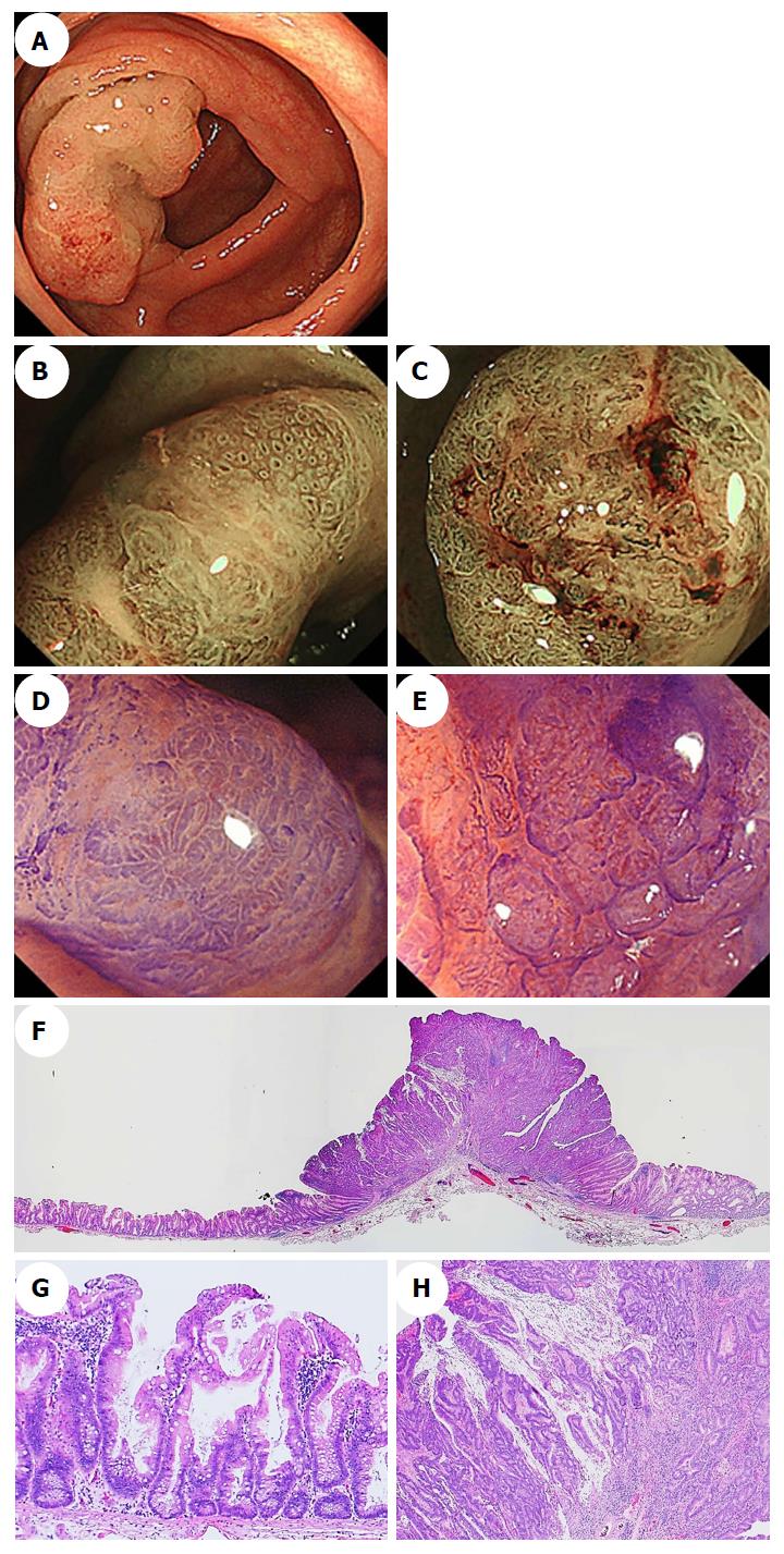
Figure 6 Endoscopic images of a sessile serrated adenoma/polyp with an invasive carcinoma in a representative case.
A: A conventional endoscopic image captured using white-light imaging shows a red 55-mm semipedunculated lesion in the ascending colon. B and C: Magnifying narrow-band imaging revealed dark spots inside the crypts on an edge of the lesion and irregular vessel patterns over a large part of the lesion, respectively. D and E: Magnifying chromoendoscopy using crystal violet staining; D: A high-powered view of the marginal zone, the dilated openings of the crypts have a type II-open pit pattern; E: A high-powered view of the middle region in which a type VI-severe pit pattern is evident. We endoscopically diagnosed the lesion as a carcinoma associated with an SSA/P, and achieved an en bloc resection by performing an endoscopic submucosal dissection. F-H: Histopathologic findings with hematoxylin-eosin staining of the resected specimen; G: Crypts with a serrated architecture exhibiting irregularly dilated crypts and irregularly branching crypts, corresponding to SSA/P; H: Well to moderately differentiated adenocarcinomas invade the submucosa with extracellular mucin production. The lesion was pathologically consistent with an invasive submucosal adenocarcinoma associated with an SSA/P. SSA/P: Sessile serrated adenoma/polyp.
- Citation: Murakami T, Sakamoto N, Nagahara A. Endoscopic diagnosis of sessile serrated adenoma/polyp with and without dysplasia/carcinoma. World J Gastroenterol 2018; 24(29): 3250-3259
- URL: https://www.wjgnet.com/1007-9327/full/v24/i29/3250.htm
- DOI: https://dx.doi.org/10.3748/wjg.v24.i29.3250


